Painful anal conditions jaber
•Als PPT, PDF herunterladen•
15 gefällt mir•9,354 views

This document discusses several painful anal conditions including anal fissures, proctalgia fugax, anorectal abscesses, perianal hematomas, complicated hemorrhoids, and anal cancer. It provides information on the typical causative microorganisms for anorectal abscesses, appropriate treatment options for various conditions which may include drainage, examination under anesthesia, antibiotics in some cases, and surgery. Sitz baths and conservative measures are recommended for treating some hemorrhoids and hematomas.

Empfohlen
Weitere ähnliche Inhalte
Was ist angesagt?
Was ist angesagt? (20)
Andere mochten auch
Andere mochten auch (17)
Ähnlich wie Painful anal conditions jaber
Ähnlich wie Painful anal conditions jaber (20)
Mehr von Jaber Manasia
Painful anal conditions jaber
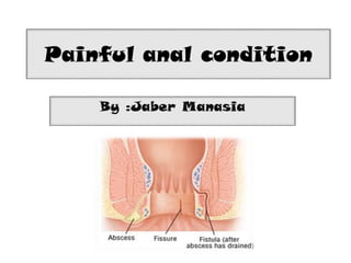
- 2. Painful anal conditions : Anal fissure Proctalgia fugax Anarectal abscess Perianal heamotoma Complicated hemorrhoids Anal cancer 2
- 6. Microrganism….?? • In 60% of cases , pus from the abscess give pure culture of E.coli • 23%...... Pure culture of staph. Aureus. • Others…… proteus , strept. Or mixed.
- 7. • fistula-in-ano (most common) • Crohn’s disesase • diabetes • immunosuppression
- 10. Anorectal abscesses should be treated by drainage as soon as the diagnosis is established . an examination under anesthesia is often the most expeditious way both to confirm the diagnosis and to treat the problem. Delayed or inadequate treatment may occasionally cause extensive and life threatening suppuration with massive tissue necrosis and septicemia
- 15. Antibiotics are only indicated if there is extensive overlying cellulitis or if the patient is immunocompromised Antibiotics alone are ineffective at treating perianal or perirectal infection.
- 16. Per i-anal Hematoma A perianal hematoma is a collection of blood under the surface of the skin at the edge of the anal opening. 1.History: Occurs at all ages. Male & female equally affected The patient often notices that the peri-anal skin is moist & itchy. It’s occasionally multiple & may be recurrent.
- 17. 2.Physical Examination • Position: – The lump may be anywhere around the anal margin – More than one may present • Color: – When the lump is close to the skin and the skin is not edematous deep red-purple color – If the skin becomes edematous the redness of the underlying blood clot can’t be seen • Tenderness: – The lump is tender due to tension – edema & ulceration of the skin ↑ tenderness
- 18. • Shape & Size: – The initial lump is spherical & up to 1 cm in diameter. – The lump becomes polypoid if the skin is lax or or becomes edematous. • Surface: – Covered by normal r edematous skin – Smooth surface • Composition: – Central lump can be felt as solid, hard, hemispherical mass.
- 19. managment • Acute phase: – Evacuate the hematoma through a small incision under LA • Discharging or absorbed hematoma hot pathes
- 20. Hemor rhoids • Painful hemorrhoids are: – 3rd degree hemorrhoid Hemorrhoids that prolapse but must be pushed back in by a finger. – Thrombosed hemorrhoid (containing blood clots) – Strangulated hemorrhoid – Ulcerated hemorrhoid
- 21. Physical Examination: Two or three tense, tender, red-purple mucosa covered swelling protruding from the anal canal Remember that major piles are at the 3,7 & 11 o’clock positions The piles that have been prolapsed & thrombosed for a long time increase possibility to be ulcerated& infected they should be differentiated from prolapsing carcinoma or other gross pathology
- 22. managment • Conservative • Avoid constipation & ensure bulky stool with increasing fiber content of the diet. • Topical preparations containing local anesthetic agents & steroids. • Thrombosed external hemorrhoids: • Bed rest. • Application of ice packs. • Oral analgesia + topical local anesthetic gel. • excision of the hemorrhoid or clot evacuation if the patient presents less than 48 hours after the onset of symptoms
- 23. Sitz Bath • Relaxes anal sphincter • Decreases inflammation w/ Epsom® salts • 10-15 minutes, 3-4 times/day
- 24. • Anal malignancy is rare and accounts for less than 2% of all large bowel cancers • Uncommon tumour, which is usually a squamous cell carcinoma • May affect the anal verge or anal canal • Associated with HPV • More prevalent in patients with HIV infection • Lymphatic spread is to the inguinal lymph nodes • Treatment is by chemoradiotherapy in the first instance • Major ablative surgery is required if the above fails
- 25. • mass, bleeding, pain, discharge, itching, and tenesmus. • more common in men
- 26. • Small, well-differentiated lesions ( < 3cm ) are treated by wide local excision. • Deep lesions that involve the sphincters require abdominoperineal resection
Hinweis der Redaktion
- Some estimate as hiegh as 90% >>>> gland in origion
- As an abscess enlarges, it spreads in one of several directions. A perianal abscess is the most common manifestation and appears as a painful swelling at the anal verge. Spread through the external sphincter below the level of the puborectalis produces an ischiorectal abscess . Intersphincteric abscesses occur in the intersphincteric space and are notoriously difficult to diagnose Pelvic and supralevator abscesses are uncommon and may result from extension of an intersphincteric or ischiorectal abscess upward, or extension of an intraperitoneal abscess downward
- The perianal space surrounds the anus and laterally becomes continuous with the fat of the buttocks. The intersphincteric space separates the internal and external anal sphincters. It is continuous with the perianal space distally and extends cephalad into the rectal wall. The ischiorectal space (ischiorectal fossa) is located lateral and posterior to the anus and is bounded medially by the external sphincter, laterally by the ischium, superiorly by the levator ani, and inferiorly by the transverse septum. The ischiorectal space contains the inferior rectal vessels and lymphatics. The two ischiorectal spaces connect posteriorly above the anococcygeal ligament but below the levator ani muscle, forming the deep postanal space. The supralevator spaces lie above the levator ani on either side of the rectum and communicate posteriorly. The anatomy of these spaces influences the location and spread of cryptoglandular infection
- As an abscess enlarges, it spreads in one of several directions. A perianal abscess is the most common manifestation and appears as a painful swelling at the anal verge. Spread through the external sphincter below the level of the puborectalis produces an ischiorectal abscess . Intersphincteric abscesses occur in the intersphincteric space and are notoriously difficult to diagnose Pelvic and supralevator abscesses are uncommon and may result from extension of an intersphincteric or ischiorectal abscess upward, or extension of an intraperitoneal abscess downward
- cruciate incision over the most fluctuant point, with excision of the skin edges to de-roof the abscess
- If the abscess is secondary to intra-abdominal disease, the primary process requires treatment and the abscess is drained via the most direct route (transabdominally, rectally, or through the ischiorectal fossa).
- Malignant lesions of the anus and anal canal