
Empfohlen
Weitere ähnliche Inhalte
Was ist angesagt?
Was ist angesagt? (20)
Ähnlich wie CHROMOSOMES
Ähnlich wie CHROMOSOMES (20)
Kürzlich hochgeladen
Kürzlich hochgeladen (20)
CHROMOSOMES
- 1. CHROMOSOME
- 2. Cell Division • The cell cycle, or cell-division cycle, is an ordered series of events that take place in a cell leading to cell growth and its division and duplication (replication into two daughter cells). • Cytokinesis is the physical division of the cytoplasm whereby the nuclei, cytoplasm, organelles and cell membrane of a single eukaryotic cell is divided into two daughter cells containing roughly equal shares. Types of Cell Division: Mitosis • Mitosis produces two daughter cells that are identical to the parent cell. • This type of cell division allows multicellular organisms to grow and repair damaged tissue. Meiosis • Meiosis (double cell division) produces daughter cells that have one half the numbers of chromosomes as the parent cell. • Meiosis is necessary in sexually-reproducing organisms because the fusion of two gametes (fertilization) doubles the number of chromosomes.
- 4. Chromosome (chroma - colour; some - body) • A chromosome is a thread-like self-replicating genetic structure containing organized DNA molecule package found in the nucleus of the cell. • Clear and detailed decriptions of mitotic chromosomes in plants and animals were published by Strasburger in 1875 and by the German scientist Walter Flemming in 1879-1882. • Heinrich Wilhelm Gottfried Waldeyer coined the term chromosome in 1888. Normally Chromosomes are of two types: – Autosomes - Control characters other than sex characters or carry genes for somatic characters. – Sex chromosomes (Synonym: Gonosomes) - Chromosomes involved in sex determination. • Humans and most other mammals have two sex chromosomes X & Y, also called heterosome, odd chromosome, or idiosome. • Females have two X chromosomes in diploid cells; males have an X and a Y chromosome. • In birds the female (ZW) is hetero-gametic and male (ZZ) is homo-gametic.
- 5. CHROMOSOME NUMBER Haploid, Diploid: • Diploid cells (2N where N- chromosome number) have two homologous copies of each chromosome. • The body cells of animals are diploid. • Haploid cells (N) have only one copy of each chromosome. • In animals, gametes (sperm and eggs) are haploid. Homologous chromosomes: • Diploid organisms have two copies of each chromosome (except the sex chromosomes). Both the copies are ordinarily identical in morphology, gene content and gene order and hence known as homologous chromosomes. • Each pair of chromosomes made up of two homologs. • Homologous chromosome is inherited from separate parents; one homolog comes from the mother and the other comes from the father. Chromosome Number: • The number of chromosomes in a given species is generally constant. • Chromosomes come in pairs. • Different organisms have different numbers of chromosomes. Normally all individual of a species have the same chromosome number
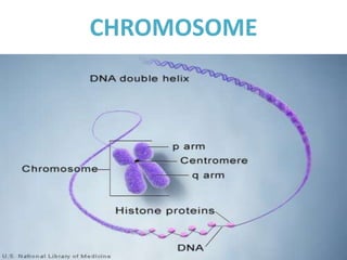
- 7. STRUCTURE OF CHROMOSOMEChromosome Morphology: • The chromosome morphology changes during cell division and mitotic metaphase is the most suitable stage for studies on chromosome morphology. • The DNA of eukaryotic cells is tightly bound to small basic proteins (histones) that package the DNA in an orderly way in the cell nucleus. • The complexes between eukaryotic DNA and proteins are called chromatin, which typically contains about twice as much protein as DNA. • The major proteins of chromatin are the histones - H1, H2A, H2B, H3, and H4 which are very similar among different species of eukaryotes. • The shape of the eukaryotic chromosomes is changeable from phase to phase in the continuous process of the cell growth and cell division. • Chromosomes are thin, coiled, elastic, thread-like structures during the interphase. • As cells enter mitosis, their chromosomes become highly condensed so that they can be distributed to daughter cells. • In mitotic metaphase chromosomes, the following structural features can be seen under the light microscope. Chromatid: • Each metaphase chromosome appears to be longitudinally divided into two identical parts each of which is called chromatid. Both the chromatids of a chromosome appear to be joined together at a point known as centromere. The two chromatids of chromosome separate from each other during mitotic anaphase (and during anaphase II of meiosis) and move towards opposite poles. • Since the two chromatids making up a chromosome are produced through replication of a single chromatid during synthesis (S) phase of interphase, they are referred to as sister chromatids. In contrast, the chromatids of homologous chromosomes are known as non-sister chromatids. Centromere (Primary constriction): • Each chromosome has a constriction point called the centromere (Synonym: Kinetochore), which divides the chromosome into two sections or arms. • The short arm of the chromosome is labeled the "p" arm. The long arm of the chromosome is labeled the "q" arm. Telomere: • The sequences at the ends of eukaryotic chromosomes, called telomeres, play critical roles in chromosome replication and maintenance. Secondary constriction: • In addition to centromere / primary constriction, one or more constrictions in the chromosome are present termed secondary constrictions. Satellite: small chromosomal segment separated from the main body of the chromosome by a secondary constriction is called Satellite.
- 8. CENTROMERE POSITIONS Size of the chromosome: • The size of the chromosome shows a remarkable variation depending upon the stage of cell division. The chromosomes are the longest and thinnest during interphase (resting stage) and hence not visible under light microscope. Chromosomes are the smallest and thickest during mitotic metaphase. • Chromosome size is not proportional to the number of genes present on the chromosome. • The location of the centromere on each chromosome gives the chromosome its characteristic shape. Centromere position: • Chromosomes are classified according to the centromere position is at one end (acrocentric), closer to one end than the other (submetacentric) or in the middle (metacentric). • Each chromosome has two arms, labeled p (the shorter of the two) and q (the longer). • The p arm is named for "petite" meaning 'small'; the q arm is named q simply because it follows p in the alphabet. (According to the NCBI, "q" refers to the French word "queue") Metacentric:: • The centromere is localized approximately midway between each end and thereby two arms are roughly equal in length. • Metacentric chromosome takes V shape during anaphase. Submetacentric: Centromere is submedian, giving one longer and one shorter arm. • Submetacentric chromosome may be J or L shaped during anaphase. Acrocentric: • The centromere is more terminally placed and forms very unequal arm length (The "acro-" in acrocentric refers to the Greek word for "peak"). • The p (short) arm is so short that is hard to observe, but still present. • Acrocentric chromosome may be rod shaped during anaphase. Telocentric: • Centromere lies at one end. • Telocentic chromosome may be rod shaped during anaphase. • According to the number of the centromere the eukaryotic chromosomes may be acentric (without any centromere), mono centric (with one centromere), dicentric (with two centromeres) or polycentric (with more than two centromeres).
- 9. KARYOTYPE AND IDEOGRAM Karyotype: • The general morphology (size of chromosomes, position of centromere, presence of secondary constriction and size of satellite bodies) of somatic chromosomal complement of an individual constitutes its karyotype. • In a karyotype, chromosomes are arranged and numbered by size, from largest to smallest. • The karyotype of a normal somatic cell of a normal individual represents the karyotype of the concerned species. • This arrangement helps scientists quickly identify chromosomal alterations that may result in a genetic disorder. • To make a karyotype, picture of someone's chromosomes taken, cut them out and match them up using size, banding pattern and centromere position as guides. • Avian karyotype is different from mammalian karyotype because of presence of very small autosomes called microchromosomes. Ideogram: • The karyotype of a species can be represented diagrammatically showing all the morphological features of chromosomes. Such a diagram is known as ideogram or ideotype.
- 10. SPECIAL TYPES OF CHROMOSOMES Polytene Chromosomes: • Polytene chromosomes are giant chromosomes common to many dipteran (two-winged) flies. • These were first discovered by E. G. Balbiani in 1882 in Dipteran salivary glands and hence commonly called salivary gland chromosomes. • They begin as normal chromosomes, but through repeated rounds of DNA replication without any cell division (called endoreplication), they become large, banded chromosomes (see figure). • For unknown reasons, the centromeric regions of the chromosomes do not endoreplicate very well. • As a result, the centromeres of all the chromosomes bundle together in a mass called the chromocenter. • Polytene chromosomes are usually found in the larvae, where it is believed these many-replicated chromosomes allow for much faster larval growth than if the cells remained diploid. • Simply because each cell now has many copies of each gene, it can transcribe at a much higher rate than with only two copies in diploid cells. • The polytene chromosomes at the right are from the salivary glands of the fruit fly Drosophila melanogaster. • The bands on each chromosome are like a road map, unique to each chromosome and well defined enough to allow high resolution mapping of each chromosome. • The Drosophila Genome Project uses polytene chromosomes as a framework for the map. Lampbrush chromosomes : • It was first observed by W. Flemming in 1882 and was described in detail in oocytes of sharks by Rukert in 1892. • It consists of an axis from which paired loops extend in opposite directions, giving the appearance of a lamp brush. Hence the name Lamp Brush Chromosomes. • It is found in the Oocytes of amphibians and in some insects. • They are formed during the active synthesis of mRNA molecules for the future use by the egg during cleavage when no synthesis of mRNA molecules is possible due to active involvement of chromosomes in the mitotic cell division. • It is larger in size. Hence it is called a giant chromosome. B-chromosomes : • B-Chromosomes (also called supernumerary chromosomes, accessory chromosomes, accessory fragments, etc.) are without obvious genetic function and usually have a normal structure, are somewhat smaller than the autosomes. Holokinetic chromosomes : • The chromosomes with a non-localized centromere are called as either holocentric or holokinetic chromosomes.