SlideShare ist ein Scribd-Unternehmen logo
1 von 75
Downloaden Sie, um offline zu lesen
NON-PCR BASED MOLECULAR
APPROACHES FOR DETECTION AND
IDENTIFICATION OF PLANT PATHOGENS
DOCTORAL SEMINAR II
PUJA PANDEY
41124
1
INTRODUCTION
Plant Pathology
Molecular Plant
Pathology
2
‱ Visual observations
‱ Cultural
‱ Microscopy
CONVENTIONAL
TECHNIQUES
3
VISUAL OBSERVATION :
SIGNS AND SYMPTOMS
Rot - Sclerotium rolfsii
Late blight of potato – Phytophthora infestans
Citrus canker
4
Problem with biotic and abiotic factors ??????????
5
CULTURAL : LABORATORY TECHNIQUES
Bacterial ooze
Cultures of Ralstonia solanacearum
6
Fungal cultures
..
Alternaria culture Fusarium culture
7
MICROSCOPY : An Aid To
The Eyes
Microscope
Stereoscopic
Microscope
Fluorescence
Microscope
Light
Microscope
Bright Field Dark Field
Phase
Contrast
Confocal
Electron
Microscope
Scanning
electron
Microscope
Transmission
electron
Microscope
8
Types of Microscope
1) Stereoscopic microscope
Function: Visible light to illuminate the surface
of a sample (2000x ), without disrupting them
2) Compound microscope (light)
Function: Visible light to illuminate a thin
section of sample, cells and tissues
3) Confocal laser scanning fluorescence
microscope
Function: Thin ‘slices’ in a sample while
keeping sample intact;
Specifically at parts of a cell (such as
individual proteins) by labelling them with
fluorescence
9
Scanning Electron Microscope
Function: Surface of objects at high resolution
(3D image - 500 000x )
Principle : Beam of electrons being knocked off
the surface of the sample and then picked up by a
detector .
Why we use SEM ???????????
The SEM probably gives the best depth of field
out of any microscope
10
Transmission Electron Microscope
Function: very thin cross-section of an
object (cell), internal structure of objects,
high resolution (500 000x )
Principle : Electrons pass through the
sample and some are deflected and some
pass right through, and that forms our
image, which is focussed on objective
lens
11
Microscopy Techniques Applied to the Study of Phytoplasma
Diseases: Traditional and Innovative Methods
Rita Musetti and Maria Augusta Favali
12
Light Microscopy
 Dienes’stain was first developed
as a specific stain for animal
mycoplasma colonies .
 Phloem tissues of stems infected
by phytoplasmas stained dark
blue while xylem was turquoise
and cortex light blue.
Musetti and Favali, 2004
13
Fluorescence Microscopy
DAPI staining of hand cut
sections of healthy
Antibody plus fluorochrome,
such as fluorescein
isothiocyanate (FITC) and
stained
 Phytoplasma-infected :
the fluorescent bright spots,
visible at phloem level
Musetti and Favali, 2004
14
Transmission electron microscopy (TEM)
Phytoplasmas in the phloem
cells of Catharanthus roseus
Musetti and Favali, 2004
15
Phytoplasmas in the phloem of
apple tissues
Immuno-electron microscopy (IEM) of thin sections
Phytoplasmas in phloem tissues of Catharanthus roseus L. embeded with primary
monoclonal antibody and gold Labelledsecondary antibody
Gold partcle (15 nm) few
particles are visible on
phytoplasma membrane
Gold partcle (5 nm) particles are
well distributed over the periphery
of the phytoplasmas
Musetti and Favali, 2004
16
High resolution autoradiography
Phytoplasmas in phloem cells of white clover (Trifolium repens L.), after 3
hours labelling with thymidine-3H. The silver grains were seen on the
dividing phytoplasmas
17
Staining Technique : For Histopathological Tests
‱ Staining is an auxiliary technique used in microscopy to
enhance contrast in the microscopic image
e.g. Crystal violet stains only Gram positive bacteria
18
19
Histopathological analysis of infected tissues
20
Valencia sweet
oranges infected
with
Colletotrichum
acutatum.
Valencia
sweet orange
fruits infected
with Guignardia
citricarpa.
Toluidine blue staining Toluidine blue plus safranin staining Marques et al., 2013
21
Limitations Faced due to conventional techniques !!!!!!!
 Latent infection : eg. Potato ring rot
 Misleading infection : eg. Black lesions (Alternaria) and bacterial
blight of carrot (Xanthomonas)
 Co-infection : Alteration of symptoms
SEROLOGICAL METHODS – De Vorac
Antigen Antibody
Antigen Antibody
Positive
Result 22
Antigen
 A molecule usually a protein, when it is injected into a
warm blooded animal produces antibody (immune
response)
Antibody
 A molecule produced in a warm
blooded serum of animal in
response to the stimulus antigens
 Antibodies are immune system-
related proteins called
immunoglobulin
23
 Variable region : composed of 110-
130 amino acids, give the antibody
its specificity for binding antigen.
 Variable region includes the ends of
the light and heavy chains.
 Constant region : determines the
mechanism used to destroy antigen.
Structure of Antibody
24
MONOCLONAL ANTIBODY
POLYCLONAL ANTIBODY
Composed of a variety of antibody
Have multiple epitopes
Antibody derived from a single
clone and specific for a single
epitope
Consist of single type of antibody
Produced by hybridoma technique
Small quantity of antigen is enough
for development
25
Production of Mabs by Hybridoma Technique
26
Antigen antibody based technique
Direct test
 Precipitation test
1. Tube precipitation
2. Ring precipitation
 Micro Precipitation test
 Agglutination tests
1. Chloroplast agglutination
2. Latex agglutination
 Gel diffusion test
 Immuno-electrophoresis
Indirect test
 ELISA test
1. Direct ELISA (DAS ELISA)
2. Indirect (DAC ELISA)
3. DIBA ELISA
4. Lateral flow device
 Immunofluorescence
 Immuno Sorbent Electron
Microsopy(ISEM)
 Flow cytometry
27
Tube precipitation test
Widely used
Reactants diluted in 8.5g/l
NaCl, followed by
incubation at 37°C in water
bath
Observations :
If elongated virus particle -
floccular
If spherical virus particle -
granular
Precipitation test
28
Done on a micro-scale to
economize on antiserum
Drop of dilution mixture (antiserum
& virus suspension) are mixed at
bottom of a Petri plate
The precipitates produced are
observed with a microscope with
dark-ground illumination
Precipitation varies depending on
the ratio of concentration of antigen
and antibody
Micro precipitition test
29
Latex agglutination
30
Chloroplast agglutination
Crude fresh
leaf sap from
diseased plant
Antiserum
Chloroplast
fragments
clump
together
31
 The reactants, antiserum and virus solution are placed in well cut
in the agar (containg 0.85% NaCl and 0.02% sodium azide) in
Petri plate
 Antibody and virus diffuse into the agar from the adjacent wells
 Where they meet precipitation zones in the form of white band
are formed
Gel diffusion test
Oservation :
a) Bands : Identical or closely related
b) Spurs : Distantly Related
c) Intersect : Unrelated 32
Radial or single diffusion
Double or Ouchterlony diffusion
33
Immuno-electrophoresis
34
ELISA : Enzyme- Linked Immuno-Sorbent Assay
ELISA was initially applied for plant viruses by M.F. Clark and
Adams (1976)
Sensitive, detects at concentration of 1-10 ng/ml
It involves an enzyme-mediated colour change reaction to detect
antibody binding
Degree of colour change, usually measured quantitatively in
spectrophotometer at 405 nm
Ward et al., 2003
35
DAS ELISA : Double antibody sandwich ELISA
 Direct ELISA
 First time describe by Clark and Adams in 1977
p-nitrophenyl
phosphate
p-nitrophenol Ward et al., 2003
36
DAC ELISA : direct antigen coating ELISA
Indirect ELISA
Stand for Easy to rapid assay
Ward et al., 2003 37
Combination of electron microscopy
and serology
First time described by Derrick in1973
Virus and antiserum are reacted
together
Antigen are trapped onto grid coated
with specific antiserum, negatively
stained (Uranyl acetate -1%) and the
result viewed in the EM
Immuno Sorbernt electron microscope (ISEM)
Tubular particle of beet
necrotic yellow vein virus
38
Dot Immunoblotting Assay (DIBA) OR Dot ELISA
Substrate : Nitro tetrazolium
BCIP
39
Lateral flow technique
‱ The principles used for rapid lateral flow devices are primarily
those of ELISA
‱ Various types of filters are used as the solid support for the
initial binding reaction
A lateral flow device test kit developed by Central Science
Laboratory, U.K., permits detection of R. solanacearum in a 3-
minute
40
41
Ouchterlonie’s double diffusion test.
I – Healthy cane
extract
II – Control blood
serum of rabbit
III, IV, V – Antigen
of host pathogen
A – Antibody
raised against host
pathogen
Lingayya and Naik, 2002
42
Detection of Colletotrichum falcatum infection in sugarcane
tissue by DAC - ELISA
Lingayya and Naik, 2002 43
TDA = 3 X standard deviation of
healthy sample + mean value for
healthy sample
44
FUNGUS
Advantage of ELISA
It is sensitive
Semiautomatic technique
Application against large number of sample
Reproducible
Qualitative & Quantitative
Suitable for automation high speed
No radiation hazards
45
Immunofluorescence
The intercellular location &
distribution of viruses
Globulins mixed with a
fluorescent dye (Fluorescein
isothiocyanate and Rhodamine
B)
Introduced into the infected
cells/tissue, with antigen and
antibody reaction, fluorescence
takes place
46
Flow cytometry
Cell suspensions are filtered to remove large particles then
stained with fluorochrome-labelled antibodies
Fluorescent markers for viability
Stains, such as propidium and
hexidium iodide for red fluorescent
staining of dead cells
Carboxy fluorescein diacetate and calcein
AM for green fluorescent staining of
viable cells can be used to differentiate
live from dead cells
Detection of C. Michiganensis subsp. Michiganensis in tomato seed extracts
Detection of X. Axonopodis pv. Dieffenbachiae, causal agent of anthurium blight (Alvarez
et.al., 1999)
Determine viability of R. Solanacearum in seed potatoes (van derwolf et.al., 2004)
47
HYBRIDIZATION BASED METHODS
48
49
Southern blotting
‱ Professor Sir Edwin Southern
developed this method in 1975
‱ This method Involves separation,
transfer and hybridization
‱ Detection of a specific DNA
sequence in DNA samples
‱ Combines agarose gel
electrophoresis for size
separation of DNA and
hybridization with probe
Professor Sir Edwin Southern
50
51
Northern Blotting
 Northern blotting is a technique for detection of specific RNA
sequences
 Northern blotting was developed by James Alwine and George
Stark at Stanford University (1979)
 Electrophoresed RNA is blotted on membrane and hybridized
52
53
Western blotting
‱ Western blotting (1981) is an immunoblotting technique which
rely on the specificity of binding between a protein of interest
and a probe (antibody raised against that particular protein) to
allow detection of the protein of interest in a mixture of many
other similar molecules.
‱ The SDS (Sodium dodecyl sulphate) page technique is a
prerequisite for western blotting .
54
Steps in western blotting
Detected
through
auto-
radiography
55
DNA Microarray
56
Microarrays for Rapid Identification of Plant
Viruses
Neil Boonham, Jenny Tomlinson,and Rick Mumford
Central Science Laboratory, Sand Hutton, York, YO41 1LZ,
United Kingdom
57
Boonham et al., 2007
A schematic diagram detailing a simple approach to virus
detection using a microarray.
58
A microarray designed to detect and
discriminate a range of small spherical
viruses. Eg. Broad bean wilt virus 2
Indicator host Chenopodium quinoa A small spherical virus was identified
using electron microscopy.
Boonham et al., 200759
Positive control spots
Detection of virus
Sensitivity comparison between ELISA and microarray
Boonham et al., 2003
Dilution
end
point
Histogram showing local
background fluorescence for the
1/1600 dilution of RNA,
BIOCHEMICAL METHODS
‱ FAME analysis
‱ BIOLOG
‱ Volatile compound
Biochemical
techniques
61
What is a BIOLOG?
 First and only bacterial identification system to identify both gram positive
and gram negative bacteria with a single universal test kit.
Add cells
96 wells contains different carbon sources and other
test. If the cells are metabolically active, they reduce
the redox dye and a purple colour is formed in al the
positive well
62
Fatty Acid Methyl Ester ( FAME ) analysis
 Change in the fatty acid profile represent a change in the
microbial population
63
Detection of Diseased Plants by Analysis of Volatile
Organic Compound Emission
R.M.C. Jansen, J. Wildt, I.F. Kappers,
H.J. Bouwmeester, J.W. Hofstee,
and E.J. van Henten
64
Emission of volatile organic compounds (VOCs)
from non-infected and Botrytis cinerea–infected
tomato plants.
Jansen et al., 2011 65
Damaged cell membranes
Local emission of several
lipoxygenase (LOX)
oxidative cleavage of C18fatty
acids
(oxygen and lipoxygenases )
Characterize diseases due to
release of VOCs
Surface Plasmon Resonance (SPR)
 The Surface Plasmon Resonance
(SPR) sensor is used for label free
detection and real-time monitoring
 Their simplicity and sensitivity make
biosensors an effective means of
disease diagnosis and monitoring
 In SPR technique, shift of the
resonance angle is observed, when
the intended species is captured by
the immobilized antibody on the
sensor surface
66
Is it the specific protein, the virus fragment or the virion itself?
Development of Surface Plasmon Resonance (SPR) Based Immuno-
Sensing System for Detection of Fungal Teliospores of Karnal Bunt
(Tilletia Indica), a Quarantined Disease of Wheat
Sadhna Singh1, Manoj Singh1, Gohar Taj1, Sanjay Gupta2 and Anil Kumar1*
1Department of Molecular Biology & Genetic Engineering, College of Basic Sciences
& Humanities, G. B. Pant University of Agriculture & Technology, Pantnagar,
Uttaranchal, India
2Department of Biotechnology, SBS PG Institute of Biomedical Sciences, Balawala,
Dehradun, Uttarakhand, India
Journal of Biosensors &
Bioelectronics
67
Experiment conducted : Interaction of teliosporic wall
antigen with the anti-teliosporic antibody immobilized on sensor
chip
 The interaction of antigen at a concentration of 80, 40, 20, 10, 5.0, 2.5,
1.25, 0.625, 0.312, 0.156, .078 and 0.039 ng/ÎŒl with immobilized antibody
on sensor chip was examined
Observation
‱ The responses increased in proportion to the concentration of teliosporic
antigen due to the change of the refractive index near the SPR sensor chip
68
SPR sensor response after the interaction of different concentrations of antigen over
the immobilized antibody (1: 500)
Singh et al., 2012
(a) 0.312
ng/ÎŒl
(b)1.25 ng/ÎŒl
(c) 5 ng/ÎŒl
(d) 20 ng/ÎŒl
(e) 80 ng/ÎŒl
Advantages of SPR
Major advantages :
‱ Rapid, real-time
‱ Non-labeling analysis
‱ Miniaturization for portable application
70
71
Phytophthora and Pythium Test Kits : 0.5 per cent of a
plant roots are infected
Tests for Phytophthora, Pythium, and Rhizoctonia root and crown decay
fungi can be performed on-site by growers in about 10 minutes
(A) Collect and grind samples using abrasive pads
(B) Fold pads and insert them into the extraction solution
(C) Apply solutions to detector
(D) Examine detector dots for color change.
72
Molecular methods for detection of plant
pathogens—What is the future?
 Strategies are needed on how to exploit deduced genomics and
proteomics supported by in silico analysis for establishing
rational disease control measures.
 The reliability of each specific on-the-spot diagnostic method
needs to be validated before results are used exclusively to
implement costly disease control strategies and/or regulatory
actions.
73
74
75

Weitere Àhnliche Inhalte

Ähnlich wie detectionofplantpathogensusingnon-pcrbasedtechniques-170112144243.pdf

Advances in Microbiological diagnosis of Periodontal diseases
Advances in Microbiological diagnosis of Periodontal diseasesAdvances in Microbiological diagnosis of Periodontal diseases
Advances in Microbiological diagnosis of Periodontal diseasesDr Fariya Ashraf
 
single chain fragment variable antibody(ScFv)
single chain fragment variable antibody(ScFv) single chain fragment variable antibody(ScFv)
single chain fragment variable antibody(ScFv) sivasankar. P
 
Advanced Microbiological Diagnostics.pptx
Advanced Microbiological Diagnostics.pptxAdvanced Microbiological Diagnostics.pptx
Advanced Microbiological Diagnostics.pptxGururam MDS
 
Uncultivable bacteria and recent trends towards their identification
Uncultivable bacteria and recent trends towards their identificationUncultivable bacteria and recent trends towards their identification
Uncultivable bacteria and recent trends towards their identificationabhishek yadav
 
Immunofluorescence
ImmunofluorescenceImmunofluorescence
ImmunofluorescenceFarhan ali
 
Viruses. Methods of Indication & Identification. Diagnosis of Viral diseases
Viruses. Methods of Indication & Identification. Diagnosis of Viral diseasesViruses. Methods of Indication & Identification. Diagnosis of Viral diseases
Viruses. Methods of Indication & Identification. Diagnosis of Viral diseasesEneutron
 
Microbiology lecture presentation-1.pptx
Microbiology lecture presentation-1.pptxMicrobiology lecture presentation-1.pptx
Microbiology lecture presentation-1.pptxkitati1
 
Microbiology
MicrobiologyMicrobiology
MicrobiologySaif Jawad
 
ajcmi-02-03-29691 edited by SC
ajcmi-02-03-29691 edited by SCajcmi-02-03-29691 edited by SC
ajcmi-02-03-29691 edited by SCKOBRA SALIMIYAN rizi
 
Microbiological Culture sensitivity tests
Microbiological Culture sensitivity testsMicrobiological Culture sensitivity tests
Microbiological Culture sensitivity testsDr Vimali
 
Serological tests with labelled antibody theoretical questions
Serological tests with labelled antibody theoretical questionsSerological tests with labelled antibody theoretical questions
Serological tests with labelled antibody theoretical questionsEneutron
 
Isolation of mrsa isolates
Isolation of mrsa isolatesIsolation of mrsa isolates
Isolation of mrsa isolatesOshin Raj
 
6 laboratory diagnosis of bacterial infection
6 laboratory diagnosis  of bacterial infection6 laboratory diagnosis  of bacterial infection
6 laboratory diagnosis of bacterial infectionPrabesh Raj Jamkatel
 
The Sensitivity Of 99mTc-Ciprofloxacin (Infecton) Scintigraphy Imaging To Det...
The Sensitivity Of 99mTc-Ciprofloxacin (Infecton) Scintigraphy Imaging To Det...The Sensitivity Of 99mTc-Ciprofloxacin (Infecton) Scintigraphy Imaging To Det...
The Sensitivity Of 99mTc-Ciprofloxacin (Infecton) Scintigraphy Imaging To Det...iosrphr_editor
 
Modern Techniques for Detection of Seed Born Fungi.
Modern Techniques for Detection of Seed Born Fungi.Modern Techniques for Detection of Seed Born Fungi.
Modern Techniques for Detection of Seed Born Fungi.ParanjayRohiwala
 
Microbiological tests of periodontal significance
Microbiological tests of periodontal significanceMicrobiological tests of periodontal significance
Microbiological tests of periodontal significanceMehul Shinde
 
nanosensor is rapid. Sensitive. Detecting multiple bacterial pathogens simul...
nanosensor is rapid.  Sensitive. Detecting multiple bacterial pathogens simul...nanosensor is rapid.  Sensitive. Detecting multiple bacterial pathogens simul...
nanosensor is rapid. Sensitive. Detecting multiple bacterial pathogens simul...M. WAQAS AKRAM
 

Ähnlich wie detectionofplantpathogensusingnon-pcrbasedtechniques-170112144243.pdf (20)

Microscope
MicroscopeMicroscope
Microscope
 
Advances in Microbiological diagnosis of Periodontal diseases
Advances in Microbiological diagnosis of Periodontal diseasesAdvances in Microbiological diagnosis of Periodontal diseases
Advances in Microbiological diagnosis of Periodontal diseases
 
Tb diagnosis
Tb diagnosisTb diagnosis
Tb diagnosis
 
single chain fragment variable antibody(ScFv)
single chain fragment variable antibody(ScFv) single chain fragment variable antibody(ScFv)
single chain fragment variable antibody(ScFv)
 
Advanced Microbiological Diagnostics.pptx
Advanced Microbiological Diagnostics.pptxAdvanced Microbiological Diagnostics.pptx
Advanced Microbiological Diagnostics.pptx
 
Uncultivable bacteria and recent trends towards their identification
Uncultivable bacteria and recent trends towards their identificationUncultivable bacteria and recent trends towards their identification
Uncultivable bacteria and recent trends towards their identification
 
Immunofluorescence
ImmunofluorescenceImmunofluorescence
Immunofluorescence
 
Bacillus anthracis
Bacillus anthracisBacillus anthracis
Bacillus anthracis
 
Viruses. Methods of Indication & Identification. Diagnosis of Viral diseases
Viruses. Methods of Indication & Identification. Diagnosis of Viral diseasesViruses. Methods of Indication & Identification. Diagnosis of Viral diseases
Viruses. Methods of Indication & Identification. Diagnosis of Viral diseases
 
Microbiology lecture presentation-1.pptx
Microbiology lecture presentation-1.pptxMicrobiology lecture presentation-1.pptx
Microbiology lecture presentation-1.pptx
 
Microbiology
MicrobiologyMicrobiology
Microbiology
 
ajcmi-02-03-29691 edited by SC
ajcmi-02-03-29691 edited by SCajcmi-02-03-29691 edited by SC
ajcmi-02-03-29691 edited by SC
 
Microbiological Culture sensitivity tests
Microbiological Culture sensitivity testsMicrobiological Culture sensitivity tests
Microbiological Culture sensitivity tests
 
Serological tests with labelled antibody theoretical questions
Serological tests with labelled antibody theoretical questionsSerological tests with labelled antibody theoretical questions
Serological tests with labelled antibody theoretical questions
 
Isolation of mrsa isolates
Isolation of mrsa isolatesIsolation of mrsa isolates
Isolation of mrsa isolates
 
6 laboratory diagnosis of bacterial infection
6 laboratory diagnosis  of bacterial infection6 laboratory diagnosis  of bacterial infection
6 laboratory diagnosis of bacterial infection
 
The Sensitivity Of 99mTc-Ciprofloxacin (Infecton) Scintigraphy Imaging To Det...
The Sensitivity Of 99mTc-Ciprofloxacin (Infecton) Scintigraphy Imaging To Det...The Sensitivity Of 99mTc-Ciprofloxacin (Infecton) Scintigraphy Imaging To Det...
The Sensitivity Of 99mTc-Ciprofloxacin (Infecton) Scintigraphy Imaging To Det...
 
Modern Techniques for Detection of Seed Born Fungi.
Modern Techniques for Detection of Seed Born Fungi.Modern Techniques for Detection of Seed Born Fungi.
Modern Techniques for Detection of Seed Born Fungi.
 
Microbiological tests of periodontal significance
Microbiological tests of periodontal significanceMicrobiological tests of periodontal significance
Microbiological tests of periodontal significance
 
nanosensor is rapid. Sensitive. Detecting multiple bacterial pathogens simul...
nanosensor is rapid.  Sensitive. Detecting multiple bacterial pathogens simul...nanosensor is rapid.  Sensitive. Detecting multiple bacterial pathogens simul...
nanosensor is rapid. Sensitive. Detecting multiple bacterial pathogens simul...
 

Mehr von DawitGetahun6

Keystone_Biology_Review_Tree_of_Life.ppt
Keystone_Biology_Review_Tree_of_Life.pptKeystone_Biology_Review_Tree_of_Life.ppt
Keystone_Biology_Review_Tree_of_Life.pptDawitGetahun6
 
FFA Judging Plant pathology study guide.ppt
FFA Judging Plant pathology study guide.pptFFA Judging Plant pathology study guide.ppt
FFA Judging Plant pathology study guide.pptDawitGetahun6
 
General_Properties_of_fungi fidrawa.pptx
General_Properties_of_fungi fidrawa.pptxGeneral_Properties_of_fungi fidrawa.pptx
General_Properties_of_fungi fidrawa.pptxDawitGetahun6
 
plant pathology and plant disease 1516297.ppt
plant pathology and plant disease 1516297.pptplant pathology and plant disease 1516297.ppt
plant pathology and plant disease 1516297.pptDawitGetahun6
 
Lecture 3- Bacterial Nutrition and Growth-.ppt
Lecture 3- Bacterial Nutrition and Growth-.pptLecture 3- Bacterial Nutrition and Growth-.ppt
Lecture 3- Bacterial Nutrition and Growth-.pptDawitGetahun6
 
microbial cycling p3ANutrientCycling.ppt
microbial cycling p3ANutrientCycling.pptmicrobial cycling p3ANutrientCycling.ppt
microbial cycling p3ANutrientCycling.pptDawitGetahun6
 
Rhizobacteria_and_their_Application.pptx
Rhizobacteria_and_their_Application.pptxRhizobacteria_and_their_Application.pptx
Rhizobacteria_and_their_Application.pptxDawitGetahun6
 
NUTRIENT CYCLING (biogeochemical_cycles).ppt
NUTRIENT CYCLING (biogeochemical_cycles).pptNUTRIENT CYCLING (biogeochemical_cycles).ppt
NUTRIENT CYCLING (biogeochemical_cycles).pptDawitGetahun6
 
culturing microorganisims chp 3 ppt.ppt
culturing microorganisims  chp 3 ppt.pptculturing microorganisims  chp 3 ppt.ppt
culturing microorganisims chp 3 ppt.pptDawitGetahun6
 
metabolsim of microorganism 1664107090.ppt
metabolsim of microorganism 1664107090.pptmetabolsim of microorganism 1664107090.ppt
metabolsim of microorganism 1664107090.pptDawitGetahun6
 
agri microbiology for Micro Hort PPT.pptx
agri microbiology for Micro Hort PPT.pptxagri microbiology for Micro Hort PPT.pptx
agri microbiology for Micro Hort PPT.pptxDawitGetahun6
 
method of studing microbial 13114527.ppt
method of studing microbial 13114527.pptmethod of studing microbial 13114527.ppt
method of studing microbial 13114527.pptDawitGetahun6
 
growth of microorganism chp 7-8 ppt.ppt
growth of microorganism  chp 7-8 ppt.pptgrowth of microorganism  chp 7-8 ppt.ppt
growth of microorganism chp 7-8 ppt.pptDawitGetahun6
 
history of microorganisim 14624202.ppt
history of microorganisim   14624202.ppthistory of microorganisim   14624202.ppt
history of microorganisim 14624202.pptDawitGetahun6
 
in the soil micriobial bioremediation2.ppt
in the soil micriobial bioremediation2.pptin the soil micriobial bioremediation2.ppt
in the soil micriobial bioremediation2.pptDawitGetahun6
 
soil and plant growth 12423241.ppt
soil and plant growth       12423241.pptsoil and plant growth       12423241.ppt
soil and plant growth 12423241.pptDawitGetahun6
 
Lecture 2- on Cell Structures.ppt biology
Lecture 2-  on Cell Structures.ppt biologyLecture 2-  on Cell Structures.ppt biology
Lecture 2- on Cell Structures.ppt biologyDawitGetahun6
 
Basic Plant Pathology in-2011-Rockland.pdf
Basic Plant Pathology in-2011-Rockland.pdfBasic Plant Pathology in-2011-Rockland.pdf
Basic Plant Pathology in-2011-Rockland.pdfDawitGetahun6
 
KOUSIK_GHOSHEVOLUTION-FUNGI2020-04-05EVOLUTION-FUNGI.ppt
KOUSIK_GHOSHEVOLUTION-FUNGI2020-04-05EVOLUTION-FUNGI.pptKOUSIK_GHOSHEVOLUTION-FUNGI2020-04-05EVOLUTION-FUNGI.ppt
KOUSIK_GHOSHEVOLUTION-FUNGI2020-04-05EVOLUTION-FUNGI.pptDawitGetahun6
 
what is plant pathology and plant disease .ppt
what is plant pathology and plant disease .pptwhat is plant pathology and plant disease .ppt
what is plant pathology and plant disease .pptDawitGetahun6
 

Mehr von DawitGetahun6 (20)

Keystone_Biology_Review_Tree_of_Life.ppt
Keystone_Biology_Review_Tree_of_Life.pptKeystone_Biology_Review_Tree_of_Life.ppt
Keystone_Biology_Review_Tree_of_Life.ppt
 
FFA Judging Plant pathology study guide.ppt
FFA Judging Plant pathology study guide.pptFFA Judging Plant pathology study guide.ppt
FFA Judging Plant pathology study guide.ppt
 
General_Properties_of_fungi fidrawa.pptx
General_Properties_of_fungi fidrawa.pptxGeneral_Properties_of_fungi fidrawa.pptx
General_Properties_of_fungi fidrawa.pptx
 
plant pathology and plant disease 1516297.ppt
plant pathology and plant disease 1516297.pptplant pathology and plant disease 1516297.ppt
plant pathology and plant disease 1516297.ppt
 
Lecture 3- Bacterial Nutrition and Growth-.ppt
Lecture 3- Bacterial Nutrition and Growth-.pptLecture 3- Bacterial Nutrition and Growth-.ppt
Lecture 3- Bacterial Nutrition and Growth-.ppt
 
microbial cycling p3ANutrientCycling.ppt
microbial cycling p3ANutrientCycling.pptmicrobial cycling p3ANutrientCycling.ppt
microbial cycling p3ANutrientCycling.ppt
 
Rhizobacteria_and_their_Application.pptx
Rhizobacteria_and_their_Application.pptxRhizobacteria_and_their_Application.pptx
Rhizobacteria_and_their_Application.pptx
 
NUTRIENT CYCLING (biogeochemical_cycles).ppt
NUTRIENT CYCLING (biogeochemical_cycles).pptNUTRIENT CYCLING (biogeochemical_cycles).ppt
NUTRIENT CYCLING (biogeochemical_cycles).ppt
 
culturing microorganisims chp 3 ppt.ppt
culturing microorganisims  chp 3 ppt.pptculturing microorganisims  chp 3 ppt.ppt
culturing microorganisims chp 3 ppt.ppt
 
metabolsim of microorganism 1664107090.ppt
metabolsim of microorganism 1664107090.pptmetabolsim of microorganism 1664107090.ppt
metabolsim of microorganism 1664107090.ppt
 
agri microbiology for Micro Hort PPT.pptx
agri microbiology for Micro Hort PPT.pptxagri microbiology for Micro Hort PPT.pptx
agri microbiology for Micro Hort PPT.pptx
 
method of studing microbial 13114527.ppt
method of studing microbial 13114527.pptmethod of studing microbial 13114527.ppt
method of studing microbial 13114527.ppt
 
growth of microorganism chp 7-8 ppt.ppt
growth of microorganism  chp 7-8 ppt.pptgrowth of microorganism  chp 7-8 ppt.ppt
growth of microorganism chp 7-8 ppt.ppt
 
history of microorganisim 14624202.ppt
history of microorganisim   14624202.ppthistory of microorganisim   14624202.ppt
history of microorganisim 14624202.ppt
 
in the soil micriobial bioremediation2.ppt
in the soil micriobial bioremediation2.pptin the soil micriobial bioremediation2.ppt
in the soil micriobial bioremediation2.ppt
 
soil and plant growth 12423241.ppt
soil and plant growth       12423241.pptsoil and plant growth       12423241.ppt
soil and plant growth 12423241.ppt
 
Lecture 2- on Cell Structures.ppt biology
Lecture 2-  on Cell Structures.ppt biologyLecture 2-  on Cell Structures.ppt biology
Lecture 2- on Cell Structures.ppt biology
 
Basic Plant Pathology in-2011-Rockland.pdf
Basic Plant Pathology in-2011-Rockland.pdfBasic Plant Pathology in-2011-Rockland.pdf
Basic Plant Pathology in-2011-Rockland.pdf
 
KOUSIK_GHOSHEVOLUTION-FUNGI2020-04-05EVOLUTION-FUNGI.ppt
KOUSIK_GHOSHEVOLUTION-FUNGI2020-04-05EVOLUTION-FUNGI.pptKOUSIK_GHOSHEVOLUTION-FUNGI2020-04-05EVOLUTION-FUNGI.ppt
KOUSIK_GHOSHEVOLUTION-FUNGI2020-04-05EVOLUTION-FUNGI.ppt
 
what is plant pathology and plant disease .ppt
what is plant pathology and plant disease .pptwhat is plant pathology and plant disease .ppt
what is plant pathology and plant disease .ppt
 

KĂŒrzlich hochgeladen

Q4 English4 Week3 PPT Melcnmg-based.pptx
Q4 English4 Week3 PPT Melcnmg-based.pptxQ4 English4 Week3 PPT Melcnmg-based.pptx
Q4 English4 Week3 PPT Melcnmg-based.pptxnelietumpap1
 
Earth Day Presentation wow hello nice great
Earth Day Presentation wow hello nice greatEarth Day Presentation wow hello nice great
Earth Day Presentation wow hello nice greatYousafMalik24
 
MULTIDISCIPLINRY NATURE OF THE ENVIRONMENTAL STUDIES.pptx
MULTIDISCIPLINRY NATURE OF THE ENVIRONMENTAL STUDIES.pptxMULTIDISCIPLINRY NATURE OF THE ENVIRONMENTAL STUDIES.pptx
MULTIDISCIPLINRY NATURE OF THE ENVIRONMENTAL STUDIES.pptxAnupkumar Sharma
 
Field Attribute Index Feature in Odoo 17
Field Attribute Index Feature in Odoo 17Field Attribute Index Feature in Odoo 17
Field Attribute Index Feature in Odoo 17Celine George
 
How to do quick user assign in kanban in Odoo 17 ERP
How to do quick user assign in kanban in Odoo 17 ERPHow to do quick user assign in kanban in Odoo 17 ERP
How to do quick user assign in kanban in Odoo 17 ERPCeline George
 
ECONOMIC CONTEXT - PAPER 1 Q3: NEWSPAPERS.pptx
ECONOMIC CONTEXT - PAPER 1 Q3: NEWSPAPERS.pptxECONOMIC CONTEXT - PAPER 1 Q3: NEWSPAPERS.pptx
ECONOMIC CONTEXT - PAPER 1 Q3: NEWSPAPERS.pptxiammrhaywood
 
THEORIES OF ORGANIZATION-PUBLIC ADMINISTRATION
THEORIES OF ORGANIZATION-PUBLIC ADMINISTRATIONTHEORIES OF ORGANIZATION-PUBLIC ADMINISTRATION
THEORIES OF ORGANIZATION-PUBLIC ADMINISTRATIONHumphrey A Beña
 
HỌC TỐT TIáșŸNG ANH 11 THEO CHÆŻÆ NG TRÌNH GLOBAL SUCCESS ĐÁP ÁN CHI TIáșŸT - Cáșą NĂ...
HỌC TỐT TIáșŸNG ANH 11 THEO CHÆŻÆ NG TRÌNH GLOBAL SUCCESS ĐÁP ÁN CHI TIáșŸT - Cáșą NĂ...HỌC TỐT TIáșŸNG ANH 11 THEO CHÆŻÆ NG TRÌNH GLOBAL SUCCESS ĐÁP ÁN CHI TIáșŸT - Cáșą NĂ...
HỌC TỐT TIáșŸNG ANH 11 THEO CHÆŻÆ NG TRÌNH GLOBAL SUCCESS ĐÁP ÁN CHI TIáșŸT - Cáșą NĂ...Nguyen Thanh Tu Collection
 
ECONOMIC CONTEXT - LONG FORM TV DRAMA - PPT
ECONOMIC CONTEXT - LONG FORM TV DRAMA - PPTECONOMIC CONTEXT - LONG FORM TV DRAMA - PPT
ECONOMIC CONTEXT - LONG FORM TV DRAMA - PPTiammrhaywood
 
Computed Fields and api Depends in the Odoo 17
Computed Fields and api Depends in the Odoo 17Computed Fields and api Depends in the Odoo 17
Computed Fields and api Depends in the Odoo 17Celine George
 
call girls in Kamla Market (DELHI) 🔝 >àŒ’9953330565🔝 genuine Escort Service đŸ”âœ”ïžâœ”ïž
call girls in Kamla Market (DELHI) 🔝 >àŒ’9953330565🔝 genuine Escort Service đŸ”âœ”ïžâœ”ïžcall girls in Kamla Market (DELHI) 🔝 >àŒ’9953330565🔝 genuine Escort Service đŸ”âœ”ïžâœ”ïž
call girls in Kamla Market (DELHI) 🔝 >àŒ’9953330565🔝 genuine Escort Service đŸ”âœ”ïžâœ”ïž9953056974 Low Rate Call Girls In Saket, Delhi NCR
 
GRADE 4 - SUMMATIVE TEST QUARTER 4 ALL SUBJECTS
GRADE 4 - SUMMATIVE TEST QUARTER 4 ALL SUBJECTSGRADE 4 - SUMMATIVE TEST QUARTER 4 ALL SUBJECTS
GRADE 4 - SUMMATIVE TEST QUARTER 4 ALL SUBJECTSJoshuaGantuangco2
 
How to Add Barcode on PDF Report in Odoo 17
How to Add Barcode on PDF Report in Odoo 17How to Add Barcode on PDF Report in Odoo 17
How to Add Barcode on PDF Report in Odoo 17Celine George
 
Gas measurement O2,Co2,& ph) 04/2024.pptx
Gas measurement O2,Co2,& ph) 04/2024.pptxGas measurement O2,Co2,& ph) 04/2024.pptx
Gas measurement O2,Co2,& ph) 04/2024.pptxDr.Ibrahim Hassaan
 
Karra SKD Conference Presentation Revised.pptx
Karra SKD Conference Presentation Revised.pptxKarra SKD Conference Presentation Revised.pptx
Karra SKD Conference Presentation Revised.pptxAshokKarra1
 
DATA STRUCTURE AND ALGORITHM for beginners
DATA STRUCTURE AND ALGORITHM for beginnersDATA STRUCTURE AND ALGORITHM for beginners
DATA STRUCTURE AND ALGORITHM for beginnersSabitha Banu
 
Like-prefer-love -hate+verb+ing & silent letters & citizenship text.pdf
Like-prefer-love -hate+verb+ing & silent letters & citizenship text.pdfLike-prefer-love -hate+verb+ing & silent letters & citizenship text.pdf
Like-prefer-love -hate+verb+ing & silent letters & citizenship text.pdfMr Bounab Samir
 
ACC 2024 Chronicles. Cardiology. Exam.pdf
ACC 2024 Chronicles. Cardiology. Exam.pdfACC 2024 Chronicles. Cardiology. Exam.pdf
ACC 2024 Chronicles. Cardiology. Exam.pdfSpandanaRallapalli
 

KĂŒrzlich hochgeladen (20)

FINALS_OF_LEFT_ON_C'N_EL_DORADO_2024.pptx
FINALS_OF_LEFT_ON_C'N_EL_DORADO_2024.pptxFINALS_OF_LEFT_ON_C'N_EL_DORADO_2024.pptx
FINALS_OF_LEFT_ON_C'N_EL_DORADO_2024.pptx
 
Q4 English4 Week3 PPT Melcnmg-based.pptx
Q4 English4 Week3 PPT Melcnmg-based.pptxQ4 English4 Week3 PPT Melcnmg-based.pptx
Q4 English4 Week3 PPT Melcnmg-based.pptx
 
Earth Day Presentation wow hello nice great
Earth Day Presentation wow hello nice greatEarth Day Presentation wow hello nice great
Earth Day Presentation wow hello nice great
 
Model Call Girl in Tilak Nagar Delhi reach out to us at 🔝9953056974🔝
Model Call Girl in Tilak Nagar Delhi reach out to us at 🔝9953056974🔝Model Call Girl in Tilak Nagar Delhi reach out to us at 🔝9953056974🔝
Model Call Girl in Tilak Nagar Delhi reach out to us at 🔝9953056974🔝
 
MULTIDISCIPLINRY NATURE OF THE ENVIRONMENTAL STUDIES.pptx
MULTIDISCIPLINRY NATURE OF THE ENVIRONMENTAL STUDIES.pptxMULTIDISCIPLINRY NATURE OF THE ENVIRONMENTAL STUDIES.pptx
MULTIDISCIPLINRY NATURE OF THE ENVIRONMENTAL STUDIES.pptx
 
Field Attribute Index Feature in Odoo 17
Field Attribute Index Feature in Odoo 17Field Attribute Index Feature in Odoo 17
Field Attribute Index Feature in Odoo 17
 
How to do quick user assign in kanban in Odoo 17 ERP
How to do quick user assign in kanban in Odoo 17 ERPHow to do quick user assign in kanban in Odoo 17 ERP
How to do quick user assign in kanban in Odoo 17 ERP
 
ECONOMIC CONTEXT - PAPER 1 Q3: NEWSPAPERS.pptx
ECONOMIC CONTEXT - PAPER 1 Q3: NEWSPAPERS.pptxECONOMIC CONTEXT - PAPER 1 Q3: NEWSPAPERS.pptx
ECONOMIC CONTEXT - PAPER 1 Q3: NEWSPAPERS.pptx
 
THEORIES OF ORGANIZATION-PUBLIC ADMINISTRATION
THEORIES OF ORGANIZATION-PUBLIC ADMINISTRATIONTHEORIES OF ORGANIZATION-PUBLIC ADMINISTRATION
THEORIES OF ORGANIZATION-PUBLIC ADMINISTRATION
 
HỌC TỐT TIáșŸNG ANH 11 THEO CHÆŻÆ NG TRÌNH GLOBAL SUCCESS ĐÁP ÁN CHI TIáșŸT - Cáșą NĂ...
HỌC TỐT TIáșŸNG ANH 11 THEO CHÆŻÆ NG TRÌNH GLOBAL SUCCESS ĐÁP ÁN CHI TIáșŸT - Cáșą NĂ...HỌC TỐT TIáșŸNG ANH 11 THEO CHÆŻÆ NG TRÌNH GLOBAL SUCCESS ĐÁP ÁN CHI TIáșŸT - Cáșą NĂ...
HỌC TỐT TIáșŸNG ANH 11 THEO CHÆŻÆ NG TRÌNH GLOBAL SUCCESS ĐÁP ÁN CHI TIáșŸT - Cáșą NĂ...
 
ECONOMIC CONTEXT - LONG FORM TV DRAMA - PPT
ECONOMIC CONTEXT - LONG FORM TV DRAMA - PPTECONOMIC CONTEXT - LONG FORM TV DRAMA - PPT
ECONOMIC CONTEXT - LONG FORM TV DRAMA - PPT
 
Computed Fields and api Depends in the Odoo 17
Computed Fields and api Depends in the Odoo 17Computed Fields and api Depends in the Odoo 17
Computed Fields and api Depends in the Odoo 17
 
call girls in Kamla Market (DELHI) 🔝 >àŒ’9953330565🔝 genuine Escort Service đŸ”âœ”ïžâœ”ïž
call girls in Kamla Market (DELHI) 🔝 >àŒ’9953330565🔝 genuine Escort Service đŸ”âœ”ïžâœ”ïžcall girls in Kamla Market (DELHI) 🔝 >àŒ’9953330565🔝 genuine Escort Service đŸ”âœ”ïžâœ”ïž
call girls in Kamla Market (DELHI) 🔝 >àŒ’9953330565🔝 genuine Escort Service đŸ”âœ”ïžâœ”ïž
 
GRADE 4 - SUMMATIVE TEST QUARTER 4 ALL SUBJECTS
GRADE 4 - SUMMATIVE TEST QUARTER 4 ALL SUBJECTSGRADE 4 - SUMMATIVE TEST QUARTER 4 ALL SUBJECTS
GRADE 4 - SUMMATIVE TEST QUARTER 4 ALL SUBJECTS
 
How to Add Barcode on PDF Report in Odoo 17
How to Add Barcode on PDF Report in Odoo 17How to Add Barcode on PDF Report in Odoo 17
How to Add Barcode on PDF Report in Odoo 17
 
Gas measurement O2,Co2,& ph) 04/2024.pptx
Gas measurement O2,Co2,& ph) 04/2024.pptxGas measurement O2,Co2,& ph) 04/2024.pptx
Gas measurement O2,Co2,& ph) 04/2024.pptx
 
Karra SKD Conference Presentation Revised.pptx
Karra SKD Conference Presentation Revised.pptxKarra SKD Conference Presentation Revised.pptx
Karra SKD Conference Presentation Revised.pptx
 
DATA STRUCTURE AND ALGORITHM for beginners
DATA STRUCTURE AND ALGORITHM for beginnersDATA STRUCTURE AND ALGORITHM for beginners
DATA STRUCTURE AND ALGORITHM for beginners
 
Like-prefer-love -hate+verb+ing & silent letters & citizenship text.pdf
Like-prefer-love -hate+verb+ing & silent letters & citizenship text.pdfLike-prefer-love -hate+verb+ing & silent letters & citizenship text.pdf
Like-prefer-love -hate+verb+ing & silent letters & citizenship text.pdf
 
ACC 2024 Chronicles. Cardiology. Exam.pdf
ACC 2024 Chronicles. Cardiology. Exam.pdfACC 2024 Chronicles. Cardiology. Exam.pdf
ACC 2024 Chronicles. Cardiology. Exam.pdf
 

detectionofplantpathogensusingnon-pcrbasedtechniques-170112144243.pdf

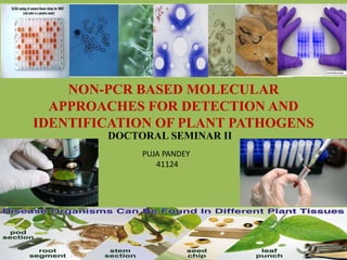
  • 1. NON-PCR BASED MOLECULAR APPROACHES FOR DETECTION AND IDENTIFICATION OF PLANT PATHOGENS DOCTORAL SEMINAR II PUJA PANDEY 41124 1
  • 3. ‱ Visual observations ‱ Cultural ‱ Microscopy CONVENTIONAL TECHNIQUES 3
  • 4. VISUAL OBSERVATION : SIGNS AND SYMPTOMS Rot - Sclerotium rolfsii Late blight of potato – Phytophthora infestans Citrus canker 4
  • 5. Problem with biotic and abiotic factors ?????????? 5
  • 6. CULTURAL : LABORATORY TECHNIQUES Bacterial ooze Cultures of Ralstonia solanacearum 6
  • 8. MICROSCOPY : An Aid To The Eyes Microscope Stereoscopic Microscope Fluorescence Microscope Light Microscope Bright Field Dark Field Phase Contrast Confocal Electron Microscope Scanning electron Microscope Transmission electron Microscope 8
  • 9. Types of Microscope 1) Stereoscopic microscope Function: Visible light to illuminate the surface of a sample (2000x ), without disrupting them 2) Compound microscope (light) Function: Visible light to illuminate a thin section of sample, cells and tissues 3) Confocal laser scanning fluorescence microscope Function: Thin ‘slices’ in a sample while keeping sample intact; Specifically at parts of a cell (such as individual proteins) by labelling them with fluorescence 9
  • 10. Scanning Electron Microscope Function: Surface of objects at high resolution (3D image - 500 000x ) Principle : Beam of electrons being knocked off the surface of the sample and then picked up by a detector . Why we use SEM ??????????? The SEM probably gives the best depth of field out of any microscope 10
  • 11. Transmission Electron Microscope Function: very thin cross-section of an object (cell), internal structure of objects, high resolution (500 000x ) Principle : Electrons pass through the sample and some are deflected and some pass right through, and that forms our image, which is focussed on objective lens 11
  • 12. Microscopy Techniques Applied to the Study of Phytoplasma Diseases: Traditional and Innovative Methods Rita Musetti and Maria Augusta Favali 12
  • 13. Light Microscopy  Dienes’stain was first developed as a specific stain for animal mycoplasma colonies .  Phloem tissues of stems infected by phytoplasmas stained dark blue while xylem was turquoise and cortex light blue. Musetti and Favali, 2004 13
  • 14. Fluorescence Microscopy DAPI staining of hand cut sections of healthy Antibody plus fluorochrome, such as fluorescein isothiocyanate (FITC) and stained  Phytoplasma-infected : the fluorescent bright spots, visible at phloem level Musetti and Favali, 2004 14
  • 15. Transmission electron microscopy (TEM) Phytoplasmas in the phloem cells of Catharanthus roseus Musetti and Favali, 2004 15 Phytoplasmas in the phloem of apple tissues
  • 16. Immuno-electron microscopy (IEM) of thin sections Phytoplasmas in phloem tissues of Catharanthus roseus L. embeded with primary monoclonal antibody and gold Labelledsecondary antibody Gold partcle (15 nm) few particles are visible on phytoplasma membrane Gold partcle (5 nm) particles are well distributed over the periphery of the phytoplasmas Musetti and Favali, 2004 16
  • 17. High resolution autoradiography Phytoplasmas in phloem cells of white clover (Trifolium repens L.), after 3 hours labelling with thymidine-3H. The silver grains were seen on the dividing phytoplasmas 17
  • 18. Staining Technique : For Histopathological Tests ‱ Staining is an auxiliary technique used in microscopy to enhance contrast in the microscopic image e.g. Crystal violet stains only Gram positive bacteria 18
  • 19. 19
  • 20. Histopathological analysis of infected tissues 20 Valencia sweet oranges infected with Colletotrichum acutatum. Valencia sweet orange fruits infected with Guignardia citricarpa. Toluidine blue staining Toluidine blue plus safranin staining Marques et al., 2013
  • 21. 21 Limitations Faced due to conventional techniques !!!!!!!  Latent infection : eg. Potato ring rot  Misleading infection : eg. Black lesions (Alternaria) and bacterial blight of carrot (Xanthomonas)  Co-infection : Alteration of symptoms
  • 22. SEROLOGICAL METHODS – De Vorac Antigen Antibody Antigen Antibody Positive Result 22
  • 23. Antigen  A molecule usually a protein, when it is injected into a warm blooded animal produces antibody (immune response) Antibody  A molecule produced in a warm blooded serum of animal in response to the stimulus antigens  Antibodies are immune system- related proteins called immunoglobulin 23
  • 24.  Variable region : composed of 110- 130 amino acids, give the antibody its specificity for binding antigen.  Variable region includes the ends of the light and heavy chains.  Constant region : determines the mechanism used to destroy antigen. Structure of Antibody 24
  • 25. MONOCLONAL ANTIBODY POLYCLONAL ANTIBODY Composed of a variety of antibody Have multiple epitopes Antibody derived from a single clone and specific for a single epitope Consist of single type of antibody Produced by hybridoma technique Small quantity of antigen is enough for development 25
  • 26. Production of Mabs by Hybridoma Technique 26
  • 27. Antigen antibody based technique Direct test  Precipitation test 1. Tube precipitation 2. Ring precipitation  Micro Precipitation test  Agglutination tests 1. Chloroplast agglutination 2. Latex agglutination  Gel diffusion test  Immuno-electrophoresis Indirect test  ELISA test 1. Direct ELISA (DAS ELISA) 2. Indirect (DAC ELISA) 3. DIBA ELISA 4. Lateral flow device  Immunofluorescence  Immuno Sorbent Electron Microsopy(ISEM)  Flow cytometry 27
  • 28. Tube precipitation test Widely used Reactants diluted in 8.5g/l NaCl, followed by incubation at 37°C in water bath Observations : If elongated virus particle - floccular If spherical virus particle - granular Precipitation test 28
  • 29. Done on a micro-scale to economize on antiserum Drop of dilution mixture (antiserum & virus suspension) are mixed at bottom of a Petri plate The precipitates produced are observed with a microscope with dark-ground illumination Precipitation varies depending on the ratio of concentration of antigen and antibody Micro precipitition test 29
  • 31. Chloroplast agglutination Crude fresh leaf sap from diseased plant Antiserum Chloroplast fragments clump together 31
  • 32.  The reactants, antiserum and virus solution are placed in well cut in the agar (containg 0.85% NaCl and 0.02% sodium azide) in Petri plate  Antibody and virus diffuse into the agar from the adjacent wells  Where they meet precipitation zones in the form of white band are formed Gel diffusion test Oservation : a) Bands : Identical or closely related b) Spurs : Distantly Related c) Intersect : Unrelated 32
  • 33. Radial or single diffusion Double or Ouchterlony diffusion 33
  • 35. ELISA : Enzyme- Linked Immuno-Sorbent Assay ELISA was initially applied for plant viruses by M.F. Clark and Adams (1976) Sensitive, detects at concentration of 1-10 ng/ml It involves an enzyme-mediated colour change reaction to detect antibody binding Degree of colour change, usually measured quantitatively in spectrophotometer at 405 nm Ward et al., 2003 35
  • 36. DAS ELISA : Double antibody sandwich ELISA  Direct ELISA  First time describe by Clark and Adams in 1977 p-nitrophenyl phosphate p-nitrophenol Ward et al., 2003 36
  • 37. DAC ELISA : direct antigen coating ELISA Indirect ELISA Stand for Easy to rapid assay Ward et al., 2003 37
  • 38. Combination of electron microscopy and serology First time described by Derrick in1973 Virus and antiserum are reacted together Antigen are trapped onto grid coated with specific antiserum, negatively stained (Uranyl acetate -1%) and the result viewed in the EM Immuno Sorbernt electron microscope (ISEM) Tubular particle of beet necrotic yellow vein virus 38
  • 39. Dot Immunoblotting Assay (DIBA) OR Dot ELISA Substrate : Nitro tetrazolium BCIP 39
  • 40. Lateral flow technique ‱ The principles used for rapid lateral flow devices are primarily those of ELISA ‱ Various types of filters are used as the solid support for the initial binding reaction A lateral flow device test kit developed by Central Science Laboratory, U.K., permits detection of R. solanacearum in a 3- minute 40
  • 41. 41
  • 42. Ouchterlonie’s double diffusion test. I – Healthy cane extract II – Control blood serum of rabbit III, IV, V – Antigen of host pathogen A – Antibody raised against host pathogen Lingayya and Naik, 2002 42
  • 43. Detection of Colletotrichum falcatum infection in sugarcane tissue by DAC - ELISA Lingayya and Naik, 2002 43
  • 44. TDA = 3 X standard deviation of healthy sample + mean value for healthy sample 44 FUNGUS
  • 45. Advantage of ELISA It is sensitive Semiautomatic technique Application against large number of sample Reproducible Qualitative & Quantitative Suitable for automation high speed No radiation hazards 45
  • 46. Immunofluorescence The intercellular location & distribution of viruses Globulins mixed with a fluorescent dye (Fluorescein isothiocyanate and Rhodamine B) Introduced into the infected cells/tissue, with antigen and antibody reaction, fluorescence takes place 46
  • 47. Flow cytometry Cell suspensions are filtered to remove large particles then stained with fluorochrome-labelled antibodies Fluorescent markers for viability Stains, such as propidium and hexidium iodide for red fluorescent staining of dead cells Carboxy fluorescein diacetate and calcein AM for green fluorescent staining of viable cells can be used to differentiate live from dead cells Detection of C. Michiganensis subsp. Michiganensis in tomato seed extracts Detection of X. Axonopodis pv. Dieffenbachiae, causal agent of anthurium blight (Alvarez et.al., 1999) Determine viability of R. Solanacearum in seed potatoes (van derwolf et.al., 2004) 47
  • 49. 49
  • 50. Southern blotting ‱ Professor Sir Edwin Southern developed this method in 1975 ‱ This method Involves separation, transfer and hybridization ‱ Detection of a specific DNA sequence in DNA samples ‱ Combines agarose gel electrophoresis for size separation of DNA and hybridization with probe Professor Sir Edwin Southern 50
  • 51. 51
  • 52. Northern Blotting  Northern blotting is a technique for detection of specific RNA sequences  Northern blotting was developed by James Alwine and George Stark at Stanford University (1979)  Electrophoresed RNA is blotted on membrane and hybridized 52
  • 53. 53
  • 54. Western blotting ‱ Western blotting (1981) is an immunoblotting technique which rely on the specificity of binding between a protein of interest and a probe (antibody raised against that particular protein) to allow detection of the protein of interest in a mixture of many other similar molecules. ‱ The SDS (Sodium dodecyl sulphate) page technique is a prerequisite for western blotting . 54
  • 55. Steps in western blotting Detected through auto- radiography 55
  • 57. Microarrays for Rapid Identification of Plant Viruses Neil Boonham, Jenny Tomlinson,and Rick Mumford Central Science Laboratory, Sand Hutton, York, YO41 1LZ, United Kingdom 57
  • 58. Boonham et al., 2007 A schematic diagram detailing a simple approach to virus detection using a microarray. 58
  • 59. A microarray designed to detect and discriminate a range of small spherical viruses. Eg. Broad bean wilt virus 2 Indicator host Chenopodium quinoa A small spherical virus was identified using electron microscopy. Boonham et al., 200759 Positive control spots Detection of virus
  • 60. Sensitivity comparison between ELISA and microarray Boonham et al., 2003 Dilution end point Histogram showing local background fluorescence for the 1/1600 dilution of RNA,
  • 61. BIOCHEMICAL METHODS ‱ FAME analysis ‱ BIOLOG ‱ Volatile compound Biochemical techniques 61
  • 62. What is a BIOLOG?  First and only bacterial identification system to identify both gram positive and gram negative bacteria with a single universal test kit. Add cells 96 wells contains different carbon sources and other test. If the cells are metabolically active, they reduce the redox dye and a purple colour is formed in al the positive well 62
  • 63. Fatty Acid Methyl Ester ( FAME ) analysis  Change in the fatty acid profile represent a change in the microbial population 63
  • 64. Detection of Diseased Plants by Analysis of Volatile Organic Compound Emission R.M.C. Jansen, J. Wildt, I.F. Kappers, H.J. Bouwmeester, J.W. Hofstee, and E.J. van Henten 64
  • 65. Emission of volatile organic compounds (VOCs) from non-infected and Botrytis cinerea–infected tomato plants. Jansen et al., 2011 65 Damaged cell membranes Local emission of several lipoxygenase (LOX) oxidative cleavage of C18fatty acids (oxygen and lipoxygenases ) Characterize diseases due to release of VOCs
  • 66. Surface Plasmon Resonance (SPR)  The Surface Plasmon Resonance (SPR) sensor is used for label free detection and real-time monitoring  Their simplicity and sensitivity make biosensors an effective means of disease diagnosis and monitoring  In SPR technique, shift of the resonance angle is observed, when the intended species is captured by the immobilized antibody on the sensor surface 66 Is it the specific protein, the virus fragment or the virion itself?
  • 67. Development of Surface Plasmon Resonance (SPR) Based Immuno- Sensing System for Detection of Fungal Teliospores of Karnal Bunt (Tilletia Indica), a Quarantined Disease of Wheat Sadhna Singh1, Manoj Singh1, Gohar Taj1, Sanjay Gupta2 and Anil Kumar1* 1Department of Molecular Biology & Genetic Engineering, College of Basic Sciences & Humanities, G. B. Pant University of Agriculture & Technology, Pantnagar, Uttaranchal, India 2Department of Biotechnology, SBS PG Institute of Biomedical Sciences, Balawala, Dehradun, Uttarakhand, India Journal of Biosensors & Bioelectronics 67
  • 68. Experiment conducted : Interaction of teliosporic wall antigen with the anti-teliosporic antibody immobilized on sensor chip  The interaction of antigen at a concentration of 80, 40, 20, 10, 5.0, 2.5, 1.25, 0.625, 0.312, 0.156, .078 and 0.039 ng/ÎŒl with immobilized antibody on sensor chip was examined Observation ‱ The responses increased in proportion to the concentration of teliosporic antigen due to the change of the refractive index near the SPR sensor chip 68
  • 69. SPR sensor response after the interaction of different concentrations of antigen over the immobilized antibody (1: 500) Singh et al., 2012 (a) 0.312 ng/ÎŒl (b)1.25 ng/ÎŒl (c) 5 ng/ÎŒl (d) 20 ng/ÎŒl (e) 80 ng/ÎŒl
  • 70. Advantages of SPR Major advantages : ‱ Rapid, real-time ‱ Non-labeling analysis ‱ Miniaturization for portable application 70
  • 71. 71
  • 72. Phytophthora and Pythium Test Kits : 0.5 per cent of a plant roots are infected Tests for Phytophthora, Pythium, and Rhizoctonia root and crown decay fungi can be performed on-site by growers in about 10 minutes (A) Collect and grind samples using abrasive pads (B) Fold pads and insert them into the extraction solution (C) Apply solutions to detector (D) Examine detector dots for color change. 72
  • 73. Molecular methods for detection of plant pathogens—What is the future?  Strategies are needed on how to exploit deduced genomics and proteomics supported by in silico analysis for establishing rational disease control measures.  The reliability of each specific on-the-spot diagnostic method needs to be validated before results are used exclusively to implement costly disease control strategies and/or regulatory actions. 73
  • 74. 74
  • 75. 75