Muscle senses reflex organization
•Als PPT, PDF herunterladen•
28 gefällt mir•10,736 views

Intro lecture to muscle spindles, golgi tendon organs and reflex organization.

Empfohlen
Weitere ähnliche Inhalte
Was ist angesagt?
Was ist angesagt? (20)
Andere mochten auch
Andere mochten auch (20)
Ähnlich wie Muscle senses reflex organization
Ähnlich wie Muscle senses reflex organization (20)
Mehr von Csilla Egri
Kürzlich hochgeladen
Kürzlich hochgeladen (20)
Muscle senses reflex organization
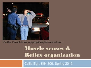
- 1. Muscle senses & Reflex organization Csilla Egri, KIN 306, Spring 2012 Ociffer, I’m not drunk, my proprioceptors are askew…
- 2. Outline Proprioception Muscle spindles Golgi tendon organs Spinal reflexes + clinical importance Stretch reflex Inverse myotatic reflex Flexion reflexes Withdrawal reflex Crossed extensor reflex 2
- 3. Proprioception 3 Sense the position of body parts in relation to each other and in space, as well as relative force applied to movements Balance Vestibular system Muscle length and force Muscle spindles Golgi tendon organs B&L Figure 9-1
- 4. Muscle Spindles - intro 4 Non-force generating intrafusal muscle fibers within a fluid filled capsule (spindle) Lie in parallel with extrafusal muscle fibers Stretch or shorten along with extrafusal fibers Innervated by both motor (efferent) and sensory (afferent) axons Efferent innervation contracts intrafusal fiber to match length of extrafusal fiber Afferent innervation sends info on relative Kandel Figure 36-3
- 5. Muscle spindles - structure 5 Three types of intrafusal fibers Central regions are non-contractile Mechanoreceptive sensory innervation Primary Ia afferents all 3 fibers Secondary II afferents static nuclear bag and nuclear chain fibers Motor innervation Dynamic γ efferent Dynamic nuclear bag Static γ efferent Combination of chain and static nuclear bag Kandel Figure 36-3
- 6. Muscle spindles – afferent function6 Each afferent has a tonic, baseline level of firing, and responds to relative stretch Ia Static and dynamic firing responses Firing proportional to amount of and rate of muscle stretch II Only static firing response Firing proportional to amount of stretch Firing of which afferent is being assessed during a tendon tap? B&L Figure 9-2
- 7. Muscle spindles – efferent function7 γ motor neurons maintain sensitivity of spindle over a range of muscle lengths α-γ coactivation Descending input can change relative dynamic vs. static γ activation to modulate spindle sensitivity http://www.ncbi.nlm.nih.gov/books/NBK11119/bin/ch16f10.jpg Activation of only dynamic γ motor neurons increases responsiveness of _____ afferents
- 8. Golgi Tendon Organs (GTOs) 8 collagen fibers located within a capsule near tendon, in series with extrafusal muscle innervated by mechanoreceptive Ib afferent fibers intertwined between collagen Activated by muscle contraction or stretch Sense changes in tendon tension/force Kandel Figure 36-6
- 9. Reflex organization 9 A reflex is a predictable, involuntary and stereotyped response to an eliciting stimulus Can be modulated by stimulus intensity and descending CNS input Testing reflexes is an important clinical tool in assessing neurological and spinal function
- 10. Myotatic or stretch reflex 10 Monosynaptic reflex mediated by muscle spindles Contraction in response to lengthening Reflex arc: 1. Muscle stretches 2. Ia afferent of muscle spindle increase firing 3. Synapse on α motor neuron and inhibitory interneuron in spinal cord 4. α motor neuron of homonymous muscle excited, and of antagonist muscle inhibited 5. Homonymous muscle contracts to oppose lengthening, antagonist muscle relaxes B&L Figure 9-6
- 11. Myotatic or stretch reflex 11 Stretch reflex has two phases: Phasic (Ia) phase dynamic change in muscle length (ex. tendon tap) triggers a transient phasic contraction Physiological importance: reflex contraction prevents overstretch of extrafusal muscle fiber beyond physiological limits Clinical importance: tendon tap used to determine integrity of spinal cord at different segmental levels
- 12. Myotatic or stretch reflex 12 Tonic phase Static stretching of muscles produces a weaker, longer lasting, tonic contraction Type II afferents also involved Physiological importance: maintains muscle tone/posture via negative feedback Ex. Soldier standing at attention legs begin to fatigue and flex quadriceps slowly begin lengthening tonic stretch reflex maintains tone and prevents collapse (to an extent) Clinical importance: assessing presence of hypertonia Ex. Patients with cerebral palsy have very rigid, tight muscles resistant to stretch overactive tonic stretch reflex due to upper motor neuron lesion
- 13. Motor neuron lesions 13 Upper motor neuron lesion of the neural pathway inside the CNS (not including the ventral horn of the spinal cord or motor nuclei of the cranial nerves) stroke, traumatic brain injury or cerebral palsy Lower motor neuron lesion affects nerve fibers within the ventral horn of the spinal cord travelling to the relevant muscle(s) Nerve trauma, polio Upper motor neuron lesion Lower motor neuron lesion Reflexes Increased, may have pathological reflex signs (Babinski sign) Decreased, Muscle tone Increased, contralateral Decreased, ipsilateral Weakness Yes, contralateral Yes, ipsilateral
- 14. Inverse myotatic or Ib reflex 14 Disynaptic reflex mediated by GTOs Relaxation in response to increased tension Reflex arc: 1. Muscle contracts 2. Ib afferent of GTO increase firing 3. Synapse on one inhibitory and one excitatory interneuron 4. α motor neuron of homonymous muscle inhibited, and of antagonist muscle excited 5. Homonymous muscle relaxes to oppose increased force in tendon, antagonist muscle contracts B&L Figure 9-7 Ib
- 15. Inverse myotatic or Ib reflex 15 Physiological importance: reflex relaxation thought to prevent excessive force from damaging muscle tissue. Acts synonymously with the myotatic stretch reflex to maintain posture and balance Clinical importance: Clasp knife reflex: seen in patients with upper motor neuron lesions muscle has increased tone and resistance to stretch if sufficient force is applied, limb resistance suddenly decreases thought to be mediated by high threshold firing of GTO afferents (but other receptors may be involved as well)
- 16. Flexion withdrawal reflex16 Polysynaptic reflex mediated by FRAs (flexion reflex afferents: nociceptors, mechanoreceptors etc.) flexion in response to painful stimuli FRAs synapse on inhibitory and excitatory interneurons which excite ipsilateral flexor motorneurons & inhibit extensor motorneurons Physiological importance: Rapid flexion away from painful stimuli Clinical importance: upper motor neuron lesion impairs flexion reflex pathalogical Babinski sign B&L Figure 9-8
- 17. Upper motor neuron lesion: Babinski sign17 (Type of flexion reflex) (pathological reflex)
- 18. Crossed extension reflex 18 occurs in lower limbs as part of reflex arc for flexion reflex FRAs synapse on interneurons which elicit contralateral limb extension to help maintain balance Similar neuronal circuits involved in central pattern generators governing locomotion (next lecture) B&L Figure 9-8
- 19. Summary of reflexes 19 REFLEX STIMULU S (CLINICA L TEST) RESPONS E SENSORY RECEPTO R SYNAPSE S EFFECT ON MUSCLE OTHER EFFECTS FUNCTIO N Stretch (Myotatic) Reflex Rapid Stretch of muscle (test: tap on muscle tendon) Stretched muscle contracts rapidly (ex. knee jerk) Muscle Spindle Primary (Ia) and Secondary (II) sensory neurons (tonic phase) Ia: Mono- synaptic II: (tonic phase) monosynapt ic and polysnaptic Excite Homonymo us (same muscle) Also Excite synergist muscles; Inhibit antagonist muscles (Reciprocal Inhibition) Aid in maintaining posture, counter sudden stretch Inverse Myotatic Reflex Large force on tendon (pull on muscle when resisted) Muscle tension decreases Golgi Tendon Organ (Ib) Disynaptic (via interneuron) Inhibit Homonymo us (same muscle) Also Inhibit synergist muscles; Excite antagonist muscles Protective, prevent damage to tendon Flexor Reflex Sharp, painful stimulus (as in stepping on nail) Limb is rapidly withdrawn from stimulus Cutaneous (skin) and pain receptors Poly- synaptic (via interneuron) Excite Flexor muscle Also Inhibit extensor muscle of same limb; Excite extensor muscles and Inhibit flexors of opposite limb (Crossed Extensor Reflex) Protective, withdraw from painful stimulus; Cross extension aids in maintaining posture when leg is lifted http://musom.marshall.edu/anatomy/grosshom/spinalreflexes.html
- 20. Objectives After this lecture you should be able to: Compare and contrast the structure and function of muscle spindles with golgi tendon organs Describe the importance of αγ coactivation Describe the reflex pathway for the myotatic, inverse myotatic, and flexion reflexes Give an example of a physiological and clinical importance for each reflex Distinguish between upper and lower motor neuron lesions 20
- 21. 21 1. Type __________ spindle afferents are responsive to the rate of change of muscle length. 2. A Babisnki sign is often associated with _____________ motor neuron lesions. 3. Relaxation of the quadriceps muscle increases/decreases firing of Ia afferents and increases/decreases firing of Ib afferents. Test your knowledge
Hinweis der Redaktion
- Readings Berne and Levy: Chapter 9, pages 157 – 179 Chapter 6, pages 82 – 91 (Focus on Neuromuscular Junction and End Plate Potential) Chapter 12, pages 233-246
- Cutaneous mechanoreceptors because we already talked about mechanically gated receptors: Hair cells in vestibular and auditory system
- size of spindle: 4-10 mm long Intrafusal fibers are specialized for sensory function and do not contribute significantly to force produced by extrafusal muscle fibers. (Extrafusal muscle fibers are normal skeletal muscle fiber that produce force). Spindles lie in parallel with extrafusal muscle fibers since the ends of the muscle spindle attach to connective tissue in muscle.
- Tap – tendon tap, the firing of which afferent is being assessed during a tendon tap?
- Descending tracts can affect whether static or dynamic gamma motor neurons preferrentially fire, affecting the nature of reflex activity in the spinal cord. Static gamma stimulated: static and dynamic response of Ia and II enhanced. Dynamic gamma stimulated: only dynamic response of Ia enhanced http://www.ncbi.nlm.nih.gov/books/NBK11119/bin/ch16f10.jpg
- Stretch of collagen fibers occurs when force is applied to tendon either by: stretching muscle or by contraction of extrafusal muscle fibers that are connected directly (in series) with tendon organ Provides complementary information about the mechanical state of the muscle Input from the GTO’s relays info about the tension in the muscle. This input can be useful in a variety of motor acts such as maintaining a steady grip on an object or compensating for the effects of fatigue.
- aka the monosynaptic stretch reflex or myotatic reflex elicited by: e.g., tendon tap or rapid movement of joint monosynaptic stretch reflex response can be used as diagnostic tool to determine integrity of spinal cord at different segmental levels Tonic phase is not apparent in intact animals (only in the decerebrate preparation) because the steady state discharge of muscle spindles is not strong enough to raise the resting potential of motor neurons above threshold for firing.
- aka the monosynaptic stretch reflex or myotatic reflex elicited by: e.g., tendon tap or rapid movement of joint monosynaptic stretch reflex response can be used as diagnostic tool to determine integrity of spinal cord at different segmental levels Tonic phase is not apparent in intact animals (only in the decerebrate preparation) because the steady state discharge of muscle spindles is not strong enough to raise the resting potential of motor neurons above threshold for firing.
- aka the monosynaptic stretch reflex or myotatic reflex elicited by: e.g., tendon tap or rapid movement of joint monosynaptic stretch reflex response can be used as diagnostic tool to determine integrity of spinal cord at different segmental levels An upper motor neuron lesion is a lesion of the neural pathway above the anterior horn cell (grey matter in front of spinal cord containing cell bodies of alpha motor neurons) of the spinal cord or motor nuclei of the cranial nerves. This is in contrast to a lower motor neuron lesion, which affects nerve fibers traveling from the anterior horn of the spinal cord to the relevant muscle(s).[1] Upper motor neuron lesions occur in conditions affecting motor neurons in the brain or spinal cord such as stroke, traumatic brain injury and cerebral palsy.
- Cerebral palsy (CP) is an umbrella term encompassing a group of non-progressive,[1][2] non-contagious motor conditions that cause physical disability in human development, chiefly in the various areas of body movement.[3] Cerebral refers to the cerebrum, which is the affected area of the brain (although the disorder most likely involves connections between the cortex and other parts of the brain such as the cerebellum), and palsy refers to disorder of movement. Upper motor neuron lesions cause spastic paralysis of muscles on the opposite side of the body. Muscle tone is increased, reflexes exaggerated, and babinski sign (pathalogical reflexes) Spasticity is probably caused by the removal of inhibitory influences exerted by the cortex on the postural centers of the vestibular nuclei and reticular formation Lower motor neuron lesions: flaccid paralysis of muscles on the same side of the body. NO voluntary or reflex action of the innervated muscle fibers, muscle tone is decreased or lost, and the muscle remains limp or flaccid.
- Prevents tearing of muscle fibers in response to force overload Vis versa: reflex contraction in response to lowered tension: explain how this is actually acting synonymously with the myotatic stretch reflex: soldier at attention, quads fatigue, force in patellar tendon decreases, inhibition of homonymous muscle prevented, homonymous muscle can contract, the lengthening of quads also stimulates muscle spindles to activate same muscle to contract
- Prevents tearing of muscle fibers in response to force overload Vis versa: reflex contraction in response to lowered tension: explain how this is actually acting synonymously with the myotatic stretch reflex: soldier at attention, quads fatigue, force in patellar tendon decreases, inhibition of homonymous muscle prevented, homonymous muscle can contract, the lengthening of quads also stimulates muscle spindles to activate same muscle to contract
- The flexion reflex afferents include group II and group III afferents from the skin, joints, and muscles, and the group II afferents from the secondary endings of muscle spindles. opposite response pattern = flexor inhibition; extensor excitation produces coordinated response of multiple joints, e.g., ankle, knee and hip mediated by polysynaptic pathway (multiple interneurons) stimulus strong enough to activate nociceptors elicits flexion withdrawal, which causes stimulated limb to be quickly withdrawn from stimulus flexion withdrawal can inhibit activation of extensor muscles of injured limb (limping)
- The flexion reflex afferents include group II and group III afferents from the skin, joints, and muscles, and the group II afferents from the secondary endings of muscle spindles. opposite response pattern = flexor inhibition; extensor excitation produces coordinated response of multiple joints, e.g., ankle, knee and hip mediated by polysynaptic pathway (multiple interneurons) stimulus strong enough to activate nociceptors elicits flexion withdrawal, which causes stimulated limb to be quickly withdrawn from stimulus flexion withdrawal can inhibit activation of extensor muscles of injured limb (limping)
- Type 1a Upper motor neuron lesions Increases Ia, decreases Ib