SlideShare ist ein Scribd-Unternehmen logo
1 von 17
1
Lecture 10
When blood sugar (glucose) levels are low, the liver converts
amino acids into glucose for energy. Amino acids come from the
breakdown of protein in the GI tract. The conversion of amino
acids to glucose results in the production of the nitrogenous
waste product, ammonia (NH3).
 Ammonia is extremely toxic to cells, so the liver converts
NH3 to the less toxic nitrogenous waste product, urea.
 The URINARY SYSTEM removes urea and other metabolic
waste products, toxins, excess water, and excess electrolytes
(salts) from the bloodstream and eliminates them from the
body in the form of urine. The process of eliminating metabolic
wastes is called excretion.
 The KIDNEYS are the excretory organs of the urinary
system. As the kidneys perform their excretory function, they
also regulate blood volume and the chemical makeup of the
blood.
 They continuously maintain the proper balance between
water and salts in the blood and they help to maintain the blood
pH at 7.35-7.45.
 Kidneys contribute to homeostasis of body fluids by
regulation of blood ionic composition, regulation of pH, ,
regulation of blood volume, regulation of blood pressure,
maintenance of blood osmolarity, producing hormones produced
by the kidneys stimulates red blood cell production, regulating
blood glucose level, and excreting wastes and foreign
substances.
The Urinary System
2
 The urinary system consists of two kidneys, two ureters,
one urinary bladder, and one urethra.
 Urine is excreted from each kidney through its ureter and
is stored in the urinary bladder until it is expelled from the
body through the urethra.
 The specialized branch of medicine that deals with
structure, function, and diseases of the male and female
urinary systems and the male reproductive system is known as
nephrology.
 The branch of surgery related to male and female urinary
systems and the male reproductive system is called urology.
3
4
EXTERNAL ANATOMY
 Kidneys are kidney bean-shaped and reddish in color. The adult
kidney weighs 150 grams, is 10-12 cm (4-5 inches) long, 5.0-7.5 cm
(2-3 in.) wide and 2.5 cm (1 in.) thick.
 The kidneys are located between the posterior abdominal wall and
parietal peritoneum (they are retroperitoneal), extending from
the level of the T12 vertebra to the level of the L3 vertebra. The
kidneys receive some protection from the lower part of the rib
cage. The right kidney is slightly lower than left kidney.
 The lateral surface of each kidney is convex. The medial surface
is concave and has an indentation called the HILUS. The ureters,
renal blood vessels, lymphatic vessels, and nerves enter or exit
the kidney at the hilus.
 There are 3 layers of supportive tissue surrounding each kidney:
1. RENAL CAPSULE: a transparent dense irregular connective
tissue covering the kidney surface. The renal capsule prevents
infections in surrounding regions from spreading to the kidneys. It
also protects the kidney from physical trauma.
 2. ADIPOSE CAPSULE: a mass of adipose tissue outside the renal
capsule. The adipose capsule helps hold the kidney in place against
the posterior abdominal wall and cushions the kidneys against
external blows.
 3. RENAL FASCIA: a layer of dense irregular connective tissue
outside the adipose capsule that anchors the kidney to the
abdominal wall.
5
KIDNEY: INTERNAL ANATOMY
A frontal section of the kidney reveals 3 distinct regions
inside the renal capsule and they are the RENAL CORTEX,
RENAL MEDULLA, & RENAL PELVIS.
1. RENAL CORTEX: the outer region just inside the renal
capsule; light red in color and granular in appearance.
2. RENAL MEDULLA: deep to the cortex; a darker, reddish
brown color. Arranged in cone-shaped regions called RENAL
PYRAMIDS, which have a striated appearance.
Extensions of the RENAL CORTEX between the renal pyramids
are called RENAL COLUMNS. Together, the RENAL CORTEX &
MEDULLA form the parenchyma (functional portion) of the
kidney where urine is formed.
3. Just inside the hilus is the cavity called the RENAL
PELVIS. The renal pelvis collects urine that is formed in the
renal cortex and medulla.
6
Nephrons
• A nephron consists of a renal corpuscle where fluid is filtered, and a
renal tubule into which the filtered fluid passes.
• Nephrons perform three basic functions: glomerular filtration,
tubular reabsorption, and tubular secretion.
• A renal tubule consists of a proximal convoluted tubule (PCT), loop
of Henle (nephron loop), and distal convoluted tubule (DCT). Distal
convoluted tubules of several nephrons drain into to a single
collecting duct and many collecting ducts drain into a small number
of papillary ducts.
• The loop of Henle consists of a descending limb, a thin ascending
limb, and a thick ascending limb.
• There are two types of nephrons that have differing structure and
function.
– A cortical nephron usually has its glomerulus in the outer
portion of the cortex and a short loop of Henle that penetrates
only into the outer region of the medulla
– A juxtamedullary nephron usually has its glomerulus deep in the
cortex close to the medulla; its long loop of Henle stretches
through the medulla and almost reaches the renal papilla.
7
In the renal cortex, the ascending limb of the loop of Henle
becomes twisted again as the DISTAL CONVOLUTED
TUBULE. The distal convoluted tubules of several nephrons
empty into each COLLECTING DUCT. The collecting ducts
absorb water from the filtrate. Several collecting ducts
empty into a PAPILLARY DUCT, which delivers urine to the
RENAL PELVIS.
The filtrate is processed as it passes through these parts of
the nephron. Needed substances, such as water, glucose and
electrolytes (salts), are returned to the bloodstream. After
the filtrate is processed by the nephron, it is called URINE.
Urine contains urea and other metabolic wastes, toxins,
excess salts, and excess water, as well as drugs and excess
vitamins.
8
9
10
11
BLOOD SUPPLY OF THE KIDNEYS
While the body is at rest, the large left and right RENAL ARTERIES
deliver about about 1.2 liters of blood through the kidneys every
minute. The renal arteries are branches of the ABDOMINAL AORTA.
After each renal artery enters a kidney, it branches into numerous
small arteries, which carry most of the blood to the renal cortex.
The arteries in the renal cortex branch into numerous AFFERENT
ARTERIOLES, one for each nephron. Each afferent arteriole, divides to
form a capillary tuft called the GLOMERULUS. The glomerulus is
surrounded by the glomerular capsule and is specialized for filtration of
blood. The glomerular capillaries merge to form the EFFERENT
ARTERIOLE, which leads away from the glomerular capsule.
The efferent arteriole's diameter is smaller than the afferent
arteriole. This raises the blood pressure in the glomerulus, which aids in
forcing water and solutes into the glomerular capsule.
12
Each efferent arteriole branches into a second capillary network
called the PERITUBULAR CAPILLARIES. These capillaries cling
closely to the PROXIMAL and DISTAL CONVOLUTED TUBULES
of both cortical and juxtamedullary nephrons.
The efferent arterioles of juxtamedullary nephrons also branch
into the VASA RECTA, capillaries that surround the LOOP OF
HENLE in the medulla. The peritubular capillaries and vasa recta
are adapted for absorption. They absorb water and needed
substances from the filtrate in the nephron, so that much of the
water and solutes that the body requires are returned to the
bloodstream rather than flushed out in the urine.
Toxins and excess
water and solutes
remain in the
filtrate and are
eliminated as urine.
The peritubular
capillaries and vasa
recta merge into
venules that join
veins which empty
into the RENAL
VEIN.
The RENAL VEIN
of each kidney
leaves the kidney
through the hilus.
The renal vein of
each kidney
returns freshly
cleansed blood to
the INFERIOR
VENA CAVA.
13
URETERS
The URETERS are slender tubes about 25-30 cm (10-12 in) long that
convey urine from the kidneys to the urinary bladder. Each ureter runs
posterior to the parietal peritoneum (retroperitoneal) from the renal
hilus to the level of the urinary bladder.
At the urinary bladder, each ureter makes a medial turn toward the
bladder and enters the posterior bladder wall at an oblique angle, which
prevents backflow of urine from the bladder into the ureters.
The inner lining of the ureter wall is MUCOSA that consists of
transitional epithelium that stretches as urine passes through. Mucus
secreted by the mucosa protects the epithelial cells from coming into
contact with the urine, which contains toxins and is slightly acidic.
Beneath the mucosa is the MUSCULARIS, which is composed of smooth
muscle arranged in longitudinal and circular layers. The muscularis
creates peristaltic contractions, similar to those in the GI tract.
When the renal pelvis fills with urine, smooth muscle in the renal pelvis
wall contracts rhythmically, sending urine into the ureter. Peristalsis
propels the urine through the ureter to the urinary bladder.
14
URINARY BLADDER
The URINARY BLADDER is a hollow muscular organ located in the pelvic
cavity posterior to the pubic symphysis. Like the kidneys and ureters,
the urinary bladder is retroperitoneal. In males, the urinary bladder lies
anterior to the rectum. In females, the bladder is anterior to the vagina
and inferior to the uterus. The function of the urinary bladder is the
temporary storage of urine.
The inner wall of the urinary bladder consists of a MUCOSA made of
transitional epithelium, which is able to stretch a great deal. Mucus
produced by the bladder mucosa helps protect the wall from urine.
Beneath the mucosa is smooth muscle, arranged in longitudinal and
circular layers. The smooth muscle in the wall allows the bladder to
stretch a great deal.
15
When empty, the bladder is 5.0-7.5 cm (2-3 in) long. Its walls are
thick and its mucosa is thrown into folds called RUGAE. When urine
accumulates in the bladder, it expands and becomes pear-shaped as it
rises upward in the pelvic cavity.
The transitional epithelium and smooth muscle stretches and thins.
This increases the internal volume of the bladder by decreasing the
thickness of its wall. A moderately full bladder is about 12.5 cm (5 in)
long and holds about 500 ml of urine. The average capacity of the
bladder is 700-800 ml. When the volume of urine in the bladder
exceeds 200-400 ml, stretch receptors in the bladder transmit nerve
impulses to the brain, initiating the conscious desire to urinate.
16
URETHRA
The URETHRA is a muscular tube that drains urine from the
urinary bladder and conveys it out of the body.
The INTERNAL URETHRAL SPHINCTER is an involuntary smooth
muscle sphincter at the base of urinary bladder, surrounding
the opening called the INTERNAL URETHRAL ORIFICE.
The EXTERNAL URETHRAL SPHINCTER is a voluntary skeletal
muscle sphincter in the UROGENITAL DIAPHRAGM. The
urogenital diaphragm is formed by skeletal muscles of the
pelvic floor;.
In FEMALES, the urethra is 4 cm (1.5 in) long and 6 mm in
diameter. It lies posterior to the pubic symphysis and anterior to
the vagina. The EXTERNAL URETHRAL ORIFICE lies anterior to
the vaginal opening.
In MALES , the urethra is 15-20 cm (6-8 in) long. The urethra
passes through the PROSTATE GLAND, then through the PENIS
to the outside through the EXTERNAL URETHRAL ORIFICE.
17
WASTE MANAGEMENT IN OTHER BODY
SYSTEMS
• One of the many functions of the urinary
system is to rid the body of waste
materials.
• Other organs, tissues and processes
contribute to waste management.
• Buffers prevent an increase in the acidity
of body fluids.
• The blood transports wastes.
• The liver is the primary site for metabolic
recycling.
• The lungs excrete CO2. H2O, and heat.
• Sweat glands eliminate excess heat, water,
and CO2, plus small quantities of salts and
urea.
• The GI tract eliminates solid, undigested
foods, waste, some CO2, H2O, salts and
heat.

Weitere ähnliche Inhalte

Was ist angesagt?

Dokumen.tips litiaza biliara-pdf-568126fda8ecf
Dokumen.tips litiaza biliara-pdf-568126fda8ecfDokumen.tips litiaza biliara-pdf-568126fda8ecf
Dokumen.tips litiaza biliara-pdf-568126fda8ecfAdiSiAncaOrban
 
MUSCULAR TISSUES.pdf
MUSCULAR TISSUES.pdfMUSCULAR TISSUES.pdf
MUSCULAR TISSUES.pdfJuliusKauki1
 
Properties of cardiac muscles
Properties of cardiac musclesProperties of cardiac muscles
Properties of cardiac musclesBobyjeet Goswami
 
Anatomy of the heart
Anatomy of the heartAnatomy of the heart
Anatomy of the heartKhalid Musa
 
Skeletal muscle contraction
Skeletal muscle contraction Skeletal muscle contraction
Skeletal muscle contraction Rohit Paswan
 
ANATOMY AND PHYSIOLOGY OF HEART
ANATOMY AND PHYSIOLOGY OF HEARTANATOMY AND PHYSIOLOGY OF HEART
ANATOMY AND PHYSIOLOGY OF HEARTHaripriya209498
 
Muscle tissue 2
Muscle tissue 2Muscle tissue 2
Muscle tissue 2KemUnited
 
The muscular system (slide show)
The muscular system (slide show)The muscular system (slide show)
The muscular system (slide show)William Banaag
 
Muscle physiology ,types of muscles: striated ,non striated and cardiac. ultr...
Muscle physiology ,types of muscles: striated ,non striated and cardiac. ultr...Muscle physiology ,types of muscles: striated ,non striated and cardiac. ultr...
Muscle physiology ,types of muscles: striated ,non striated and cardiac. ultr...Tiffy John
 
Appendicular muscles
Appendicular musclesAppendicular muscles
Appendicular musclesKemUnited
 
Muscles classification
Muscles classificationMuscles classification
Muscles classificationRajesh Goit
 
Anatomy of the heart
Anatomy of the heartAnatomy of the heart
Anatomy of the heartZeyad Magdy
 
Bone structure & blood supply
Bone structure & blood supplyBone structure & blood supply
Bone structure & blood supplyDr. sana yaseen
 
Anatomy of the heart
Anatomy of the heartAnatomy of the heart
Anatomy of the heartMirzaNaadir
 
Musculoskeletal system
Musculoskeletal system Musculoskeletal system
Musculoskeletal system Vamsi kumar
 

Was ist angesagt? (20)

Dokumen.tips litiaza biliara-pdf-568126fda8ecf
Dokumen.tips litiaza biliara-pdf-568126fda8ecfDokumen.tips litiaza biliara-pdf-568126fda8ecf
Dokumen.tips litiaza biliara-pdf-568126fda8ecf
 
MUSCULAR TISSUES.pdf
MUSCULAR TISSUES.pdfMUSCULAR TISSUES.pdf
MUSCULAR TISSUES.pdf
 
Properties of cardiac muscles
Properties of cardiac musclesProperties of cardiac muscles
Properties of cardiac muscles
 
Bones
BonesBones
Bones
 
Anatomy of the heart
Anatomy of the heartAnatomy of the heart
Anatomy of the heart
 
Skeletal muscle contraction
Skeletal muscle contraction Skeletal muscle contraction
Skeletal muscle contraction
 
ANATOMY AND PHYSIOLOGY OF HEART
ANATOMY AND PHYSIOLOGY OF HEARTANATOMY AND PHYSIOLOGY OF HEART
ANATOMY AND PHYSIOLOGY OF HEART
 
Muscle tissue 2
Muscle tissue 2Muscle tissue 2
Muscle tissue 2
 
The muscular system (slide show)
The muscular system (slide show)The muscular system (slide show)
The muscular system (slide show)
 
Muscle physiology ,types of muscles: striated ,non striated and cardiac. ultr...
Muscle physiology ,types of muscles: striated ,non striated and cardiac. ultr...Muscle physiology ,types of muscles: striated ,non striated and cardiac. ultr...
Muscle physiology ,types of muscles: striated ,non striated and cardiac. ultr...
 
Appendicular muscles
Appendicular musclesAppendicular muscles
Appendicular muscles
 
Vertebral column
Vertebral columnVertebral column
Vertebral column
 
Muscles classification
Muscles classificationMuscles classification
Muscles classification
 
Anatomy of the heart
Anatomy of the heartAnatomy of the heart
Anatomy of the heart
 
Bone structure & blood supply
Bone structure & blood supplyBone structure & blood supply
Bone structure & blood supply
 
Anatomy of the heart
Anatomy of the heartAnatomy of the heart
Anatomy of the heart
 
Cardiac muscle tissue
Cardiac muscle tissueCardiac muscle tissue
Cardiac muscle tissue
 
Biomechanics of knee complex 1
Biomechanics of knee complex 1Biomechanics of knee complex 1
Biomechanics of knee complex 1
 
150 Heart
150 Heart150 Heart
150 Heart
 
Musculoskeletal system
Musculoskeletal system Musculoskeletal system
Musculoskeletal system
 

Ähnlich wie Lecture 10 and 11 urinary system

Cape biology unit 2-_the_kidney_and_osmoregulation
Cape biology unit 2-_the_kidney_and_osmoregulationCape biology unit 2-_the_kidney_and_osmoregulation
Cape biology unit 2-_the_kidney_and_osmoregulationHilton Ritch
 
Anaphysio ch 12 urinary
Anaphysio ch 12   urinaryAnaphysio ch 12   urinary
Anaphysio ch 12 urinaryariannarecio
 
Excretory system 2
Excretory system 2Excretory system 2
Excretory system 2pramod kumar
 
URINARY SYSTEM.pptx
URINARY SYSTEM.pptxURINARY SYSTEM.pptx
URINARY SYSTEM.pptxRameeThj
 
urinary system (1).pptx
urinary system (1).pptxurinary system (1).pptx
urinary system (1).pptxAbidaLaghari2
 
URINARY SYSTEM PPT.pptx
URINARY SYSTEM PPT.pptxURINARY SYSTEM PPT.pptx
URINARY SYSTEM PPT.pptxnaazfarah2
 
Urinary and Excretory system
Urinary and Excretory system Urinary and Excretory system
Urinary and Excretory system harshraman1989
 
Urinary system-presentationyorkville-college-1203376567476185-2
Urinary system-presentationyorkville-college-1203376567476185-2Urinary system-presentationyorkville-college-1203376567476185-2
Urinary system-presentationyorkville-college-1203376567476185-2Ksenja Ljashenko
 
Bio 120 Presentation #9
Bio 120 Presentation #9Bio 120 Presentation #9
Bio 120 Presentation #9Daley College
 
Function/physiology of genitourinary system
Function/physiology  of genitourinary systemFunction/physiology  of genitourinary system
Function/physiology of genitourinary systemMR. JAGDISH SAMBAD
 
Urinary System Presentationyorkville College 1203376567476185 2
Urinary System Presentationyorkville College 1203376567476185 2Urinary System Presentationyorkville College 1203376567476185 2
Urinary System Presentationyorkville College 1203376567476185 2guest8b2ebd
 
Excretory system:Kidneys,ureter,urethra and urinary bladder
Excretory system:Kidneys,ureter,urethra and urinary bladderExcretory system:Kidneys,ureter,urethra and urinary bladder
Excretory system:Kidneys,ureter,urethra and urinary bladderJasleen Kaur
 

Ähnlich wie Lecture 10 and 11 urinary system (20)

Urinary system
Urinary systemUrinary system
Urinary system
 
Cape biology unit 2-_the_kidney_and_osmoregulation
Cape biology unit 2-_the_kidney_and_osmoregulationCape biology unit 2-_the_kidney_and_osmoregulation
Cape biology unit 2-_the_kidney_and_osmoregulation
 
Anaphysio ch 12 urinary
Anaphysio ch 12   urinaryAnaphysio ch 12   urinary
Anaphysio ch 12 urinary
 
Excretory system 2
Excretory system 2Excretory system 2
Excretory system 2
 
“Anatomy Renal System” Unidad Ii
“Anatomy Renal System”  Unidad Ii“Anatomy Renal System”  Unidad Ii
“Anatomy Renal System” Unidad Ii
 
URINARY SYSTEM.pptx
URINARY SYSTEM.pptxURINARY SYSTEM.pptx
URINARY SYSTEM.pptx
 
Urinary system
Urinary systemUrinary system
Urinary system
 
urinary system (1).pptx
urinary system (1).pptxurinary system (1).pptx
urinary system (1).pptx
 
Renal system
Renal systemRenal system
Renal system
 
URINARY SYSTEM PPT.pptx
URINARY SYSTEM PPT.pptxURINARY SYSTEM PPT.pptx
URINARY SYSTEM PPT.pptx
 
Urinary System
Urinary SystemUrinary System
Urinary System
 
Urinary and Excretory system
Urinary and Excretory system Urinary and Excretory system
Urinary and Excretory system
 
Urinary system-presentationyorkville-college-1203376567476185-2
Urinary system-presentationyorkville-college-1203376567476185-2Urinary system-presentationyorkville-college-1203376567476185-2
Urinary system-presentationyorkville-college-1203376567476185-2
 
Bio 120 Presentation #9
Bio 120 Presentation #9Bio 120 Presentation #9
Bio 120 Presentation #9
 
URINARY SYSTEM
URINARY  SYSTEMURINARY  SYSTEM
URINARY SYSTEM
 
Function/physiology of genitourinary system
Function/physiology  of genitourinary systemFunction/physiology  of genitourinary system
Function/physiology of genitourinary system
 
Uninary System
Uninary SystemUninary System
Uninary System
 
Urinary System Presentationyorkville College 1203376567476185 2
Urinary System Presentationyorkville College 1203376567476185 2Urinary System Presentationyorkville College 1203376567476185 2
Urinary System Presentationyorkville College 1203376567476185 2
 
Uri A
Uri AUri A
Uri A
 
Excretory system:Kidneys,ureter,urethra and urinary bladder
Excretory system:Kidneys,ureter,urethra and urinary bladderExcretory system:Kidneys,ureter,urethra and urinary bladder
Excretory system:Kidneys,ureter,urethra and urinary bladder
 

Mehr von BilalHoushaymi

Chapter 15 microbial pathogenicity
Chapter 15 microbial pathogenicityChapter 15 microbial pathogenicity
Chapter 15 microbial pathogenicityBilalHoushaymi
 
Chapter 15 microbial pathogenicity
Chapter 15 microbial pathogenicityChapter 15 microbial pathogenicity
Chapter 15 microbial pathogenicityBilalHoushaymi
 
Principles of disease and epidemiology print
Principles of disease and epidemiology printPrinciples of disease and epidemiology print
Principles of disease and epidemiology printBilalHoushaymi
 
Concepts of infection control
Concepts of infection controlConcepts of infection control
Concepts of infection controlBilalHoushaymi
 
Chapter 8 microbial genetics
Chapter 8 microbial geneticsChapter 8 microbial genetics
Chapter 8 microbial geneticsBilalHoushaymi
 
Chapter 7 microbial control
Chapter 7 microbial controlChapter 7 microbial control
Chapter 7 microbial controlBilalHoushaymi
 
Chapter 6 microbial growth partial
Chapter 6 microbial growth partialChapter 6 microbial growth partial
Chapter 6 microbial growth partialBilalHoushaymi
 
Chapter 4 functional anatomy of prok and euk partial
Chapter 4 functional anatomy of prok and euk partialChapter 4 functional anatomy of prok and euk partial
Chapter 4 functional anatomy of prok and euk partialBilalHoushaymi
 
Chapter 3 observing microrganisms partial
Chapter 3 observing microrganisms partialChapter 3 observing microrganisms partial
Chapter 3 observing microrganisms partialBilalHoushaymi
 
Chapter 1 the microbial world partial
Chapter 1  the microbial world partialChapter 1  the microbial world partial
Chapter 1 the microbial world partialBilalHoushaymi
 
Lecture 11 and 12 reproductive system final
Lecture 11 and 12 reproductive system finalLecture 11 and 12 reproductive system final
Lecture 11 and 12 reproductive system finalBilalHoushaymi
 
Lecture 5 and 6 digestive system
Lecture 5 and 6 digestive systemLecture 5 and 6 digestive system
Lecture 5 and 6 digestive systemBilalHoushaymi
 
Lecture 3 the cardiovascular system
Lecture 3 the cardiovascular systemLecture 3 the cardiovascular system
Lecture 3 the cardiovascular systemBilalHoushaymi
 
Lecture 7 the integumentary system
Lecture 7 the integumentary systemLecture 7 the integumentary system
Lecture 7 the integumentary systemBilalHoushaymi
 
Lecture 4 lymphatic system
Lecture 4 lymphatic systemLecture 4 lymphatic system
Lecture 4 lymphatic systemBilalHoushaymi
 
Lecture 1 Anatomy and physiology Introduction
Lecture 1 Anatomy and  physiology IntroductionLecture 1 Anatomy and  physiology Introduction
Lecture 1 Anatomy and physiology IntroductionBilalHoushaymi
 

Mehr von BilalHoushaymi (18)

Chapter 15 microbial pathogenicity
Chapter 15 microbial pathogenicityChapter 15 microbial pathogenicity
Chapter 15 microbial pathogenicity
 
Chapter 15 microbial pathogenicity
Chapter 15 microbial pathogenicityChapter 15 microbial pathogenicity
Chapter 15 microbial pathogenicity
 
Principles of disease and epidemiology print
Principles of disease and epidemiology printPrinciples of disease and epidemiology print
Principles of disease and epidemiology print
 
Concepts of infection control
Concepts of infection controlConcepts of infection control
Concepts of infection control
 
Chapter 13 viruse
Chapter 13 viruseChapter 13 viruse
Chapter 13 viruse
 
Chapter 8 microbial genetics
Chapter 8 microbial geneticsChapter 8 microbial genetics
Chapter 8 microbial genetics
 
Chapter 7 microbial control
Chapter 7 microbial controlChapter 7 microbial control
Chapter 7 microbial control
 
Chapter 6 microbial growth partial
Chapter 6 microbial growth partialChapter 6 microbial growth partial
Chapter 6 microbial growth partial
 
Chapter 4 functional anatomy of prok and euk partial
Chapter 4 functional anatomy of prok and euk partialChapter 4 functional anatomy of prok and euk partial
Chapter 4 functional anatomy of prok and euk partial
 
Chapter 3 observing microrganisms partial
Chapter 3 observing microrganisms partialChapter 3 observing microrganisms partial
Chapter 3 observing microrganisms partial
 
Chapter 1 the microbial world partial
Chapter 1  the microbial world partialChapter 1  the microbial world partial
Chapter 1 the microbial world partial
 
Lecture 11 and 12 reproductive system final
Lecture 11 and 12 reproductive system finalLecture 11 and 12 reproductive system final
Lecture 11 and 12 reproductive system final
 
Lecture 2 blood
Lecture 2 bloodLecture 2 blood
Lecture 2 blood
 
Lecture 5 and 6 digestive system
Lecture 5 and 6 digestive systemLecture 5 and 6 digestive system
Lecture 5 and 6 digestive system
 
Lecture 3 the cardiovascular system
Lecture 3 the cardiovascular systemLecture 3 the cardiovascular system
Lecture 3 the cardiovascular system
 
Lecture 7 the integumentary system
Lecture 7 the integumentary systemLecture 7 the integumentary system
Lecture 7 the integumentary system
 
Lecture 4 lymphatic system
Lecture 4 lymphatic systemLecture 4 lymphatic system
Lecture 4 lymphatic system
 
Lecture 1 Anatomy and physiology Introduction
Lecture 1 Anatomy and  physiology IntroductionLecture 1 Anatomy and  physiology Introduction
Lecture 1 Anatomy and physiology Introduction
 

Kürzlich hochgeladen

Thyroid Physiology_Dr.E. Muralinath_ Associate Professor
Thyroid Physiology_Dr.E. Muralinath_ Associate ProfessorThyroid Physiology_Dr.E. Muralinath_ Associate Professor
Thyroid Physiology_Dr.E. Muralinath_ Associate Professormuralinath2
 
Terpineol and it's characterization pptx
Terpineol and it's characterization pptxTerpineol and it's characterization pptx
Terpineol and it's characterization pptxMuhammadRazzaq31
 
Phenolics: types, biosynthesis and functions.
Phenolics: types, biosynthesis and functions.Phenolics: types, biosynthesis and functions.
Phenolics: types, biosynthesis and functions.Cherry
 
Porella : features, morphology, anatomy, reproduction etc.
Porella : features, morphology, anatomy, reproduction etc.Porella : features, morphology, anatomy, reproduction etc.
Porella : features, morphology, anatomy, reproduction etc.Cherry
 
The Mariana Trench remarkable geological features on Earth.pptx
The Mariana Trench remarkable geological features on Earth.pptxThe Mariana Trench remarkable geological features on Earth.pptx
The Mariana Trench remarkable geological features on Earth.pptxseri bangash
 
Human & Veterinary Respiratory Physilogy_DR.E.Muralinath_Associate Professor....
Human & Veterinary Respiratory Physilogy_DR.E.Muralinath_Associate Professor....Human & Veterinary Respiratory Physilogy_DR.E.Muralinath_Associate Professor....
Human & Veterinary Respiratory Physilogy_DR.E.Muralinath_Associate Professor....muralinath2
 
TransientOffsetin14CAftertheCarringtonEventRecordedbyPolarTreeRings
TransientOffsetin14CAftertheCarringtonEventRecordedbyPolarTreeRingsTransientOffsetin14CAftertheCarringtonEventRecordedbyPolarTreeRings
TransientOffsetin14CAftertheCarringtonEventRecordedbyPolarTreeRingsSérgio Sacani
 
FS P2 COMBO MSTA LAST PUSH past exam papers.
FS P2 COMBO MSTA LAST PUSH past exam papers.FS P2 COMBO MSTA LAST PUSH past exam papers.
FS P2 COMBO MSTA LAST PUSH past exam papers.takadzanijustinmaime
 
Bhiwandi Bhiwandi ❤CALL GIRL 7870993772 ❤CALL GIRLS ESCORT SERVICE In Bhiwan...
Bhiwandi Bhiwandi ❤CALL GIRL 7870993772 ❤CALL GIRLS  ESCORT SERVICE In Bhiwan...Bhiwandi Bhiwandi ❤CALL GIRL 7870993772 ❤CALL GIRLS  ESCORT SERVICE In Bhiwan...
Bhiwandi Bhiwandi ❤CALL GIRL 7870993772 ❤CALL GIRLS ESCORT SERVICE In Bhiwan...Monika Rani
 
POGONATUM : morphology, anatomy, reproduction etc.
POGONATUM : morphology, anatomy, reproduction etc.POGONATUM : morphology, anatomy, reproduction etc.
POGONATUM : morphology, anatomy, reproduction etc.Cherry
 
Biogenic Sulfur Gases as Biosignatures on Temperate Sub-Neptune Waterworlds
Biogenic Sulfur Gases as Biosignatures on Temperate Sub-Neptune WaterworldsBiogenic Sulfur Gases as Biosignatures on Temperate Sub-Neptune Waterworlds
Biogenic Sulfur Gases as Biosignatures on Temperate Sub-Neptune WaterworldsSérgio Sacani
 
PODOCARPUS...........................pptx
PODOCARPUS...........................pptxPODOCARPUS...........................pptx
PODOCARPUS...........................pptxCherry
 
biology HL practice questions IB BIOLOGY
biology HL practice questions IB BIOLOGYbiology HL practice questions IB BIOLOGY
biology HL practice questions IB BIOLOGY1301aanya
 
Call Girls Ahmedabad +917728919243 call me Independent Escort Service
Call Girls Ahmedabad +917728919243 call me Independent Escort ServiceCall Girls Ahmedabad +917728919243 call me Independent Escort Service
Call Girls Ahmedabad +917728919243 call me Independent Escort Serviceshivanisharma5244
 
Role of AI in seed science Predictive modelling and Beyond.pptx
Role of AI in seed science  Predictive modelling and  Beyond.pptxRole of AI in seed science  Predictive modelling and  Beyond.pptx
Role of AI in seed science Predictive modelling and Beyond.pptxArvind Kumar
 
GBSN - Microbiology (Unit 3)Defense Mechanism of the body
GBSN - Microbiology (Unit 3)Defense Mechanism of the body GBSN - Microbiology (Unit 3)Defense Mechanism of the body
GBSN - Microbiology (Unit 3)Defense Mechanism of the body Areesha Ahmad
 
Climate Change Impacts on Terrestrial and Aquatic Ecosystems.pptx
Climate Change Impacts on Terrestrial and Aquatic Ecosystems.pptxClimate Change Impacts on Terrestrial and Aquatic Ecosystems.pptx
Climate Change Impacts on Terrestrial and Aquatic Ecosystems.pptxDiariAli
 

Kürzlich hochgeladen (20)

Thyroid Physiology_Dr.E. Muralinath_ Associate Professor
Thyroid Physiology_Dr.E. Muralinath_ Associate ProfessorThyroid Physiology_Dr.E. Muralinath_ Associate Professor
Thyroid Physiology_Dr.E. Muralinath_ Associate Professor
 
Terpineol and it's characterization pptx
Terpineol and it's characterization pptxTerpineol and it's characterization pptx
Terpineol and it's characterization pptx
 
Phenolics: types, biosynthesis and functions.
Phenolics: types, biosynthesis and functions.Phenolics: types, biosynthesis and functions.
Phenolics: types, biosynthesis and functions.
 
Porella : features, morphology, anatomy, reproduction etc.
Porella : features, morphology, anatomy, reproduction etc.Porella : features, morphology, anatomy, reproduction etc.
Porella : features, morphology, anatomy, reproduction etc.
 
The Mariana Trench remarkable geological features on Earth.pptx
The Mariana Trench remarkable geological features on Earth.pptxThe Mariana Trench remarkable geological features on Earth.pptx
The Mariana Trench remarkable geological features on Earth.pptx
 
Human & Veterinary Respiratory Physilogy_DR.E.Muralinath_Associate Professor....
Human & Veterinary Respiratory Physilogy_DR.E.Muralinath_Associate Professor....Human & Veterinary Respiratory Physilogy_DR.E.Muralinath_Associate Professor....
Human & Veterinary Respiratory Physilogy_DR.E.Muralinath_Associate Professor....
 
TransientOffsetin14CAftertheCarringtonEventRecordedbyPolarTreeRings
TransientOffsetin14CAftertheCarringtonEventRecordedbyPolarTreeRingsTransientOffsetin14CAftertheCarringtonEventRecordedbyPolarTreeRings
TransientOffsetin14CAftertheCarringtonEventRecordedbyPolarTreeRings
 
Site Acceptance Test .
Site Acceptance Test                    .Site Acceptance Test                    .
Site Acceptance Test .
 
FS P2 COMBO MSTA LAST PUSH past exam papers.
FS P2 COMBO MSTA LAST PUSH past exam papers.FS P2 COMBO MSTA LAST PUSH past exam papers.
FS P2 COMBO MSTA LAST PUSH past exam papers.
 
Bhiwandi Bhiwandi ❤CALL GIRL 7870993772 ❤CALL GIRLS ESCORT SERVICE In Bhiwan...
Bhiwandi Bhiwandi ❤CALL GIRL 7870993772 ❤CALL GIRLS  ESCORT SERVICE In Bhiwan...Bhiwandi Bhiwandi ❤CALL GIRL 7870993772 ❤CALL GIRLS  ESCORT SERVICE In Bhiwan...
Bhiwandi Bhiwandi ❤CALL GIRL 7870993772 ❤CALL GIRLS ESCORT SERVICE In Bhiwan...
 
POGONATUM : morphology, anatomy, reproduction etc.
POGONATUM : morphology, anatomy, reproduction etc.POGONATUM : morphology, anatomy, reproduction etc.
POGONATUM : morphology, anatomy, reproduction etc.
 
Early Development of Mammals (Mouse and Human).pdf
Early Development of Mammals (Mouse and Human).pdfEarly Development of Mammals (Mouse and Human).pdf
Early Development of Mammals (Mouse and Human).pdf
 
Biogenic Sulfur Gases as Biosignatures on Temperate Sub-Neptune Waterworlds
Biogenic Sulfur Gases as Biosignatures on Temperate Sub-Neptune WaterworldsBiogenic Sulfur Gases as Biosignatures on Temperate Sub-Neptune Waterworlds
Biogenic Sulfur Gases as Biosignatures on Temperate Sub-Neptune Waterworlds
 
PODOCARPUS...........................pptx
PODOCARPUS...........................pptxPODOCARPUS...........................pptx
PODOCARPUS...........................pptx
 
biology HL practice questions IB BIOLOGY
biology HL practice questions IB BIOLOGYbiology HL practice questions IB BIOLOGY
biology HL practice questions IB BIOLOGY
 
Call Girls Ahmedabad +917728919243 call me Independent Escort Service
Call Girls Ahmedabad +917728919243 call me Independent Escort ServiceCall Girls Ahmedabad +917728919243 call me Independent Escort Service
Call Girls Ahmedabad +917728919243 call me Independent Escort Service
 
Role of AI in seed science Predictive modelling and Beyond.pptx
Role of AI in seed science  Predictive modelling and  Beyond.pptxRole of AI in seed science  Predictive modelling and  Beyond.pptx
Role of AI in seed science Predictive modelling and Beyond.pptx
 
PATNA CALL GIRLS 8617370543 LOW PRICE ESCORT SERVICE
PATNA CALL GIRLS 8617370543 LOW PRICE ESCORT SERVICEPATNA CALL GIRLS 8617370543 LOW PRICE ESCORT SERVICE
PATNA CALL GIRLS 8617370543 LOW PRICE ESCORT SERVICE
 
GBSN - Microbiology (Unit 3)Defense Mechanism of the body
GBSN - Microbiology (Unit 3)Defense Mechanism of the body GBSN - Microbiology (Unit 3)Defense Mechanism of the body
GBSN - Microbiology (Unit 3)Defense Mechanism of the body
 
Climate Change Impacts on Terrestrial and Aquatic Ecosystems.pptx
Climate Change Impacts on Terrestrial and Aquatic Ecosystems.pptxClimate Change Impacts on Terrestrial and Aquatic Ecosystems.pptx
Climate Change Impacts on Terrestrial and Aquatic Ecosystems.pptx
 

Lecture 10 and 11 urinary system

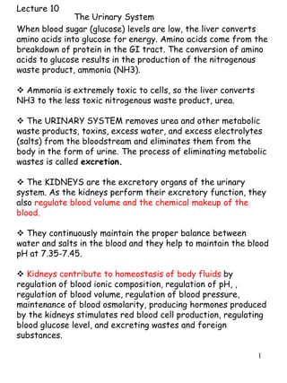
  • 1. 1 Lecture 10 When blood sugar (glucose) levels are low, the liver converts amino acids into glucose for energy. Amino acids come from the breakdown of protein in the GI tract. The conversion of amino acids to glucose results in the production of the nitrogenous waste product, ammonia (NH3).  Ammonia is extremely toxic to cells, so the liver converts NH3 to the less toxic nitrogenous waste product, urea.  The URINARY SYSTEM removes urea and other metabolic waste products, toxins, excess water, and excess electrolytes (salts) from the bloodstream and eliminates them from the body in the form of urine. The process of eliminating metabolic wastes is called excretion.  The KIDNEYS are the excretory organs of the urinary system. As the kidneys perform their excretory function, they also regulate blood volume and the chemical makeup of the blood.  They continuously maintain the proper balance between water and salts in the blood and they help to maintain the blood pH at 7.35-7.45.  Kidneys contribute to homeostasis of body fluids by regulation of blood ionic composition, regulation of pH, , regulation of blood volume, regulation of blood pressure, maintenance of blood osmolarity, producing hormones produced by the kidneys stimulates red blood cell production, regulating blood glucose level, and excreting wastes and foreign substances. The Urinary System
  • 2. 2  The urinary system consists of two kidneys, two ureters, one urinary bladder, and one urethra.  Urine is excreted from each kidney through its ureter and is stored in the urinary bladder until it is expelled from the body through the urethra.  The specialized branch of medicine that deals with structure, function, and diseases of the male and female urinary systems and the male reproductive system is known as nephrology.  The branch of surgery related to male and female urinary systems and the male reproductive system is called urology.
  • 3. 3
  • 4. 4 EXTERNAL ANATOMY  Kidneys are kidney bean-shaped and reddish in color. The adult kidney weighs 150 grams, is 10-12 cm (4-5 inches) long, 5.0-7.5 cm (2-3 in.) wide and 2.5 cm (1 in.) thick.  The kidneys are located between the posterior abdominal wall and parietal peritoneum (they are retroperitoneal), extending from the level of the T12 vertebra to the level of the L3 vertebra. The kidneys receive some protection from the lower part of the rib cage. The right kidney is slightly lower than left kidney.  The lateral surface of each kidney is convex. The medial surface is concave and has an indentation called the HILUS. The ureters, renal blood vessels, lymphatic vessels, and nerves enter or exit the kidney at the hilus.  There are 3 layers of supportive tissue surrounding each kidney: 1. RENAL CAPSULE: a transparent dense irregular connective tissue covering the kidney surface. The renal capsule prevents infections in surrounding regions from spreading to the kidneys. It also protects the kidney from physical trauma.  2. ADIPOSE CAPSULE: a mass of adipose tissue outside the renal capsule. The adipose capsule helps hold the kidney in place against the posterior abdominal wall and cushions the kidneys against external blows.  3. RENAL FASCIA: a layer of dense irregular connective tissue outside the adipose capsule that anchors the kidney to the abdominal wall.
  • 5. 5 KIDNEY: INTERNAL ANATOMY A frontal section of the kidney reveals 3 distinct regions inside the renal capsule and they are the RENAL CORTEX, RENAL MEDULLA, & RENAL PELVIS. 1. RENAL CORTEX: the outer region just inside the renal capsule; light red in color and granular in appearance. 2. RENAL MEDULLA: deep to the cortex; a darker, reddish brown color. Arranged in cone-shaped regions called RENAL PYRAMIDS, which have a striated appearance. Extensions of the RENAL CORTEX between the renal pyramids are called RENAL COLUMNS. Together, the RENAL CORTEX & MEDULLA form the parenchyma (functional portion) of the kidney where urine is formed. 3. Just inside the hilus is the cavity called the RENAL PELVIS. The renal pelvis collects urine that is formed in the renal cortex and medulla.
  • 6. 6 Nephrons • A nephron consists of a renal corpuscle where fluid is filtered, and a renal tubule into which the filtered fluid passes. • Nephrons perform three basic functions: glomerular filtration, tubular reabsorption, and tubular secretion. • A renal tubule consists of a proximal convoluted tubule (PCT), loop of Henle (nephron loop), and distal convoluted tubule (DCT). Distal convoluted tubules of several nephrons drain into to a single collecting duct and many collecting ducts drain into a small number of papillary ducts. • The loop of Henle consists of a descending limb, a thin ascending limb, and a thick ascending limb. • There are two types of nephrons that have differing structure and function. – A cortical nephron usually has its glomerulus in the outer portion of the cortex and a short loop of Henle that penetrates only into the outer region of the medulla – A juxtamedullary nephron usually has its glomerulus deep in the cortex close to the medulla; its long loop of Henle stretches through the medulla and almost reaches the renal papilla.
  • 7. 7 In the renal cortex, the ascending limb of the loop of Henle becomes twisted again as the DISTAL CONVOLUTED TUBULE. The distal convoluted tubules of several nephrons empty into each COLLECTING DUCT. The collecting ducts absorb water from the filtrate. Several collecting ducts empty into a PAPILLARY DUCT, which delivers urine to the RENAL PELVIS. The filtrate is processed as it passes through these parts of the nephron. Needed substances, such as water, glucose and electrolytes (salts), are returned to the bloodstream. After the filtrate is processed by the nephron, it is called URINE. Urine contains urea and other metabolic wastes, toxins, excess salts, and excess water, as well as drugs and excess vitamins.
  • 8. 8
  • 9. 9
  • 10. 10
  • 11. 11 BLOOD SUPPLY OF THE KIDNEYS While the body is at rest, the large left and right RENAL ARTERIES deliver about about 1.2 liters of blood through the kidneys every minute. The renal arteries are branches of the ABDOMINAL AORTA. After each renal artery enters a kidney, it branches into numerous small arteries, which carry most of the blood to the renal cortex. The arteries in the renal cortex branch into numerous AFFERENT ARTERIOLES, one for each nephron. Each afferent arteriole, divides to form a capillary tuft called the GLOMERULUS. The glomerulus is surrounded by the glomerular capsule and is specialized for filtration of blood. The glomerular capillaries merge to form the EFFERENT ARTERIOLE, which leads away from the glomerular capsule. The efferent arteriole's diameter is smaller than the afferent arteriole. This raises the blood pressure in the glomerulus, which aids in forcing water and solutes into the glomerular capsule.
  • 12. 12 Each efferent arteriole branches into a second capillary network called the PERITUBULAR CAPILLARIES. These capillaries cling closely to the PROXIMAL and DISTAL CONVOLUTED TUBULES of both cortical and juxtamedullary nephrons. The efferent arterioles of juxtamedullary nephrons also branch into the VASA RECTA, capillaries that surround the LOOP OF HENLE in the medulla. The peritubular capillaries and vasa recta are adapted for absorption. They absorb water and needed substances from the filtrate in the nephron, so that much of the water and solutes that the body requires are returned to the bloodstream rather than flushed out in the urine. Toxins and excess water and solutes remain in the filtrate and are eliminated as urine. The peritubular capillaries and vasa recta merge into venules that join veins which empty into the RENAL VEIN. The RENAL VEIN of each kidney leaves the kidney through the hilus. The renal vein of each kidney returns freshly cleansed blood to the INFERIOR VENA CAVA.
  • 13. 13 URETERS The URETERS are slender tubes about 25-30 cm (10-12 in) long that convey urine from the kidneys to the urinary bladder. Each ureter runs posterior to the parietal peritoneum (retroperitoneal) from the renal hilus to the level of the urinary bladder. At the urinary bladder, each ureter makes a medial turn toward the bladder and enters the posterior bladder wall at an oblique angle, which prevents backflow of urine from the bladder into the ureters. The inner lining of the ureter wall is MUCOSA that consists of transitional epithelium that stretches as urine passes through. Mucus secreted by the mucosa protects the epithelial cells from coming into contact with the urine, which contains toxins and is slightly acidic. Beneath the mucosa is the MUSCULARIS, which is composed of smooth muscle arranged in longitudinal and circular layers. The muscularis creates peristaltic contractions, similar to those in the GI tract. When the renal pelvis fills with urine, smooth muscle in the renal pelvis wall contracts rhythmically, sending urine into the ureter. Peristalsis propels the urine through the ureter to the urinary bladder.
  • 14. 14 URINARY BLADDER The URINARY BLADDER is a hollow muscular organ located in the pelvic cavity posterior to the pubic symphysis. Like the kidneys and ureters, the urinary bladder is retroperitoneal. In males, the urinary bladder lies anterior to the rectum. In females, the bladder is anterior to the vagina and inferior to the uterus. The function of the urinary bladder is the temporary storage of urine. The inner wall of the urinary bladder consists of a MUCOSA made of transitional epithelium, which is able to stretch a great deal. Mucus produced by the bladder mucosa helps protect the wall from urine. Beneath the mucosa is smooth muscle, arranged in longitudinal and circular layers. The smooth muscle in the wall allows the bladder to stretch a great deal.
  • 15. 15 When empty, the bladder is 5.0-7.5 cm (2-3 in) long. Its walls are thick and its mucosa is thrown into folds called RUGAE. When urine accumulates in the bladder, it expands and becomes pear-shaped as it rises upward in the pelvic cavity. The transitional epithelium and smooth muscle stretches and thins. This increases the internal volume of the bladder by decreasing the thickness of its wall. A moderately full bladder is about 12.5 cm (5 in) long and holds about 500 ml of urine. The average capacity of the bladder is 700-800 ml. When the volume of urine in the bladder exceeds 200-400 ml, stretch receptors in the bladder transmit nerve impulses to the brain, initiating the conscious desire to urinate.
  • 16. 16 URETHRA The URETHRA is a muscular tube that drains urine from the urinary bladder and conveys it out of the body. The INTERNAL URETHRAL SPHINCTER is an involuntary smooth muscle sphincter at the base of urinary bladder, surrounding the opening called the INTERNAL URETHRAL ORIFICE. The EXTERNAL URETHRAL SPHINCTER is a voluntary skeletal muscle sphincter in the UROGENITAL DIAPHRAGM. The urogenital diaphragm is formed by skeletal muscles of the pelvic floor;. In FEMALES, the urethra is 4 cm (1.5 in) long and 6 mm in diameter. It lies posterior to the pubic symphysis and anterior to the vagina. The EXTERNAL URETHRAL ORIFICE lies anterior to the vaginal opening. In MALES , the urethra is 15-20 cm (6-8 in) long. The urethra passes through the PROSTATE GLAND, then through the PENIS to the outside through the EXTERNAL URETHRAL ORIFICE.
  • 17. 17 WASTE MANAGEMENT IN OTHER BODY SYSTEMS • One of the many functions of the urinary system is to rid the body of waste materials. • Other organs, tissues and processes contribute to waste management. • Buffers prevent an increase in the acidity of body fluids. • The blood transports wastes. • The liver is the primary site for metabolic recycling. • The lungs excrete CO2. H2O, and heat. • Sweat glands eliminate excess heat, water, and CO2, plus small quantities of salts and urea. • The GI tract eliminates solid, undigested foods, waste, some CO2, H2O, salts and heat.