2. 2
Terminology
• Genetics: Study of what genes are, how they carry information,
how information is expressed, and how genes are replicated.
• Gene: Segment of DNA that encodes a functional product,
usually a protein.
• Genome: All of the genetic material in a cell
• Genotype: Genes of an organism
• Phenotype: Expression of the genes
3. 3
Flow of Genetic Information
Genetic information can be transferred between generations of the cells,
through replication and Occasionally between cells of the same generation.
4. 4
The structure of the DNA molecule explains
how it carries and replicates genetic
information.
1. Nucleic acids are polymers made up of
nucleotides.
What is a nucleotide?
• Consists of a 5 carbon sugar (deoxyribose
or ribose)
• Consists of a nitrogenous base (adenine,
thymine, guanine and cytosine in DNA and in
RNA uracil replaces thymine.
• Consists of a phosphate group that was
converted from a triphosphate to a
monophosphate in forming the chain.
• Nucleotides linked together by
phosphodiester bonds.
• NOTE that one end of this chain will have a
free 5' OH group (referred to as the 5'
end) and the other end of the chain will
have a fee 3' OH group (referred to as the
3' end).
6. 6
DNA STRUCTURE
• hydrogen bonded
nucleotides on opposite
helices
• DNA helices are
antiparallel
• pyrimidines bond with
purines
- T A
- C G
7. 7
DNA is the Genetic Material
• Of all eukaryotic and
prokaryotic organisms
• Of many viruses
• Some viruses have RNA
as the genetic material
• DNA is made up of two
chains (strands) each
consisting of a series of
nucleotides linked
together by
phosphodiester bonds.
8. 8
DNA
• Eukaryotic organisms have DNA in the mitochondria and
chloroplasts
• There are differences between the DNA of the nucleus and
the mitochondria and chloroplasts.
• Genes are specific sequences of the nucleotides.
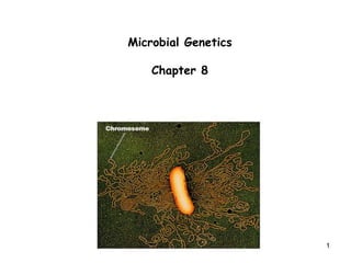
• In the cell, nucleotides are arranged into structures called
chromosomes
• Most prokaryotes have a single circular chromosome
• Eukaryotic nuclei have a number of linear chromosomes
9. 9
The complementary structure of the DNA provides a basis for copying a
complementary strand from each strand of the DNA molecule to make two
molecules identical to the original strand.
DNA replication is semi-conservative
• In order to replicate a DNA molecule, it is possible to imagine three ways,
this would be done.
• Method 1 - the two original DNA strands separate, new copies of each are
made, and the old copies; then reform the double helix, and the new copies
reform a second double helix. This would be called conservative replication.
• Method 2 - the two original DNA strands separate, new complementary
copies of each strand are made forming two new molecules each containing
one of the original strands and one of the new strands. This would be called
semiconservative replication.
• Method 3 – the parental and the newly synthesized strands become
randomly mixed during the replication process, so that two strand arise
which contain both old and newly made DNA. This would be called a
dispersive mode of replication.
11. 11
The details of DNA replication
2. DNA strands in the double helix must be unwound during replication.
- If the two strands of the double helix are wrapped around each other
like the strands of a rope the strands must be unwound for replication to
occur.
Note that if you pull the strands of a rope apart, the rope must either
rotate or twist into tighter coils. So we would expect similar things to
happen when complementary DNA strands are separated for replication.
- Separating the two strands of DNA accomplished by DNA helicases
enzymes that travel along the helix, opening the double helix as they move.
Once the strands are separated, helix-destabilizing proteins bind to single
DNA strands preventing re-formation of the double helix until the strands
are copied (Fig 5.15).
- Another group of enzymes called topoisomerases produce breaks in the
DNA molecules and then rejoin the strands, relieving strain and
effectively preventing the formation of knots which can block the DNA
replication (Fig 5.24, 5.25).
12. 12
DNA
• Helicase – forces open strands of DNA
• Single stranded binding proteins - keeps strands single
• Primase - makes RNA primer
• DNA polymerase - extends DNA from RNA primer
• All known DNA polymerases synthesize DNA from the 5’ end to the 3’ end.
13. 13
Fig 5.24. The “winding problem” that arises during DNA replication.
For a bacterial replication fork moving at 500 nucleotides per second,
the parental DNA helix a head of the fork must rotate at 50
revolutions per second.
This is achieved by
Topoisomerases
Helicase
14. 14
Fig 5.25 The reversible nicking reaction catalyzed by a eukaryotic DNA
topoisomerase I enzyme.
16. 16
3. DNA synthesis always proceeds 5' to 3'.
- The enzyme that actually duplicates the DNA strand
is called DNA polymerase. DNA polymerase can only
make DNA molecules starting at the 5' end and
proceeding to the 3' end.
4. DNA synthesis requires an RNA primer.
- DNA polymerase cannot start making DNA without a
primer around 10 nucleotides long.
- An RNA primer is first made to start DNA synthesis.
This is around 10 nucleotide long piece of RNA that is
complementary to the DNA strand.
- The RNA primer is made by a complex of enzymes
called a primosome.
- Enzyme that remove the RNA and replace the RNA
by copying DNA nucleotides to the strand is called
RNase.
18. 18
5. DNA synthesis is continuous on one strand and discontinuous in the other.
• DNA synthesis does not proceed from the ends of strands to the middle,
but from the middle to the ends.
• During replication of DNA the two strands of the duplex molecule separate
at a special sequence called origin of replication to form a replication fork
(Y-shaped structure) . Replication forks are the point at which DNA
replication is occurring.
• At a replication fork, the DNA of both new daughter strands is synthesized
by DNA polymerase.
• Because the two template strands are anti-parallel and polymerases only
work 5‘ to 3‘.
• The strand that is continuously replicated in the same direction as the fork
is called leading strand. While the other strand (Lagging strand) in the
direction opposite to the fork is replicated discontinuously in short
fragments called Okazaki fragments. These fragments are about 300
nucleotides long in eukaryotic cells, and about 1,000-2,000 nucleotides long
in bacteria.
They are joined together soon after synthesis by DNA ligase to produce a
continuous lagging strand. (Fig 5.26)
19. 19
Fig 5.26A. DNA Synthesis is Directional (5’ to 3’ only)
Continuous
Replication
Dis-continuous
Replication
Leading Strand:
Lagging Strand:
Replication forks are the point at which DNA replication is occurring
20. 20
6. DNA synthesis is usually bidirectional.
- Experimental evidence shows that DNA replication is bidirectional.
Replication Bubble
Lagging Strands
Leading Strands
Replication Fork Replication Fork
Fig 5.26A
21. 21
Transcription
The structure, function, development, and reproduction of an organism
depend on the properties of the proteins present in each cell and tissue.
When a protein is needed by a cell, the genetic code for that protein
must must be read from the DNA and processed.
Two major steps occurring during protein synthesis:
1. Transcription = synthesis of a single-stranded RNA molecule using the
DNA template (1 strand of DNA is transcribed).
2. Translation = conversion of a messenger RNA sequence into the amino
acid sequence of a polypeptide (i.e., protein synthesis)
22. 22
Not all genes encoded protein, so not all gene transcripts are the kind of
RNA that is translated. In fact, there are four different types of RNA,
each encoded by different genes:
1. mRNA: Messenger RNA, encodes the amino acid sequence of a
polypeptide.
2. tRNA: Transfer RNA, brings amino acids to ribosomes during
translation
3. rRNA: Ribosomal RNA, with ribosomal proteins, forms complexes
called ribosomes, the structure on which mRNA is translated
How RNA chain is Synthesized?
Associated with each gene are sequences called gene regulatory
elements which are involved in regulation of transcription.
In both prokaryotes and eukaryotes, RNA polymerase catalyzes the
process of transcription.
The DNA double helix unwinds for a short region next to the gene
before transcription can begin .
One strand serves as a template strand for the synthesis of mRNA
Nucleotides are added to the growing RNA strand in a 5’-3’ direction.
23. 23
RNA synthesis
• DNA continues to be open up as transcription occurs.
• Transcription bubble of approximately 17 base pairs open up as the
RNA/DNA hybrid complex is formed.
• This bubble closes back up as the RNA/DNA hybrid passes out of
the transcription bubble.
26. 26
INTIATION OF TRANSRIPTION AT PROMOTERS
In both prokaryotes and eukaryotes, the process of transcription
occurs in three steps:
• Initiation
• Elongation
• Termination
• Elongation is conserved in prokaryotes and eukaryotes.
• Initiation and termination proceed differently.
Initiation of transcription in
E. coli :
Each gene has three regions:
1. 5’ Promoter, interacts
with the RNA
polymerase and
determines the start
point for transcription.
2. Transcribed sequence,
or RNA coding sequence
that will contain
message for mRNA.
3. 3’ Terminator, sequence
which specifies stop
Promoter.
28. 28
mRNA production is different in prokaryotes and eukaryotes:
Prokaryotes
1. mRNA transcript is mature, and used directly for translation without
modification.
2. Since prokaryotes lack a nucleus, mRNA also is translated on ribosomes
before is is transcribed completely (i.e., transcription and translation are
coupled).
3. Prokaryote mRNAs are polycistronic, they carry sequences coding for
several protein.
Eukaryotes
1. mRNA transcript is not mature (pre-mRNA) and must be modified by
processing.
2. Transcription and translation are not coupled (mRNA must first be exported
to the cytoplasm before translation occurs).
3. Eukaryote mRNAs are monocistronic, they carry sequences coding for one
protein.
30. 30
1. Synthesis of ribosomal RNA and ribosomes:
Protein synthesis takes place in ribosomes.
1. Each cell contains thousands of ribosomes.
2. Consist of two subunits (large and small) in prokaryotes and eukaryotes, in
combination with ribosomal proteins.
3. E. coli 70S model: (nt: nucleotide)
• 50S subunit = 23S (2,904 nt) + 5S (120 nt) + 34 proteins
• 30S subunit = 16S (1,542 nt) + 20 proteins
4. Mammalian 80S model:
• 60S subunit = 28S (4,700 nt) +5.8S (156 nt) + 5S (120 nt) + 50 proteins
• 40S subunit = 18S (1,900 nt) + 35 proteins
31. 31
Translation
• Gene expression involves processes of transcription and
translation which result in the production of polypeptides whose
structure is determined by genes.
• Each sequence of 3 bases (a codon) codes for a specific amino
acid or represents a stop codon. 20 different amino acids occur
in living cells
• There are 64 possible 3 nucleotide sequences for the 20 amino
acids, some amino acids are coded for by more than one codon.
We say the genetic code is redundant, therefore.
• The genetic code is universal. That is all organisms use essentially
the same genetic code.
32. 32
Fig. 6.2 Structures
of the 20 naturally
occurring amino
acids organized
according to
chemical type.
34. 34
Translation-protein synthesis:
1. Protein synthesis occurs on ribosomes.
2. mRNA is translated 5’ to 3’.
3. Protein is synthesized N-terminus to C-terminus.
4. Amino acids bound to tRNAs are transported to the ribosome.
Facilitated by:
• The binding of each amino acid to its own specific tRNAs.
• Complementary base-pairing between the mRNA codon and
the tRNA anti-codon.
• mRNA recognizes the tRNA anti-codon (not the amino acid).
35. 35
tRNA: Transfer RNA, brings amino acids to ribosomes during
translation. tRNA will bind to the tri-nucleotide.
mRNA UUU codon
tRNA AAA (with Phe) anti-codon
mRNA UCU codon
tRNA AGU (with Ser) anti-codon
mRNA CUC codon
tRNA GAG (with Leu) anti-codon
- tRNA molecules are transcribed from tRNA
genes and are about 70-80 nucleotides long.
- tRNA's can fold back on themselves and form
loops with complementary base pairing in a
stems region. Most tRNA molecules contain 3 or
4 such loops, and a stem where the 5‘ and 3'
ends of the molecule are complementary.
38. 38
Fig. 6.17 Diagram of a polysome, a
number of ribosomes each translating
the same mRNA sequentially.
• In both prokaryotes and eukaryotes, once the ribosome moves away from
the initiation site mRNA, many ribosomes around 8-10 ribosomes
simultaneously translate mRNA and synthsesing protein from it. The
complex between an mRNA molecule and all ribosomes that are translating it
simultaneously is called polyribosome or polysome (fig 6.17).
39. 39
Mutations are changes in the DNA sequence:
If an error in copying the DNA strand is made so that a strand with an altered
nucleotide sequence is created this may change the amino acid coded for by
the mRNA derived from that gene.
- We call such an event a mutation in a gene.
- Such changes may occur as accidents during DNA replication, or may be
induced by radiation or chemicals.
1. Mutations which result in the substitution of one base for another are
referred to as point mutations or missense mutations.
2. Mutations which result in the formation of a stop codon where an amino
previously was are called nonsense mutations.
- Nonsense mutations result in the premature termination of the protein
sequence, and thus an active protein is not formed.
3. When mistakes are made which either add an extra nucleotide, or delete a
nucleotide, the reading frame of the codons for the remainder of the length
of the mRNA is altered. Such mutations are called frameshift mutations.
4. Some mutations are caused by pieces of DNA which can jump around the
genome. Such jumping DNA is called a transposon.
- Transposons exist in both prokaryotes and eukaryotes.
40. 40
Regulation of Gene Expression in Bacteria
Bacteria are free-living organisms that grow by increasing in mass and
then divide by binary fission. Growth and division are controlled by genes.
1. Genes whose activity is controlled in response to the needs of a cell or
organism are called regulated genes.
2. Genes that generally are continuously expressed (always active in
growing cell; no matter what the conditions are) are known as constitutive
genes (housekeeping genes).
Examples include genes that code for enzymes needed for protein
synthesis and glucose metabolism.
All genes are regulated at some level, so that as resources impaired the
cell can respond with a different molecular strategy.
41. 41
Prokaryotic genes are often organized into operons: cluster of genes in which
expression is regulated by operator-repressor protein interactions, operator
region, and the promoter.
That is, the genes are adjacent to each other and are transcribed together to
make a single mRNA. This mRNA is said to be polycistronic, because it carries
the information for more than one type of protein.
Contents of an operon:
•Promoter
•Repressor
•Operator (controlling site)
•Coding sequences
•Terminator
Gene regulation in bacteria is similar in many ways to the emerging information
about gene regulation in eukaryotes, including humans.
Much remains to be discovered; even in E. coli, one of the most closely studied
organisms on earth, 35% of the genomic ORFs (nucleotide sequence starts with
initiation codon and ends with termination codon) have no attributed function.
42. 42
Most studied operon:
The lac Operon of E. coli
The lac operon is one example of how bacteria can turn on or turn off
genes in response to environmental conditions.
When gene expression is turned on in a bacterium by adding a substance
(such as lactose) to the medium, the genes involved are said to be
inducible.
An inducible operon responds to an inducer substance (e.g., lactose). An
inducer is a small molecule, called effectors, that joins with a regulatory
protein to control transcription of the operon.
• Inducer = chemical or environmental agent that initiates transcription
of an operon.
• Induction = synthesis of gene product(s) in response to an inducer.
43. 43
Fig. 16.1
General
organization
of an inducible
gene
The regulatory event typically occurs at a specific DNA sequence (controlling
site) near the protein-coding sequence (Figure 16.1).
Control of lactose metabolism in E. coli is an example of an inducible operon
44. 44
Lactose as a Carbon Source for E. coli
E. Coli can grow in a simple medium containing salts (including a nitrogen
source) and a carbon source such as glucose. The energy for biochemical
reactions in the cell comes from glucose metabolism. The enzymes required
for glucose metabolism are coded for by constitutive genes.
1. Metabolism of other alternative types of sugars (e.g., lactose) are regulated
specifically. In other words, the genes are regulated genes whose products are
needed only at certain time. Presence of the sugar stimulates synthesis of the
proteins needed.
2. Lactose is a disaccharide (glucose + galactose). If lactose is E. coli’s sole
carbon source, three genes are expressed:
a. β-galactosidase (lacZ) has two functions:
i. Breaking lactose into glucose and galactose. Galactose is converted
to glucose, and glucose is metabolized by constitutively produced
enzymes.
ii. Converting lactose to allolactose. Allolactose is a compound
important in regulating expression of the lac operon (Figure 16.2).
b. Lactose permease (lacY; also called M protein) is required for transport
of lactose across the cytoplasmic membrane.
c. Transacetylase (lacA) is poorly understood.
45. 45
Fig. 16.2 Reactions catalyzed by the enzyme -galactosidase. Lactose brought
into the cell by the permease is converted to glucose and galactose (top) or to
allolactose (bottom), the true inducer for the lactose operon of E. coli.
46. 46
Experimental Evidence for the Regulation of lac Genes
• The experiments of Jacob and Monod (Pasteur Institute, Paris, France)
produced an understanding of arrangement and control of the lac operon
in E.coli.
• Earned Nobel Prize in Physiology or Medicine 1965.
• They produced 2 different types of mutations in the lac operon:
– Mutations in protein-coding gene sequences.
– Mutations in regulatory sequences.
3. The lac operon shows coordinate induction: the simultaneous transcription
and translation of two or more genes brought about by the presence of an
inducer.
In glucose medium, E. coli normally has very low levels of the lac
gene products.
When lactose is the sole carbon source (in the absence of glucose),
levels of the three enzymes increase coordinately (simultaneously)
about 1,000-fold.
Allolactose (not lactose) is the inducer molecule directly
responsible for the increased production of the three
enzymes. Furthermore, the mRNA for the enzymes has a
short half-life. When lactose is gone, lac transcription
stops, and enzyme levels drop rapidly.
47. 47
Mutations in the Protein-coding (structural) Genes
1. Mutagens (chemicals that induce mutations) produced mutations in the lac
structural genes that were used to map their locations.
a. β-galactosidase is lacZ. b. Permease is lacY. c. Transacetylase is lacA
Mapping experiments showed that the three genes are tightly linked in the
order: lacZ-lacY-lacA
2. The type of mutation made a difference in expression of the downstream
genes: a. Missense mutation b. Nonsense mutations
– Nonsense mutations in the lacZ not only knocked out the function of -
galactosidase but also knocked out the function of lactose permease and
transacetylase.
– LacY nonesense mutations results in nonfunctional lactose permease and
transacetylase, but -galactosidase activity was unaffected.
– LacA nonesense mutations results in nonfunctional transacetylase, but -
galactosidase and permease activities were unaffected.
• Conclusion: 3 lac operon genes are linked in the following order:
–lacZ codes -galactosidase
–lacY codes lactose permease
–lacA codes transacetylase
48. 48
Fig. 16.3 Translation of the polygenic mRNA encoded by lac utilization genes in (a) wild type
E.coli and (b) a mutant strain with a nonsense mutation in the b-galactosidase (lacZ) gene.
3. The interpretation of gene polarity is that ribosomes translate the first
gene in the polycistronic (polygenic) mRNA, and finish in proper position to
initiate and translate the next gene. Premature translation termination
prevents this by reducing or abolishing the synthesis of enzymes encoded by
structural genes or translation of the downstream genes (Figure 16.3).
49. 49
Mutations Affecting the Regulation of Gene Expression
Jacob and Monod also isolated mutants in which all gene products of the lac
operon were synthesized constitutively; that is, they were synthesized
whether or not lactose (inducer) was present. They hypothesized that the
mutations were regulatory mutations that affect the normal mechanism
controlling the expression of the structural genes for the enzymes.
They identified 2 classes of constitutive mutations (Figure 16.4):
a. Mutations in the lac operator (lacO) just upstream from the lacZ gene.
b. Mutations further upstream in the lac repressor gene (lacI).
Fig. 16.4 Organization of the lac genes of E. coli and the associated
regulatory elements the lac operon: the operator, the promoter, and
regulatory gene.
50. 50
Jacob and Monod’s Operon Model for the Regulation of lac Genes
1. An operon is a cluster of genes that are regulated together. Expression is regulated by
operator-repressor protein interactions, operator, and a promoter.
2. The lacI gene has its own constitutive weak promoter and terminator, and repressor
protein is always present in low concentration.
i. The repressor functions as a tetramer (4 polypeptides).
ii. Repressor protein binds the operator (lacO+ ;normal operator), and prevents RNA
polymerase initiation to transcribe the operon genes.
iii. Binding of the repressor to the operator is not absolute, and so an occasional
transcript is made, resulting in low levels of the structural proteins (lacZ, lacY,
and lacA proteins are always synthesized).
iii. As soon as lactose occurs in high concentration, lac operon switches to the “on”
position.
Fig. 16.6 Molecular model of the lac
repressor tetramer. The four
monomers are colored green, violet, red,
and yellow.
Fig. 16.5 Functional state of the lac operon in wild-
type E. coli growing in the absence of lactose
51. 51
When wild-type E. coli grows in the presences of lactose as the sole carbon
source , some lactose is converted by β-galactosidase into allolactose.
i. Repressor protein bound with allolactose and changes its shape; this is called
allosteric shift. As a result, the repressor looses its affinity for the lac
operator, and it dissociates from the site (lac operator). Free repressor-
allolactose complexes are unable to bind the operator.
ii. Allolactose induces expression of the lac operon, by removing the repressor
and allowing transcription to occur.
Fig. 16.7 Functional state
of the lac operon in wild-
type E. coli growing in the
presence of lactose as
the sole carbon source
iii. SO with no repressor
bound to the operator,
RNA polymerase initiates
synthesis of a single
polygenic mRNA containing
mRNA for lacZ, lacY, and
lacA.
52. 52
Jacob and Monod’s Operon Model for the Regulation of lac Genes (cont’d)
Different types of mutations occur in lacO and lacI :
lacO - change repressor binding site (repressor does not bind)
- continuously expressed
lacI - change repressor conformation results of amino acid changes
in the repressor (repressor cannot bind operator)
- continuously expressed
Other classes of lac I mutant:
- super-repressors mutants shows no production of lac enzymes
in the presence or absence of lactose.
- the mutant repressor gene produces a super-repressor protein
that can bind to the operator but cannot recognize the inducer
(allolactose).
56. 56
8. The mutants of lacI gene point out the different functions of the repressor.
Specifically, the repressor is involved in three different recognition
interactions:
a. Binding of the repressor protein to the operator region.
b. Binding of the inducer (allolactose) to the repressor protein.
c. Binding of individual repressor polypeptides to each other to form an
active tetramer.
The lac operon is one example of how bacteria can turn on or off genes
in response to the environment conditions. The presence of lactose
induces the synthesis of enzymes necessary to convert lactose into
glucose. Mutations in this operon demonstrate how the different regions
are controlled.
57. 57
Tryptophan Operon of E. coli
• A bacterium has certain operons and other genes systems that enable it
to manufacture any amino acid that is lacking in the medium so that it
can grow and develop (genes for amino acid synthesis are expressed;
turned on).
• When the amino acid is present in the growth medium, though, the genes
encoding the enzymes for that amino acid’s biosynthetic pathway are
turned off ( Genes for amino acid synthesis are repressed, repressible
operons).
• This is the opposite of the lac operon in which the presence of the
inducer turned on the pathway. Here the presence of an amino acid
(tryptophan) turns off transcription.
• The tryptophan (Trp) operon of E. coli is one of the most extensively
studied repressible operons.