2. 420 Jordan
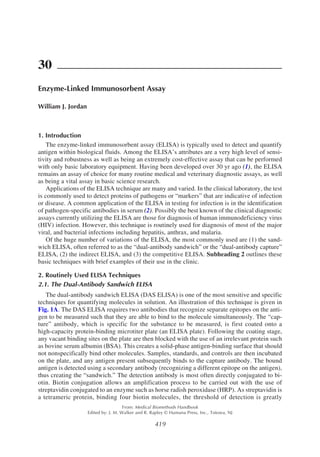
Fig. 1. (A) The dual-antibody sandwich ELISA. (B) Estimating antigen concentration using
a typical DAS ELISA standard curve. In parallel with samples, a titration series of known
amounts of antigen are also examined and the optical density (OD) readings are used to give a
“standard curve.” OD readings from samples can then be compared directly to this standard
curve in order to gain an estimate of the amount of antigen in the sample. Samples often require
testing at several dilution s so that the reading can be made within or as close to this part of the
curve as possible.
3. Enzyme-Linked Immunosorbent Assay 421
enhanced. Developing the assay into a readable format involves the addition of a substrate
such as 3,3',5,5'-tetramethylbenzidine (TMB) for the HRP enzyme. In the presence of the HRP
enzyme, TMB begins a colorimetric reaction that can then be measured using a spectrophotom-
eter. The resulting color (optical density [OD]) relates directly to the amount of antigen present
within the sample. Comparison of the OD within a sample to those obtained using a standard
curve of known concentrations allows an estimate of antigen concentration within that sample
to be gained (see Fig. 1B)
2.1.1. Basic DAS ELISA Protocol
1. Coating with capture antibody: Capture antibody diluted in coating buffer is added to a high-
capacity-protein binding 96-well microtiter plate. The plate is then incubated, allowing the
antibody to bind to the plate; subsequently, the plate is washed to remove any excess or un-
bound antibody.
2. Blocking vacant binding sites: Vacant binding sites on the plate are then blocked with an irrel-
evant protein such as BSA. Following incubation, the plate is washed once more to remove
excess unbound protein.
3. Addition of samples/standards: A titration series of known standards must be prepared. Ide-
ally, these should be diluted in a matrix representing that of the samples to help identify false
positives (e.g., if there are any substances in the matrix that bind nonspecifically to the plate
that have enzyme activity). A negative control must also be included (e.g., culture medium
only or serum known to be negative for the antigen to be measured). Samples and antigen
standards are then incubated on the ELISA plate, allowing any antigen present to bind to the
coating antibody.
4. Addition of detection antibody: Following the addition of samples, the biotinylated detection
antibody is added, which binds to any antigen bound to the plate. Following incubation, the
plate is once again washed thoroughly to remove unbound reagents.
5. Addition of enzyme conjugate: Streptavidin conjugated to an enzyme such as HRP is then
incubated onto the plate. This binds to biotin molecules on the detection antibody. The plate is
once again washed thoroughly to remove unbound reagents.
6. Development and analysis: The ELISA is then developed using a suitable substrate (e.g., TMB
to detect the HRP enzyme). The OD can be measured using a spectrophotometer. The OD
values from the standard titration of antigen are then used to determine an estimate of antigen
within samples.
An example of the DAS ELISA in the clinic is the AlzheimAlert™ ELISA kit (Nymox
Corporation) for measuring a neural thread protein (AD7c-NTP) in the urine of patients. The
presence of high levels of AD7c-NTP has been shown to be correlated with Alzheimer’s dis-
ease and is used for early diagnosis and subsequent monitoring of the progression of this dis-
ease. During the assay, microtiter plates are first coated with a monoclonal antibody reacting
against the neural protein. After blocking vacant binding sites and washing the plate to remove
unbound proteins, a urine sample from the patient is incubated onto the plate. Any neural pro-
tein present binds to the antibody. As this assay is quantitative, a standard titration of known
amounts of recombinant neural protein is set up in parallel. The readings from these are com-
pared to that obtained from the sample(s) to estimate the level of neural protein in the urine.
The plate is then washed to remove any excess protein and a secondary (enzyme-conjugated)
antibody that recognizes a different epitope of the neural peptide is added to the plate. The
presence of the protein in the urine sample is thus reflected by the presence of the enzyme, and
this enzyme is detected using a substrate that completes a colorimetric reaction that is read
using a spectrophotometer. The presence of high levels of the neural protein AD7c-NTP is
indicative of Alzheimer’s disease.
2.2. The Indirect ELISA
In many instances, only one specific antibody might be available with which to create an
assay to detect antigen and so a DAS ELISA is unsuitable. In such a situation, an indirect
ELISA or a competitive ELISA can be established. During the indirect ELISA, the sample
4. 422 Jordan
itself is coated directly onto the microtiter plate and is then detected using the specific anti-
body. An illustration of this technique is presented in Fig. 2.
The indirect ELISA is often used to detect antibody in samples. An example of such an
indirect ELISA used in the clinic is the Murex HIV-1.2.0 ELISA kit (Abbott Laboratories).
HIV-infected individuals commonly produce antibodies against the HIV protein and the pres-
ence of such antibodies in the serum is indicative of infection. The basis of this ELISA is a
microtiter plate coated with a mixture of HIV proteins. This mixture includes a synthetic pep-
tide representing an immunodominant region of HIV-1, a recombinant HIV envelope protein,
and an HIV core protein. Blood is taken from patients and the serum fraction used for the test.
By allowing the red blood cells from a blood sample to “settle” in a test tube, the serum layer
can then be taken with a pipet. This serum sample is normally tested against control sera (both
positive and negative to compare to the sample and thus give a diagnosis). During incubation of
the serum on the microtiter plate, antibodies reacting against the HIV in the sample bind to the
antigens. The plate is then washed to remove excess antibodies and a secondary antibody (con-
jugated to the enzyme HRP) that specifically recognizes human antibodies is added to the plate.
This will, therefore, only bind if antibodies against the HIV proteins were originally present in
the serum sample. Samples not containing specific antibody will not cause the conjugate to
bind to the well. A further wash of the plate is then performed to clear any unbound secondary
antibody and the substrate is then added (TMB). The wells that had sera containing specific
anti-HIV antibodies develop a blue color, which is converted to yellow when the reaction is
stopped with sulfuric acid. The color is read spectrophotometrically at 450 nm and is directly
related to the concentration of antibody to HIV in the sample. Thus, a strong positive signal
indicates that the individual has been infected with HIV.
2.3. The Competitive ELISA
There are many variations and adaptations of the competitive ELISA, although the general
principle for all of these remains the same. A typical representation of this assay is shown in
Fig. 2. The basic direct ELISA.
5. Enzyme-Linked Immunosorbent Assay 423
Fig. 3A. As with the ELISAs described above, the initial stage of the competitive ELISA gen-
erally involves coating a high-capacity protein-binding microtiter plate with an antibody di-
rected against the antigen to be measured. However, during a competitive ELISA, a sample that
is to be analyzed is first mixed with a known amount of antigen that has been enzyme-conju-
gated. This mixture is then added to the coated ELISA plate. The two forms of the antigen
compete for binding sites to the antibody-coated plate and this binding is proportional to their
respective molar ratios in the mixture. Because only the conjugated form of the antigen allows
for the colorimetric reaction to develop in the presence of the substrate, the maximal reading
occurs when there is no antigen in the sample. The more antigen that is present in the sample,
the lower the resulting OD reading. Thus, unlike a standard sandwich ELISA, the readout is
inversely associated with the amount of antigen (see Fig. 3B).
In practice, the competitive ELISA is the least commonly used of the ELISA variations,
mainly because of the increased workload and expertise required. This technique is most often
used to detect antigens that are very small, such as hormones. An example of the competitive
ELISA in the clinic is the BQ T4 ELISA kit (Bioquant Corporation). This ELISA is used for the
quantitative measurement of total thyroxine (T4) in human serum or plasma and is used for the
diagnosis of hypothyroidism and hyperthyroidism. The level of T4 is decreased in hypothyroid
patients and is increased in hyperthyroid patients. The BQ T4 is a solid-phase competitive
ELISA. The samples are mixed with T4 that has been enzyme-conjugated and are then added to
a microtiter plate that has been precoated with an anti-T4 monoclonal antibody. T4 in the
patient’s serum competes with a T4 enzyme-conjugated recombinant T4 for binding sites. Plates
are then washed to remove unbound T4 and T4 enzyme conjugate. Upon the addition of the
substrate, the intensity of color is inversely proportional to the concentration of T4 in the
samples. A standard curve is prepared relating color intensity to the concentration of the T4 and
a diagnosis can be made.
A variation of this competitive ELISA is often used to measure levels of antibody in solu-
tion. In this technique, the antigen itself is coated onto the ELISA plate and an enzyme-conju-
gated “detection antibody” is used to generate the OD reading. Any antigen-specific antibody
in the sample competes with the enzyme-conjugated antibody for binding to the plate. Again,
the reading is inversely proportional to the amount of antigen present and can be cross-refer-
enced with readings from a standard curve to gain a quantitative estimate of antibody in the
sample.
2.4. The Blocking ELISA
In this variation of the competitive ELISA, the sample to be measured is not mixed with the
enzyme-conjugated antigen, but is preincubated onto the coated plate prior to washing and the
addition of the conjugated antigen. Thus, the sample “blocks” rather than “competes” for the
sites on the plate. This can result in a greater degree of sensitivity, although it is more time-
consuming because it relies on an additional step. The principle of the assay, however, remains
the same as the competitive ELISA.
3. Establishing an ELISA Protocol
In order to set up a reliable and durable ELISA, it is essential to first optimize a number of
the parameters mentioned above. The level of optimization will, of course, depend on exactly
what is required from the assay. In some cases, a simple “yes or no” answer is desired and a
simple standard procedure might be sufficient. If, however, high sensitivity is the aim with
accurate quantification of the molecule in question, then carefully setting up the optimal condi-
tions in advance saves a great deal of time in the long term.
Optimization of the following parameters is most often required: (1) pH of coating buffer,
(2) concentration of capture antibody, and (3) concentration of streptavidin–HRP conjugate.
Another important aspect of optimization that must be considered, especially when it is antigen
rather than antibody that is to be coated onto the plate, is the type of ELISA plate used. Recent
6. 424 Jordan
Fig. 3. (A) Principles of a typical competitive ELISA. (B) Estimating antigen concentration
using a typical competitive ELISA standard curve. A constant amount of conjugated standard
antigen is used to give the background OD reading (a). This reading is competed out with
increasing concentrations of unconjugated antigen added, creating the standard curve. Antigen
within samples competes with the constant conjugated antigen in the same manner, giving an
OD reading (b) that can be read off using the standard curve to estimate the concentration
within the sample (c). The most sensitive part of the curve (d) is where the smallest difference
in concentration of the competing antigen has the greatest impact on the OD. Samples often
require testing at several dilutions so that the reading can be made within or as close to this part
of the curve as possible.
7. Enzyme-Linked Immunosorbent Assay 425
advances in ELISA-plate-binding surface technology now allow adhesion of a number of
important molecules other than protein, including carbohydrates and lipids, thus expanding
the array of microbial antigen that can be identified and quantified using this technique (3).
If the major aim of the ELISA is to obtain quantification of substances present in extremely
low concentrations, there are a number of adaptations to the technique that can be used. Such
techniques often use alkaline phosphatase (AP) enzyme systems rather than HRP, providing
greater levels of sensitivity. Other technological advances that increase ELISA sensitivity can
be found in the color-development stage of the technique. For example, the AP enzyme has
been used to lock into a circular redox cycle producing an end product such as red formazan,
which is hugely amplified in comparison to standard amplification methods (4). Chemilumi-
nescent amplified ELISA principles have also been shown to give very high sensitivity (5). In
one example, an ELISA to measure proinsulin in serum was optimized using chemilumines-
cence to increase the sensitivity to measure as little as 1 zmol (about 350 molecules) of alkaline
phosphatase (6). Although extremely sensitive, such techniques are time-consuming to set up
and optimize and more expensive than the simple colorimetric ELISAs described in this chap-
ter, making them unsuitable for many routine diagnostic assays.
4. Clinical Applications
The ELISA is used for a huge number of clinical applications. Of these, perhaps the best
known are those used for the diagnosis of HIV infection. The basic routine diagnosis of HIV
infection using ELISA detects the antibodies that are produced by a patient that react against
proteins of the virus. The presence of such antibodies in serum or saliva is indicative of infec-
tion (7–9). The ELISA is considered the best available screening because of its low cost, stan-
dardization, high reliability, and relatively quick turnaround. The price of each ELISA test kit
can be less than $2. The ELISA’s reliability and accuracy has been shown to have a sensitivity
of 99.7% (i.e., 99.7% of test samples were correctly diagnosed as positive when antibodies
were present). There are currently 18 Food and Drug Administration (FDA)-approved ELISAs
for the detection of antibodies reacting against either HIV-1, HIV-2, or both.
The ELISA is used to identify infections from most common viral, bacterial, parasitic, and
some fungal infections. Other infections that ELISA is commonly used to diagnose include
hepatitis B and C viruses (10–13), parasitic infections such as Giardia and Strongyloides (14,15)
and a number of bacterial infections, including anthrax (16). In most cases, these assays iden-
tify antibodies against the pathogen in the serum. However, some ELISAs are used to measure
proteins from the organism themselves that might be in the blood. ELISAs are used to directly
identify proteins from the circumsporozoite stage of the malaria parasite within the mosquito to
identify those insects carrying the disease. The technique is also useful in early prediction,
diagnosis, and tracking of the course of autoimmune disease through measurement of “rheuma-
toid factors” and other autoantibodies in the serum. High levels of these autoantibodies in the
serum help diagnose and monitor diseases such as systemic lupus erythematosus and rheuma-
toid arthritis (17–19).
Other than in disease diagnosis, ELISA has a host of other important applications. The assay
is used to detect hormone levels in serum—for example to examine levels of luteinizing hor-
mone in order to determine the time of ovulation (20) or to measure human growth hormone in
order to identify deficient individuals who could benefit from administration of the hormone.
In food science, the ELISA is used to detect products of genes that are produced through GM
technology and also to identify the presence or absence of allergens in food (21–23). In sports
science, ELISAs have been developed that can detect recombinant hormones such as recombi-
nant growth hormones or anabolic steroids that can be used illicitly by athletes or administrated
to animals such as racing horses (24–26). The ELISA is also widely used in forensic drug
analysis—for example to identify the presence of tetrahydrocannabinol, the active ingredient
in marijuana (27,28).
8. 426 Jordan
References
1. Engvall, E. and Perlman, P. (1971) Enzyme-linked immunosorbent assay (ELISA). Quantitative
assay of immunoglobulin G. Immunochemistry 8(9), 871–874.
2. Peterson, E. M. (1981) ELISA: a tool for the clinical microbiologist. Am. J. Med. Technol. 47(11),
905–908.
3. Gervay, J. and McReynolds, K. D. (1999) Utilization of ELISA technology to measure biological
activities of carbohydrates relevant in disease status. Curr. Med. Chem. 6(2), 129–153.
4. Johannsson, A., Stanley, C. J. and Self, C. H. (1985) A fast highly sensitive colorimetric enzyme
immunoassay system demonstrating benefits of enzyme amplification in clinical chemistry. Clin.
Chim. Acta 148(2), 119–124.
5. Bronstein, I., et al. (1989) Chemiluminescent assay of alkaline phosphatase applied in an
ultrasensitive enzyme immunoassay of thyrotropin. Clin. Chem. 35(7), 1441–1446.
6. Cook, D. B. and Self, C. H. (1993) Determination of one thousandth of an attomole (1 zeptomole) of
alkaline phosphatase: application in an immunoassay of proinsulin. Clin. Chem. 39(6), 965–971.
7. Chassany, O., et al. (1994) Testing of anti-HIV antibodies in saliva. Aids 8(5), 713–714.
8. Akanmu, A. S., et al. (2001) Evaluation of saliva-based diagnostic test kit for routine detection of
antibodies to HIV. Afr. J. Med. Med. Sci. 30(4), 305–308.
9. Emmons, W. W., et al. (1995) A modified ELISA and western blot accurately determine anti-human
immunodeficiency virus type 1 antibodies in oral fluids obtained with a special collecting device. J.
Infect. Dis. 171(6), 1406–1410.
10. Siddiqi, M. A. and Abdullah. S. (1988) An “antigen capture” ELISA for secretory immunoglobulin A
antibodies to hepatitis B surface antigen in human saliva. J. Immunol. Methods 114(1–2), 207–211.
11. Ukkonen, P., Koistinen, V., and Penttinen, K. (1977) Enzyme-immunoassay in the detection of
hepatitis B surface antigen. J. Immunol. Methods 15(4), 343–353.
12. Wolters, G., et al. (1976) Solid-phase enzyme-immunoassay for detection of hepatitis B surface
antigen. J. Clin. Pathol. 29(10), 873–879.
13. Wu, C. L., et al. (1999) Hepatitis C virus core protein fused to hepatitis B virus core antigen for
serological diagnosis of both hepatitis C and hepatitis B infections by ELISA. J. Med. Virol. 57(2),
104–110.
14. Conway, D. J., et al. (1993) Immunodiagnosis of Strongyloides stercoralis infection: a method for
increasing the specificity of the indirect ELISA. Trans. R. Soc. Trop. Med. Hyg. 87(2), 173–176.
15. Hopkins, R. M., et al. (1993) A field and laboratory evaluation of a commercial ELISA for the detec-
tion of Giardia coproantigens in humans and dogs. Trans. R. Soc. Trop. Med. Hyg. 87(1), 39–41.
16. Sastry, K. S., et al. (2003) Identification of Bacillus anthracis by a simple protective antigen-spe-
cific mAb dot-ELISA. J. Med. Microbiol. 52(Pt 1), 47–49.
17. Bayer, P. M., Fabian, B., and Hubl, W. (2001) Immunofluorescence assays (IFA) and enzyme-
linked immunosorbent assays (ELISA) in autoimmune disease diagnostics—technique, benefits,
limitations and applications. Scand. J. Clin. Lab. Invest. 235, 68–76.
18. Bonagura, V. R., et al. (1989) The major rheumatoid factor cross-reactive idiotype in rheumatic
disease. Int. Rev. Immunol. 5(2), 139–151.
19. Griesmacher, A. and Peichl, P. (2001) Autoantibodies associated with rheumatic diseases. Clin.
Chem. Lab. Med. 39(3), 189–208.
20. Desai, M. P., Donde, U. M., and Khatkhatay, M. I. (2002) Improved performance of ELISAs for
fertility assessment using common reagents and assay protocol as evidence from quality control
studies. J. Immunoassay Immunochem. 23(2), 163–180.
21. Arilla, M. C., et al. (2001) Quantification in mass units of group 1 grass allergens by a monoclonal
antibody-based sandwich ELISA. Clin. Exp. Allergy 31(8), 1271–1278.
22. Wei, Y., et al. (2003) A sensitive sandwich ELISA for the detection of trace amounts of cashew
(Anacardium occidentale L.) nut in foods. J. Agric. Food Chem. 51(11), 3215–3221.
23. Yamashita, H., et al. (2001) Sandwich enzyme-linked immunosorbent assay system for micro-
detection of the wheat allergen, Tri a Bd 17 K. Biosci. Biotechnol. Biochem. 65(12), 2730–2734.
24. Snow, D. H. (1993) Anabolic steroids. Vet. Clin. North Am. Equine Pract. 9(3), 563–576.
25. Pescovitz, O. H., et al. (1986) Production of monoclonal antibodies against human growth hormone
releasing hormone and their use in an enzyme-linked immunosorbent assay (ELISA). J. Immunol.
Methods 94(1–2), 257–262.
26. Meyer, H. H. and Hoffmann, S. (1987) Development of a sensitive microtitration plate enzyme-
immunoassay for the anabolic steroid trenbolone. Food Addit. Contam. 4(2), 149–160.
9. Enzyme-Linked Immunosorbent Assay 427
27. Tanaka, H. and Shoyama, Y. (1999) Monoclonal antibody against tetrahydrocannabinolic acid dis-
tinguishes Cannabis sativa samples from different plant species. Forensic Sci. Int. 106(3), 135–146.
28. Kerrigan, S. and Phillips, W. H. Jr. (2001) Comparison of ELISAs for opiates, methamphetamine,
cocaine metabolite, benzodiazepines, phencyclidine, and cannabinoids in whole blood and urine.
Clin. Chem. 47(3) 540–547.