The Cell Cycle.pptx
•
0 gefällt mir•29 views

The cell cycle is orderly sequence of events that occur during cell division

Empfohlen
Weitere ähnliche Inhalte
Ähnlich wie The Cell Cycle.pptx
Ähnlich wie The Cell Cycle.pptx (20)
Mehr von Alisha Shaikh
Mehr von Alisha Shaikh (16)
Kürzlich hochgeladen
Kürzlich hochgeladen (20)
The Cell Cycle.pptx
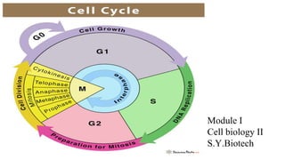
- 1. Module I Cell biology II S.Y.Biotech
- 2. Introduction to Cell Cycle ● New cell arises by the division of cell which already exists. ● Definition: A cell reproduces by performing an orderly sequence of events in which it duplicates its contents and then divides in two. This cycle of duplication and division, known as the cell cycle, is the essential mechanism by which all living things reproduce. ● All living organisms, from the unicellular bacterium to the multicellular mammal, are products of repeated rounds of cell growth and division. ● In unicellular species, such as bacteria and yeasts, each cell division produces a complete new organism. In multicellular species, long and complex sequences of cell divisions are required to produce a functioning organism. Even in the adult body, cell division is usually needed to replace cells that die.
- 3. Phases of Cell Cycle Interphase M Phase G1 Phase First Gap Phase S Phase Synthesis Phase G2 Phase second Gap Phase ● Prophase ● Prometaphase ● Metaphase ● Anaphase ● Telophase ● Cytokinesis
- 5. Eukaryotic cell cycle ● The eukaryotic cell cycle is traditionally divided into four sequential phases: G1, S, G2, and M. G1, S, and G2 together are called interphase. ● Interphase require 23 hours while M phase require 1 hour. ● Gap Phases: ➔ To allow time for growth, most cell cycles have gap phases—a G1 phase between M phase and S phase and a G2 phase between S phase and mitosis. They also provide time for the cell to monitor the internal and external environment. ➔ They ensure that conditions are suitable and preparations are complete before the cell commits itself to the major upheavals of S phase and mitosis. The G1 phase is especially important in this respect. Its length can vary greatly depending on external conditions and extracellular signals from other cells.
- 6. ➔ G0 Phase: ❖ If extracellular conditions are unfavorable, for example, cells delay progress through G1 and may even enter a specialized resting state known as G0 (G zero), in which they can remain for days, weeks, or even years before resuming proliferation. Indeed, many cells remain permanently in G0 until they or the organism dies. ❖ If extracellular conditions are favorable and signals to grow and divide are present, cells in early G1 or G0 progress through a commitment point near the end of G1 known as Start (in yeasts) or the restriction point (in mammalian cells).
- 7. ● S phase: ➔ DNA replication occurs during a period of the cell cycle termed S phase (Synthesis Phase) . ➔ The linear chromosomes of eukaryotic cells are vast and dynamic assemblies of DNA and protein, and their duplication is a complex process that takes up a major fraction of the cell cycle. ➔ Duration of S phase is about 8 hours. ➔ S phase is also the period when the cell synthesizes the additional histones that will be needed as the cell doubles the number of nucleosomes in its chromosomes ensuring that the daughter cells inherit all features of chromosome structure.
- 8. DNA synthesis: 1. Helix Unwinding 2. Extension of Primer 3. Complementary Strand Synthesis 4. Formation of new copy of DNA
- 9. ● M Phase 1. Prophase:
- 10. Prometaphase:
- 11. Metaphase
- 12. Anaphase
- 13. Telophase
- 14. Cytokinesis ● After mitosis the cell is divided into two daughter cells by a separate process called cytokinesis. ● The first hint of cytokinesis in most animal cells appears during anaphase as an indentation of the cell surface in a narrow band around the cell. As time progresses, the indentation deepens to form a furrow that moves inward toward the center of the cell. ● The plane of the furrow lies in the same plane previously occupied by the chromosomes of the metaphase plate, so that the two sets of chromosomes are ultimately partitioned into different cells.
- 16. Cell Cycle Control System ● The control system is rigidly programmed to provide a fixed amount of time for the completion of each cell-cycle event. ● The cell-cycle control system is based on a connected series of biochemical switches, each of which initiates a specific cell-cycle event. ● Features of cell cycle control system: 1. The biochemical switches are generally binary (on/off) and launch events in a complete, irreversible fashion. It would clearly be disastrous, for example, if events like chromosome condensation or nuclear-envelope breakdown were only partially initiated or started but not completed 2. Robust and reliable system,because backup mechanisms and other features allow the system to operate effectively under a variety of conditions and even if some components fail. 3. Highly adaptable and can be modified to suit specific cell types or to respond to specific intracellular or extracellular signals.
- 17. Checkpoints 1. G1-S Checkpoint: It is a Start (or the restriction) point in late G1, where the cell commits to cell-cycle entry and chromosome duplication. 2. G2-M Checkpoint: where the control system triggers the early mitotic events that lead to chromosome alignment on the mitotic spindle in metaphase. 3. Metaphase to anaphase transition: where the control system stimulates sister-chromatid separation, leading to the completion of mitosis and cytokinesis.
- 18. Key Components of Control System Cyclins Cdk (Cyclin dependent kinases) ● Central components of the cell-cycle control system are members of a family of protein kinases known as cyclin-dependent kinases (Cdks). ● The activities of these kinases rise and fall as the cell progresses through the cycle, leading to cyclical changes in the phosphorylation of intracellular proteins that initiate or regulate the major events of the cell cycle. ● Cyclical changes in Cdk activity are controlled by a complex array of enzymes and other proteins. The most important of these Cdk regulators are proteins known as cyclins. ● Cdks, as their name implies, are dependent on cyclins for their activity: unless they are bound tightly to a cyclin, they have no protein kinase activity
- 19. Fig: Two key components of the cell-cycle control system: When cyclin forms a complex with Cdk, the protein kinase is activated to trigger specific cell-cycle events. Without cyclin, Cdk is inactive. Cyclins were originally named because they undergo a cycle of synthesis and degradation in each cell cycle. The levels of the Cdk proteins, by contrast, are constant. Cyclical changes in cyclin protein levels result in the cyclic assembly and activation of cyclin–Cdk complexes at specific stages of the cell cycle.
- 20. Classes of Cyclins G1S Cyclins S Cyclins M Cyclins Cyclin E: Activate Cdks in late G1 and thereby help trigger progression through Start, resulting in a commitment to cell- cycle entry. Their levels fall in S phase. Cyclin A: Bind Cdks soon after progression through Start and help stimulate chromosome duplication. S-cyclin levels remain elevated until mitosis, and these cyclins also contribute to the control of some early mitotic events. Cyclin B: Activate Cdks that stimulate entry into mitosis at the G2/M transition. M-cyclin levels fall in mid- mitosis. G1 Cyclins Cyclin D: Helps govern the activities of the G1/S- cyclins, which control progression through Start in late G1.
- 21. Cyclin–Cdk complexes of the cell-cycle control system. The concentrations of the three major cyclin types oscillate during the cell cycle, while the concentrations of Cdks (not shown) do not change and exceed cyclin amounts. In late G1, rising G1/S-cyclin levels lead to the formation of G1/S-Cdk complexes that trigger progression through the Start transition. S-Cdk complexes form at the start of S phase and trigger DNA replication, as well as some early mitotic events. M-Cdk complexes form during G2 but are held in an inactive state; they are activated at the end of G2 and trigger entry into mitosis at the G2/M transition.
- 22. Classes of CDKs G1 Cdk G1-S Cdk S Cdk M Cdk ● Cdk 4 and Cdk 6 ● Interact with G1 cyclins ● Cdk 2 ● Interact with G1-S cyclins ● Cdk 2 and Cdk 1 ● Interact with S cyclins ● Cdk 1 ● Interact with M cyclins
- 24. Activation Mechanism of Cdk by cyclins The enzyme is shown in three states. (A) In the inactive state, without cyclin bound, the active site is blocked by a region of the protein called the T-loop (red). (B) The binding of cyclin causes the T-loop to move out of the active site, resulting in partial activation of the Cdk2. (C) Phosphorylation of Cdk2 (by CAK) at a threonine residue in the T-loop further activates the enzyme by changing the shape of the T-loop, improving the ability of the enzyme to bind its protein substrates.
- 25. Suppression of Cdk activity 1. By Inhibitory phosphorylation: The active cyclin–Cdk complex is turned off when the kinase Wee1 phosphorylates two closely spaced sites above the active site. Removal of these phosphates by the phosphatase Cdc25 activates the cyclin-Cdk complex. For simplicity, only one inhibitory phosphate is shown.
- 26. 2.By Cdk Inhibitor Proteins: Binding of Cdk inhibitor proteins (CKIs) inactivates cyclin–Cdk complexes. The three-dimensional structure of a cyclin–Cdk–CKI complex reveals that CKI binding stimulates a large rearrangement in the structure of the Cdk active site, rendering it inactive.Cells use CKIs primarily to help govern the activities of G1/S- and S-Cdks early in the cell cycle.
- 27. Regulation of Metaphase to anaphase transition ● Activation of specific cyclin–Cdk complexes drives progression through the Start and G2/M transitions, progression through the metaphase-to-anaphase transition is triggered not by protein phosphorylation but by protein destruction, leading to the final stages of cell division. ● The key regulator of the metaphase-to-anaphase transition is the anaphase promoting complex, or cyclosome (APC/C), It is a large mutisubunit complex and a member of the ubiquitin ligase family of enzymes.They polyubiquitylate specific target proteins, resulting in their destruction in proteasomes.
- 28. Targets Of APC/C Securin S and M cyclins It protects the protein linkages that hold sister-chromatid pairs together in early mitosis. Destruction of securin in metaphase activates a protease that separates the sisters and unleashes anaphase, Destroying these cyclins inactivates most Cdks in the cell. As a result, the many proteins phosphorylated by Cdks from S phase to early mitosis are dephosphorylated by various phosphatases in the anaphase cell. This dephosphorylation of Cdk targets is required for the completion of M phase, including the final steps in mitosis and then cytokinesis.
- 29. APC/C is activated by Cdc20 subunit. Then this APC/C acts on S and M cyclin for their degradation.
- 30. SCF (Skp, Cullin, F-box containing complex) ● SCF is a multisubunit protein complex. It is a ubiquitin ligase protein. ● It has many functions in the cell, but its major role in the cell cycle is to ubiquitylate certain CKI proteins in late G1, thereby helping to control the activation of S- Cdks and DNA replication. ● SCF is also responsible for the destruction of G1/S-cyclins in early S phase. ● SCF activity depends on substrate-binding subunits called F-box proteins. Unlike APC/C activity, however, SCF activity is constant during the cell cycle. Ubiquitylation by SCF is controlled instead by changes in the phosphorylation state of its target proteins, as F-box subunits recognize only specifically phosphorylated proteins.
- 31. The activity of the ubiquitin ligase SCF depends on substrate-binding subunits called F-box proteins, of which there are many different types. The phosphorylation of a target protein, such as the CKI shown, allows the target to be recognized by a specific F-box subunit.
- 32. Cell Cycle Control at S Phase The central event of chromosome duplication—DNA replication—poses two problems for the cell. 1. Replication must occur with extreme accuracy to minimize the risk of mutations in the next cell generation. 2. Every nucleotide in the genome must be copied once, and only once, to prevent the damaging effects of gene amplification.
- 34. Control of chromosome duplication ● Preparations for DNA replication begin in late mitosis and G1, when the DNA helicases are loaded by multiple proteins at the replication origin, forming the prereplicative complex (preRC). ● In late mitosis and early G1, the proteins Cdc6 and Cdt1 collaborate with the ORC(Origin Recognition Complex) to load the inactive DNA helicases around the DNA next to the origin. ● S-Cdk activation leads to activation of the DNA helicases, which unwind the DNA at origins to initiate DNA replication. Two replication forks move out from each origin until the entire chromosome is duplicated. Duplicated chromosomes are then segregated in M phase. ● S-Cdk activation in S phase also prevents assembly of new preRCs at any origin until the following G1—thereby ensuring that each origin is activated only once in each cell cycle.
- 35. Control of initiation of replication
- 36. Control of the initiation of DNA replication: ● The replication origin is bound by the ORC(Origin recognition complex) throughout the cell cycle. ● In early G1, Cdc6 associates with the ORC, and these proteins bind the DNA helicase, which contains six closely related subunits called Mcm proteins. The helicase also associates with a protein called Cdt1. ● Using energy provided by ATP hydrolysis, the ORC and Cdc6 proteins load two copies of the DNA helicase, in an inactive form, around the DNA next to the origin, thereby forming the prereplicative complex (preRC). ● At the onset of S phase, S-Cdk stimulates the assembly of several initiator proteins on each DNA helicase, while another protein kinase, DDK, phosphorylates subunits of the DNA helicase. As a result, the DNA helicases are activated and unwind the DNA. ● DNA polymerase and other replication proteins are recruited to the origin, and DNA replication begins. The ORC is displaced by the replication machinery and then rebinds. ● S-Cdk and other mechanisms also inactivate the preRC components ORC, Cdc6, and Cdt1, thereby preventing formation of new preRCs at the origins until the end of mitosis.
- 37. Ensuring that DNA is replicated once per cell cycle: 1. At the same time as S-Cdk initiates DNA replication, several mechanisms prevent assembly of new preRCs. S-Cdk phosphorylates and thereby inhibits the ORC and Cdc6 proteins. 2. Inactivation of the APC/C in late G1 also helps turn off preRC assembly. In late mitosis and early G1, the APC/C triggers the destruction of a Cdt1 inhibitor called geminin, thereby allowing Cdt1 to be active. 3. When the APC/C is turned off in late G1, geminin accumulates and inhibits the Cdt1 that is not associated with DNA. 4. Also, the association of Cdt1 with a protein at active replication forks stimulates Cdt1 destruction. 5. In these various ways, preRC formation is prevented from S phase to mitosis, thereby ensuring that each origin is fired only once per cell cycle.
- 38. Cohesin holds sister chromatids together Cohesin is a protein complex with four subunits. (A) Two subunits, Smc1 and Smc3, are coiled-coil proteins with an ATPase domain at one end; (B) two additional subunits, Scc1 and Scc3, connect the ATPase head domains, forming a ring structure that may encircle the sister chromatids as shown in (C). The ATPase domains are required for cohesin loading on the DNA.
- 39. Overview of cell cycle control system The core of the cell cycle control system consists of a series of cyclin– Cdk complexes (yellow). The activity of each complex is also influenced by various inhibitory mechanisms, which provide information about the extracellular environment, cell damage, and incomplete cell-cycle events (top). These inhibitory mechanisms are not present in all cell types; many are missing in early embryonic cell cycles, for example.