Empfohlen

Empfohlen
Weitere ähnliche Inhalte
Was ist angesagt?
Was ist angesagt? (8)
Ähnlich wie SMFMposterCPR
Ähnlich wie SMFMposterCPR (20)
SMFMposterCPR
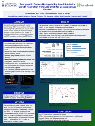
- 1. 1 Nir Melamed,1 Alex Pittini, 2 John Kingdom,1 Jon F.R. Barrett 1 Sunnybrook Health Sciences Centre, Toronto, ON, Canada. 2 Mount Sinai Hospital, Toronto, ON, Canada Sonographic Factors Distinguishing Late Intrauterine Growth Restriction from Late Small for Gestational Age Fetuses ABSTRACTABSTRACT Distinguishing between constitutionally small for gestational age (SGA) fetuses and those affected by intrauterine growth restriction (IUGR) who are at risk of adverse outcome presents an important challenge, especially in cases of late IUGR. Our aim was to identify sonographic factors that distinguish between late IUGR fetuses and constitutionally SGA fetuses. •Retrospective chart review at a single tertiary care center from 2010-2015 of women with a singleton pregnancy who delivered ≥32 weeks with a neonate birth weight <10th percentile for gestational age •Neonates were classified as IUGR if they experienced any of the following complications: perinatal mortality, urgent Caesarean section for fetal distress, 5-minutes Apgar < 7, umbilical artery pH < 7.1, need for resuscitation or admission to NICU METHODSMETHODS OBJECTIVEOBJECTIVE Identify sonographic factors that distinguish between late IUGR fetuses and constitutionally SGA fetuses. RESULTSRESULTS •548 women met inclusion criteria,194 (35.4%) were in the late IUGR group (Table 1) •CPR is the most predictive of the adverse outcome (Figure 3) •Abnormal CPR values are more frequent in the IUGR group, with the Baschat gestational age adjusted cut-offs giving the clearest distinction (Figure 4) •CPR is useful to distinguish groups after 32 weeks (Figure 5) •Multivariable model shows only CPR<5th percentile (Baschat curve) as significantly predictive with an adjusted odds ratio of 6.6 (2.8-15.6) Characteristic Late IUGR group N=194 Control group N=354 P Value Characteristics Nulliparity 140)72.2( 216) 61.0( 0.009 Pregnancy complications Gestational diabetes 19)9.8( 18) 5.1( 0.04 Hypertensive complications 39)20.1( 32) 9.0( >0.001 Placental abruption 6) 3.1( 1) 0.3( 0.005 Placenta previa 5) 2.6( 1) 0.3( 0.014 Table 1. Study population characteristics Figure 3. Adjusted odds ratios of sonographic predictors of the composite adverse outcome Figure 5. Average CPR values in control and IUGR groups over the gestational period Control IUGR Ebbings curve 5th percentile cut-off Baschat curve 5th percentile cut-off 1.08 fixed cut-off Figure 4. Frequency of abnormal CPR measurements in control (black) and IUGR (red) groups. Abnormality determined by three cut-offs based on nomograms and fixed cut-offs from previous studies CONCLUSIONSCONCLUSIONS •Low EFW is a poor tool to assess IUGR fetuses •CPR<5th percentile is the best predictor of neonatal morbidity in IUGR fetuses •Its use is effective after 32 weeks gestational age •CPR may have predictive utility in other at risk groups •Intrauterine growth restriction (IUGR) is associated with higher neonatal mortality and morbidity •Currently identified by low estimated fetal weight (EFW) •Doppler blood vessel studies have been proposed as alternate identifiers of IUGR •Umbilical artery Doppler-measures placental resistance •Middle cerebral artery Doppler-measures the “brain sparing effect” seen in IUGR fetuses •Cerebroplacental ratio (CPR) between the two is a more sensitive measure of blood flow redistribution than either alone (Figure 2) •CPR is proposed as a better identifier of IUGR but studies disagree on chosen cut-offs and utility in later gestations BACKGROUNDBACKGROUND Figure 1. Sonographic measurement of the middle cerebral artery Figure 2. Comparative rates of change for sonographic measurements MCA PI<5th % UA PI>95th % EFW<3rd% CPR<5th% AC<3rd%