
Empfohlen
Weitere ähnliche Inhalte
Was ist angesagt?
Was ist angesagt? (20)
Ähnlich wie tumor and tumor like condition of blood vessel ,Dr sanjay bhardwaj ,jnmc amu aligarh
Ähnlich wie tumor and tumor like condition of blood vessel ,Dr sanjay bhardwaj ,jnmc amu aligarh (20)
Kürzlich hochgeladen
Kürzlich hochgeladen (20)
tumor and tumor like condition of blood vessel ,Dr sanjay bhardwaj ,jnmc amu aligarh
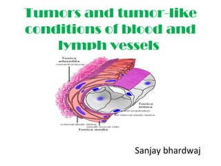
- 1. Tumors and tumor-like conditions of blood and lymph vessels Sanjay bhardwaj
- 4. BENIGN TUMORS AND TUMOR-LIKE CONDITIONS HEMANGIOMAS Hemangiomas, are the most common tumors of the head and neck in infancy and childhood, comprising approximately 7% of all benign soft tissue tumors A benign skin lesion consisting of dense, usually elevated masses of dilated blood vessels.
- 5. Types of Hemangiomas: Strawberry(capillary) Hemangioma Cavernous (Deep) Hemangioma Pyogenic granuloma
- 6. Capillary Hemangioma The most common variant Site : skin, subcutaneous tissues, and mucous membranes of the oral cavities and lips, liver, spleen, and kidneys salivary gland and breast. .
- 7. The “strawberry type” or juvenile hemangioma [benign hemangioendothelioma /hyperplastic hemangioma] ,is the most distinctive and common lesion of the skin of newborns is extremely common (1 in 200 births), fades at 1 to 3 years of age and completely regresses by age 7 in 75% to 90% of cases.
- 9. Aggregates of closely packed, thin- walled capillaries, usually blood-filled and lined by flattened endothelium; vessels are separated by scant connective tissue stroma.
- 10. immunohistochemically positivity for FVIII, CD31, CD34, FLI- 1, estrogen receptor beta, annexin II
- 11. Cavernous (Deep) Hemangioma deeply situated red-blue spongy mass of tissue filled with blood found on 2 out of 100 babies Deep hemangiomas involve muscle or visceral organs and, are more difficult to diagnose. Therefore, further diagnostic studies are required. Brain hemangiomas are most problematic, because they can cause pressure symptoms or rupture. Cavernous hemangiomas are a component of von Hippel- Lindau disease
- 13. A large cavernous hemangioma of the liver in a 28-year-old man.
- 14. large, cavernous blood-filled vascular spaces, separated by a modest connective tissue stroma
- 15. Pyogenic Granuloma. : It is rapidly growing pedunculated red nodule on the skin, or gingival or oral mucosa; it bleeds easily and is often ulcerated associated with history of trauma Pregnancy tumor (granuloma gravidarum) is a pyogenic granuloma that occurs infrequently (1% of patients) in the gingiva of pregnant women. These lesions can spontaneously regress.
- 16. Clinical appearance of typical pyogenic granuloma
- 17. capillary or vascular lobule
- 18. Newly formed capillaries embedded in a loose edematous stroma which has been infiltrated with chronic inflammatory cells Fibrosis develops and the small capillaries disappear, leaving a dense plug of moderately healthy cellular fibrous tissue
- 19. They occur in the back, gluteal region, thigh, and other sites; sometimes an entire extremity is involved. The association of varicose veins, (dysplastic) cutaneous hemangiomas, and soft tissue and bone hypertrophy is known as Klippel–Trenaunay syndrome. Large-vessel hemangiomas
- 20. composed of veins (venous hemangiomas) or a combination of veins and arteries (racemose, cirsoid, or arteriovenous hemangiomas). Venous hemangiomas of the feet have been seen in patients with Turner syndrome
- 21. The mass consists of thick-walled irregular vascular channels with smooth muscle walls of varying thickness, thus suggesting a venous hemangioma Staining for CD34, a vascular endothelial marker, reveals strong reactivity in the flattened luminal-lining endothelial cells, confirming the tumor’s vascular origin.
- 22. Skeletal muscle (intramuscular) hemangiomas venous or cavernous microscopic appearance these very cellular, with plump nuclei, mitotic figures, intraluminal papillary projections, and even infiltration of perineurial spaces
- 23. Masson heMangioMa or Masson lesion/ intravascular papillary endothelial hyperplas •It represent exuberant organization and recanalization of a thrombus •Occur in previously normal vessels or in varices, hemorrhoids, hematomas, pyogenic granulomas, hemangiomas, •Found in the extremities (particularly the fingers) and the head and neck region, whereas the type engrafted on a pre existing vascular disorder tends to be in the trunk. •It simulates malignant vascular tumors
- 24. The confinement of the lesion to the vascular lumen and the hyaline core of the papillae are characteristic features.
- 25. Spindle cell hemangioma/spindle cell hemangioendothelioma A benign endothelial neoplasm. Occur at any age, has a male predominance,occurs preferentially in the dermisand subcutaneous tissue of the distal extremities, Some cases have occurred in patients with Maffucci or Klippel–Trenaunay syndrome
- 26. characteristic ‘spongy’ low-power appearance
- 27. cavernous hemangioma-like area Kaposi sarcoma-like area
- 28. Hobnail hemangioma. The endothelial cells protrude into the vessel lumina
- 30. Clinical presentation : Hemangiomas On examination, the superficial hemangioma usually consists of a raised, reddish to purple tumor with a distinct margin. In contrast, deep subcutaneous hemangiomas often have a deep bluish hue with normal overlying skin, making diagnosis more difficult. Both the lesions are firm to palpation and do not pulsate or exhibit any thrills or bruits.
- 31. Investigations : Hemangiomas •Computed tomography (C. T .Scan) and Magnetic resonance imaging ( M. R.I) imaging techniques are used as diagnostic aids to document the extent of the deep hemangiomas "popcorn" pattern in case of cerebellaum cavernous hemengioma
- 32. •Arteriography: rarely indicated for the diagnosis of a hemangiomas. •Biopsy :for final diagnosis
- 33. Consider Treatment • Treatment should be considered if the hemangioma…. • ulcerates • bleeds • causes functional impairment • causes infection • grows rapidly and uncontrollably • causes psychological problems
- 34. Treatments of Hemangiomas Medical • steroid injection • interferon alfa-2a Surgical • resection • FPDL • YAG laser
- 35. Medical Treatments Interferon Alfa-2a benefit in inhibiting angiogenisis and stimulate endothelial cell prostacyclin formation, which prevents platelet trapping interferon alfa-2a is administered in daily subcutaneous injections of 1 to 3 million units per square meter of body surface area for an average of 7 months of therapy acute side effects, which are reversible, include fever, chills, arthralgias and retinal vasculopathy
- 36. Surgical Procedures Resection surgical excision is occasionally advocated as the primary treatment of hemangiomas resection surgically removes all or part of the tissue indicated as the management of visceral lesions unresponsive to steroids used for the cosmetic revision of redundant skin remaining after spontaneous involution of deeper hemangiomas
- 37. Surgical Procedures FPDL - flashlight-pumped pulsed dye laser treatment of choice for superficial strawberry hemangiomas with a response rate of 60 percent penetrates to a depth of 1.8mm and has a low risk of scarring local anesthetic is effective in reducing pain or discomfort and some bruising may occur several laser sessions may be needed to achieve optimal improvement
- 38. Surgical Procedures YAG Laser treatment of choice for rapidly growing deep or mixed hemangiomas with a response rate of 75 percent penetrates to a depth of 5 to 6 mm, although scar formation is more frequent than with the FPDL since the laser penetrates deeper into the skin requires local or general anesthesia not recommended in the initial treatment of cutaneous hemangiomas
- 39. Lymphangiomas Lymphangiomas are the benign lymphatic analogues of blood vessel hemangiomas Simple (Capillary) Lymphangioma. These are composed of small lymphatic channels predominantly occurring in the head, neck, and axillary subcutaneous tissues. They are slightly elevated or sometimes pedunculated lesions up to 1 to 2 cm in diameter.
- 40. Cystically dilated vascular spaces are lined by flattened endothelial cells. Red blood cells are nearly absent
- 42. Cavernous Lymphangioma (Cystic Hygroma): o These lesions are typically found in the neck or axilla of children, and rarely occur in the retroperitonem. o Cavernous lymphangiomas can occasionally be enormous (up to 15 cm) and may fill the axilla or produce gross deformities about the neck.
- 43. Large cystic hygroma in an infant.
- 44. The image shows lymphatic spaces lined by attenuated endothelium. The cyst walls contain lymphoid aggregates.
- 45. Glomus Tumor (Glomangioma) Benign, painful tumors. arising from modified smooth muscle cells of the glomus body, a specialized arteriovenous structure involved in thermoregulation. They are most commonly found in the distal portion of the digits, especially under the fingernails . Excision is curative.
- 46. Glomus tumor lesions are round, slightly elevated, red-blue, firm nodules (usually <1 cm in diameter)
- 48. aggregates, nests, and masses of specialized glomus cells intimately associated with branching vascular channels, all within a connective tissue stroma. Individual tumor cells are small, uniform, and round or cuboidal, with scant cytoplasm
- 49. Immunohistochemically, they shows reactivity for myosin, vimentin, actin,
- 50. Vascular Malformations/ Vascular ectasias • Vascular malformations are present at birth and unlike hemangiomas, do not go through a “rapid proliferation and they do not “involute”. • They grow gradualy with the patient.
- 51. Vascular Malformations - Types • Vascular malformations may be capillary, venous, arterial, or combinations of these. • Approximately 31% of these malformations are found in the head and neck region.
- 52. Vascular Malformations types • Vascular Malformations are divided into two categories: Low-flow and High-flow lesions. • Capillary, venous, and lymphatic malformations exhibit “low flow lesions”. • Arterial and arterio-venous malformations exhibit “high flow” and are capable of severe hemorrhage with significant morbidity.
- 53. Venous Malformations - Low-flow lesions • Venous malformations are bluish, soft and easily compressible, and auscultation reveals no bruits. • These malformations can vary from superficial, localized, mucosal spongy ectasis to complex invasive lesions that permeate tissue planes and alter the regional anatomy.
- 54. The masses enlarge with physical activity or a dependent position. Venous malformations can be painful in the morning due to stasis and microthrombi within the veins. Venous malformations’ localization is usually in the head and neck. Venous malformations are the most common vascular anomaly, they are 40% of all vascular malformations Venous malformation can be treated with sclerotherapy and surgical reduction
- 55. at histological examination showing disorganized muscular wall of veins blending with surrounding soft tissues
- 56. Capillary Malformations - Low-flow lesions Capillary malformation (also known as port-wine stain)/Nevus Flammeus:. flat, reddish lesions affect the skin of the head and the neck They darken with age. Syndromes associated are: Sturge-Weber syndrome and Klippel-Trenaunay syndrome .Capillary malformations can be treated with IPL-(Intensed-pulsed-light)-therapy or surgical reduction.
- 58. collections of haphazardly arranged dilated vessels in the papillary and reticular dermis
- 59. mononuclear cell infiltrate with occasional dilated blood vessels within the superficial dermis. Spider Telangiectasia.
- 60. Hereditary Hemorrhagic Telangiectasia (OslerWeber-Rendu Disease). autosomal dominant Telangiectasias are Present from birth, over the skin and oral mucous membranes, the respiratory, gastrointestinal, and urinary tracts. These lesions rupture, causing serious epistaxis , GI bleeding, or hematuria.
- 61. Lymphatic Malformations - Low- flow lesions • Lymphatic malformations are normally colorless; however, combined lesions take on the hue of the additional vessel typ. • These malformations can become invasive by dissecting along tissue planes and can cause bony hypertrophy, distortion or both. As these are lymphatic tissues, infections may result in rapid enlargement of these lesions.
- 62. Arterial / Arteriovenous Malformations • Clinically these lesions appear stained, warm and tender to palpation. There may be swelling or asymmetry. • A bruit may be detected.
- 64. Reactive vascular proliferations: Bacillary angiomatosis Skin lesions are red papules and nodules, acute neutrophilic inflammation and vascular (capillary) proliferation.
- 65. INTERMEDIATE-GRADE (BORDERLINE) TUMORS Chronic KS (also called classic or European KS) : •it is not associated with human immunodeficiency virus (HIV). •it associated with an underlying second malignancy or altered . immunity, • it presents with multiple red to purple skin plaques or nodules, usually in the distal lower extremitie. • asymptomatic and remain localized to the skin and subcutaneous tissue Kaposi Sarcoma
- 66. Lymphadenopathic KS (also called African or endemic KS): it is also not associated with HIV. No Skin lesions are sparse, and patients present instead with lymphadenopathy . the tumor occasionally involves the viscera and is aggressive. Transplant-associated KS : occurs in the setting of solid-organ transplantation mucosal, and visceral involvement; cutaneous lesions may be absent.
- 67. AIDS-associated (epidemic) KS AIDS-associated KS can involve lymph nodes or viscera and disseminates widely early in the course of the disease found in a third of AIDS patients, particularly male homosexuals Pathogenesis human herpesvirus-8 (HHV-8) or KS-associated herpesvirus (KSHV) it is transmitted sexually and by poorly understood nonsexual route—perhaps including saliva
- 68. three stages are recognized: Patches are red to purple macules typically confined to the distal lower extremities With time, lesions spread proximally and become larger, violaceous, raised plaques Eventually, lesions become nodular and more distinctly neoplastic
- 69. coalescent red-purple macules and plaques of the skin Histologic appearance of nodular form, demonstrating sheets of plump, proliferating spindle cells.
- 70. Elongated spindle cells showing minimal atypia are separated by slits containing red blood cells
- 71. Hemangioendothelioma vascular tumors of an endothelial nature that occupy an intermediate position between the benign hemangioma and the full-blown angiosarcoma. The tumor partially fills the lumen of the femoral vein Epithelioid hemangioendothelioma cytoplasm is abundant and eosinophilic, predominantly vacuolated
- 72. Malignant endovascular papillary angioendothelioma (Dabska tumor; papillary intralymphatic angioendothelioma) papillary tufts that are lined by plump endothelial cells located within dilated vascular lumina, some of which have a glomeruloid configuration
- 73. Retiform Hemangioendothelioma 'long arborizing vessels' with features reminiscent of rete-testis ; focal solid areas composed of spindle and epithelioid cells may be present.
- 74. MALIGNANT TUMORS Angiosarcoma Angiosarcomas are malignant endothelial neoplasms Older adults are more commonly affected Equal gender predilections They occur at any site but most often involve skin, soft tissue, breast, and liver. angiosarcomas are locally invasive and can metastasize readily. Angiosarcomas are aggressive tumors with current 5-year survival rates of 30%.
- 75. angiosarcoma of the heart (right ventricle) large, fleshy masses of red-tan to gray- white tissue
- 76. dense clumps of irregular, moderate anaplastic cells and distinct vascular lumens CD31 positivity
- 77. Hemangiopericytoma Hemangiopericytomas derived from pericytes-myofibroblast-like cells that are normally arranged around capillaries and venules. they can occur as slowly enlarging, painless masses, common on the lower extremities and in the retroperitoneum.. The tumors may recur after excision, and roughly half will metastasize, usually hematogenously to lungs, bone, or liver.
- 78. consist of numerous branching capillary channels and gaping sinusoidal spaces enclosed within nests of spindle-shaped to round cells.