Cell Plate Formation and Cell Wall Development
•
2 gefällt mir•2,477 views

1) The document describes the structure and composition of plant cell walls. It discusses the layers that make up primary and secondary cell walls, including the middle lamella, primary wall, and secondary wall. 2) The process of cell plate formation during cell division is summarized in 6 steps. Key structures involved include vesicles, fusion tubes, and the phragmoplast. 3) Various cell wall components are described such as cellulose, hemicellulose, pectin, and lignin. Factors influencing cell wall growth and differentiation are also outlined.

Empfohlen
Weitere ähnliche Inhalte
Was ist angesagt?
Was ist angesagt? (20)
Andere mochten auch
Andere mochten auch (20)
Ähnlich wie Cell Plate Formation and Cell Wall Development
Ähnlich wie Cell Plate Formation and Cell Wall Development (20)
Mehr von Jasper Obico
Mehr von Jasper Obico (20)
Kürzlich hochgeladen
Kürzlich hochgeladen (20)
Cell Plate Formation and Cell Wall Development
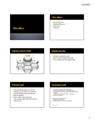
- 1. 1/13/2010 Gross Structure Detailed Structure Chemistry Features Cement; amorphous subs. Bet. P‐walls of neighboring cells Pectic substances (Ca, Mg pectate) First wall the develops on new cell Formed in the inner surface of P‐wall Same content as Pwall ( > cellulose) + lignin Cellulose, pectic cpds., non‐cellulosic In cells that ceased to grow; devoid of protoplast at maturity polysaccharides and hemicellulose * xylem ray, xylem parenchyma – still living May be lignified b l f d Mechanical support M h i l Assoc. with living protoplasts ‐‐eg. meristematic cells, parenchyma, Compound middle lamella * = 3‐layered or 5‐layered collenchyma = middle lamella + 2 P‐walls (+ 2 S‐walls) *if middle lamella is obscured 1
- 2. 1/13/2010 Preprophase band a cortical belt of microtubules and actin filaments, that predicts the plane of the future cell plate Phragmosome Ph a layer of cytoplasm which spreads across the future division contains microtubules and actin filaments (1) the arrival of Golgi‐derivedvesicles in the division plane; fusion tube starts to form Start of accumulation of cell wall materials in the lumen‐‐callose (2) the formation of fusion tubes that grow out of (3) transformation of the tubulo‐vesicular network the vesicles and fuse with others tubulo‐ into a tubular network vesicular network (interwoven) 2
- 3. 1/13/2010 (4) Formation of fenestrated plate‐like structure (5)the formation of numerous finger‐like projections the dense membrane coat and the associated at the margins of the cell plate that fuse with the phragmoplast microtubules are disassembled; plasma membrane of the mother cell wall Abundant accummulation of cell wall building materials (6) maturation of the cell plate into a new cell wall. Cell plate‐ precursor of cell wall; rich in pectins Phragmoplast‐ a complex of microtubules h l l f b l and ER that forms during late anaphase or early telophase from dissociated spindle Closing of fenestrae subunits. Formation of plasmodesmata After completion of the cell plate, additional wall material is deposited inc. in thickness New wall material deposition mosaic fasion Matrix materials Matri materials Delivered by Golgi vesicles Cellulose microfibrils Cellulose synthase complex Cellulose synthase appear as rosettes exude the microfibrils on the outer surface of the membrane. 3
- 4. 1/13/2010 A. Growth in thickness 1. Apposition 2. Intussusception Lignification f Intussusception Cutinization Intussusception B. Growth in surface (wall expansion) Rosettes are inserted in the plasma membrane and pushed forward by synthesis and crystallization of microfibrils Or extension Requires: wall stress relaxation (loosening of wall structure) and turgor pressure Controlled by: a] amt. of turgor pressure ll d b f b] extensibility *Extensibility ability to expand permanently when a force is applied to it (plastic) affected by hormones : auxin Marks cessation of growth (irreversible) Cellulose fibrils during maturation Matrix (non‐cellulosic): FACTORS: that contribute ‐ with lignin, cutin, suberin, hemicelluloses (1) a reduction in wall‐loosening processes, d ll l etc. (2) An increase in cross‐linking of cell wall components, (3) a change in wall composition (more rigid structure or less susceptible to wall loosening) 4
- 5. 1/13/2010 Long chains of linked glucose residues Micellae – bundles of cellulose molecules or ELEMENTARY FIBRIL = ~40 cellulose molecules l l Microfibril Bundles of microfibril Ml‐ middle lamella Pm‐ plasma membrance Cellulose Pectic substances Gums and mucilages Lignin Fatty substances 5
- 6. 1/13/2010 Hydrophilic crystalline compound Amorphous colloidal substances Repeating monomers of glucose Plastic and hydrophilic Appear as a result of physiological or Phenolic compounds pathological disturbances that induce May be found in middle lamella, primary wall, breakdown of walls and cell contents and secondary wall hydrophobic fi ller h h d h b f ll that replaces the wall’s l h ll water compressive strength and bending stiffness Microbial attack resistance Cutin, suberin, waxes Tensile strength (bend under compressive Waxes‐ glaucous condition; assoc. with cutin stress) and suberin Incrustation– eg. Lignification Suberin‐ cork cells of periderm; endodermis and exodermis; prevents apoplastic transport Cutin‐ cuticle layer; epidermis of aerial parts Cell wall growth A. intussusception Cutinization, suberization‐ impregnation in cell B. apposition wall C. mosaic growth Cuticularization‐ formation of layer D. multinet growth 6
- 7. 1/13/2010 Material of new wall is laid down bet. Growth is due to the centripetal addition of Particles of the existing substance of the new layers one upon the other expanding wall Fibrillar texture in certain wall areas become separation of crossed microfibrils and loosened as a result of turgor pressure and alteration in their orientation afterwards mended by deposition of new transverse longitudinal microfibrils in the gaps caused by the strain fb l h db h Primary pit fields Primordial pits/ primary pit fields Pits Certain areas of primary wall of young cells Crassulae remain thin Trabeculae b l May appear beaded in xs b d d Wart structures Cystoliths 7
- 8. 1/13/2010 Plasmodesmata‐ connnect protoplasts of neighboring cells ‐ transport; relay of stimuli * symplast‐ 2 or more interconnected l d protopolast * apoplast – cell walls, intercellular spaces and lumen desmotubule 8
- 9. 1/13/2010 Portions of the cell wall that remained thin even as secondary wall is formed Primary wall only Can develop over primary pit fields f Function? TYPES: Branched simple pits (ramiform) a. simple pit Found in parenchyma cells with thickened b. bordered pit—S‐wall develops over the pit walls, libriform fibers, sclereids, phloem fibers cavity to form an overarching roof f h f Structure: Pit cavity / pit chamber Pit aperture Pit border Pit canal, inner and outer aperture (very thick S‐ wall) water‐conducting and mechanical xylem cells (vessel elements, tracheids, etc.) 9
- 10. 1/13/2010 Angiosperm Gymnosperm Pit cavity – break in S‐wall Simple pit pair Pit membrane/ closing membrane –primary Bordered pit pair wall + middle lamella Half bordered pit pair Pit aperture Blind pit l d Unilateral compound pitting 10
- 11. 1/13/2010 pit membrane thickening; disc shaped – porous pit membrane around the torus flexible; can go median or lateral ‐‐conifer tracheids Aspirate condition (lateral)– latewood and all ‐‐ occurs through matrix dissolution heartwood Coniferales, Gnetales Round, elliptic, linear In thick cell walls: *inner aperture becomes long and narrow *outer aperture remains circular round l d * pit canal is funnel‐shaped *fiber‐tracheid feature Scalariform Linear or crescent‐shaped thickenings of the Opposite primary wall and middle lamella Alternate gymnosperms 11
- 12. 1/13/2010 Rod shape thickenings of the wall which Fahn, A. 1990. Plant Anatomy, 4th ed.. traverse the cell lumen radially Pergamon Press Esau, K. 1958. Plant Anatomy. John Wiley and Sons, Inc. Evert, R. 2006. Esau’s Plant Anatomy. John Wiley and Sons, Inc. Protopectin, pectin, pectic acid Related to pectic Plastic, amorphous colloid, hydrophilic Swelling property (hydrophilic) Hydration in young walls Impregnation starts in the intercellular Cellulose molecules micelles (crystal) lamella micellar system (porous) Microfibril f consists of micellar system f And Microcapillaries liquids, lignin, waxes, cutin, suberin, hemicellulose, pectic substances, crystals, silica 12
- 13. 1/13/2010 Plasticity ‐becoming permanently deformed when subjected to changes in shape or size Elasticity ‐property of recovery of the original size and shape after deformation Tensile strength ‐ relative to chemical composition and microscopic and submicroscopic structure 13