SlideShare ist ein Scribd-Unternehmen logo
1 von 77
Downloaden Sie, um offline zu lesen
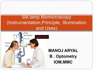
MANOJ ARYAL
B . Optometry
IOM,MMC
Slit lamp Biomicroscopy
(Instrumentation,Principle, Illumination
and Uses)
optometris
t
Introduction
 Biomicroscope derives its name from the
fact that it enables the practitioner to
observe the living tissue of eye under
magnification.
 It not only provides magnified view of every
part of eye but also allows quantitative
measurements and photography of every
part for documentation.
• The lamp facilitates an examination
which looks at anterior segment, or
frontal structures, of the human eye,
which includes the
–Eyelid
–Cornea
–Sclera
–Conjunctiva
–Iris
–Aqueous
–Natural crystalline lens and
–Anterior vitreous.
Important historical landmarks
 De Wecker 1863 devised a portable
ophthalmomicroscope .
 Albert and Greenough 1891,developed
a binocular microscope which provided
stereoscopic view.
 Gullstrand ,1911 introduced the
illumination system which had for the
first time a slit diapharm in it
 Therefore Gullstrand is credited with
the invention of slit lamp.
Large Gullstrand
Ophthalmomicroscop
e (1911)
TYPES
 There are 2 types of slit lamp
biomicroscope
1)Zeiss slit lamp biomicroscope
2)Haag streit slit lamp biomicroscope
 In Zeiss type light source is at the base of
the instrument while in Haag streit type it is
at the top of the instrument.
Zeiss slit lamp biomicroscope
Haag streit slit lamp biomicroscope
PRINCIPLE
 A "slit" beam of very bright light
produced by lamp. This beam is focused
on to the eye which is then viewed
under magnification with a microscope
Instrumentation
 Operational components of slit lamp
biomicroscope essentially consist of:
Illumination system
Observation system
Mechanical system
 Illumination system
 It consist of:
A bright ,focal source of light with a slit
mechanism
 Provides an illumination of 2*10^5 to 4*10^5
lux.
 The beam of light can be changed in
intensity,height,width,direction or angle and
color during the examination with the flick of
lever.
Condensing lens system:
Consist of a couple of planoconvex
lenses with their convex surface in
apposition.
Slit and other diapharm:
 Height and width of slit can be varied by
using knobs.
 Projection lens:
 Form an image of slit at eye.
 Advantages,
 1.keeps the aberration of lens down.
 2.increase the depth of focus of slit.
 Reflecting mirrors and prisms
 Filters
Yellow barrier filter
Red free filter
Neutral density filter
Cobalt blue filter
diffuser
Observation system(microscope)
 Observation system is essentially a compound
microscope composed of two optical elements
1.an objective ,2.an eyepiece
 It presents to the observer an enlarged image of
a near object.
 The objective lens consists of two planoconvex
lenses with their convexities put together
providing a composite power of +22D.
 Microscope is binocular i.e. it has two
eyepieces giving binocular observer a
 The eye piece has a lens of +10D.
 To overcome the problem of inverted
image produced by compound
microscope ,slit lamp microscope uses a
pair of prisms b/w the objective and
eyepiece to reinvert the image.
 Most slit lamp provide a range of
magnification from 6x to 40x
Mechanical system
 Joystick arrangement
 Movement of microscope and illumination
system towards and away from the eye
and from side and side is achieved via
joystick arrangement.
Up and down movement arrangement
Obtained via some sort or screw
devices.
Patient support arrangement
Vertically movable chin rest and the
Fixation target:
A movable fixation target greatly faciliates
the examination under some conditions.
Mechanical coupling :
Provides a coupling of microscope and
the illumination system along a common
axis of rotation that coincides their focal
planes.
 This ensures that light falls on the point
where the microscope is focused
 Has advantages when using the slit lamp
for routine examination of anterior
 Magnification control :
Including two or pair of readily
changeable objective lenses and two
sets of eyepieces.
 An on and off switch and illumination
control .
Topcon slit lamp model SL-3E
Light beam is controlled by
knobs
Joy stick arrangement
Chin rest
Reflecting mirror
biomicroscope
Illumination control
Magnification
may be changed
by
flipping a lever...
Changing filters. biomicroscope
Patient positioning
Alignmen
t mark
Microscope
and light
source rotate
indepedently
Filters used in slit lamp biomicroscopy
 Cobalt blue filter
 Used in conjunction with fluorescein stain
 Dye pods in area where the corneal epithelium
is broken or absent.
 The dye absorbs blue light and emits green.
 Uses:
 Ocular staining
 RGP lenses fitting
 Tear layer
 Red free(green)filter:
Obscure any thing that is red hence
the red free light , thus blood vessels
or haemorrhages appears black.
This increases contrast ,revealing the
path and pattern of inflammed blood
vessels.
Fleischer ring can also be viewed
satisfactorily with the red green filter.
Illumination techniques
 Includes
 Diffuse illumination
 Direct illumination
 Parallilepiped
 Optic section
 Conical(pinpoint)
 Tangential
 Specular reflection
 Indirect illumination
 Retro-illumination
 Sclerotic scatter
 Transillumination
 Proximal illumination
Diffuse illumination
 Angle between microscope and
illumination system should be 30-45
degree.
 Slit width should be widest.
 Filter to be used is diffusing filter.
 Magnification: low to medium
 Illumination: medium to high.
Applications:
General view of anterior of eye:
lids,lashes,sclera,cornea ,iris, pupil,
Gross pathology and media opacities
Contact lens fitting.
Assessment of lachrymal reflex.
Optics of diffuse illumination Diffuse illumination with slit beam and
background illumination
Direct illumination
 Involves placing the light source at an
angle of about 40-50 degree from
microscope.
 This arrangement permits both light beam
and microscope to be sharply focused on
the ocular tissue being observed.
 Wide beam direct illumination is commonly
used as a preliminary technique to
evaluate large area.
 it is particularly suitable for assessment
of cataracts,scars,nerves,vessels etc.
 It is also of great importance for the
determination of stabilization of axis of
toric contact lens.
 Parallelepiped:
Constructed by narrowing the beam to 1-
2mm in width to illuminate a rectangular
area of cornea.
Microscope is placed directly in front of
patients cornea.
Light source is approximately 45 degree
from straight ahead position.
Applications:
Used to detect and examine corneal
structures and defects.
Used to detect corneal striae that
develop when corneal edema occurs
with hydrogel lens wear and in
keratoconus.
Higher magnification than that used with
wide beam illumination is preferred to
evaluate both depth and extent of
corneal ,scarring or foreign bodies.
Conical beam(pinpoint)
 Produced by narrowing the vertical height of a
parallelepiped to produce a small circular or
square spot of light.
 Light source is 45-60 degree temporally and
directed into pupil.
 Biomicroscope: directly in front of eye.
 Magnification: high(16-25x)
 Intensity of light source to heighest setting.
 Focusing:
Beam is focused between cornea and
anterior lens surface and dark zone
between cornea and anterior lens
observed.
Principle is same as that of beam of sun
light streaming through a room
,illuminating airborne dust particles.
This occurance is called tyndall
phenomenon.
 Most useful when examining the
Tyndall phenomenon
 Cells, pigment or proteins in the
aqueous humour reflect the light like a
faint fog.
 To visualise this the slit illuminator is
adjusted to the smallest circular beam
and is projected through the anterior
chamber from a 42° to 90° angle.
 The strongest reflection is possible at
90°.
Optic section
 Optic section is a very thin parallelepiped and
optically cuts a very thin slice of the cornea.
 Axes of illuminating and viewing path intersect
in the area of anterior eye media to be
examined e.g. the individual corneal layers.
 Angle between illuminating and viewing path
is 45 degree.
 Slit length should be kept small to minimize
dazzling the patient.
 With narrow slit the depth and portion of
different objects(penetration depth of
foreign bodies, shape of lens etc) can be
resolved more easily.
 With wider slit their extension and shape
are visible more clearly.
 Magnification: maximum.
 Examination of AC depth is performed by
wider slit width .1-.3mm .
 Used to localize:
Nerve fibers
Blood vessels
Infiltrates
Cataracts
AC depth.
Optical section of lens
1.Corneal scar with wide beam illumination 2.optical section through scar
indicating scar is with in superficial layer of cornea.
Tangential illumination
 Requires that the illumination arm and the
viewing arm be separated by 90 degree.
 Medium –wide beam of moderate height is
used.
 Microscope is pointing straight ahead.
 Magnification of 10x,16x,or 25x are used.
 Observe:
Anterior and posterior cornea
Iris is best viewed without dilation by
this method.
Anterior lens (especially useful for
viewing pseudoexfolation).
Example of tangential illumination (iris).
Specular reflection
 Established by separating the microscope and
slit beam by equal angles from normal to cornea.
 Position of illuminator about 30 degree to one
side and the microscope 30 degree to otherside.
 Angle of illuminator to microscope must be equal
and opposite.
 Angle of light should be moved until a very bright
reflex obtained from corneal surface which is
called zone of specular reflection.
 Irregularities ,deposits ,or excavasation in these
smooth surface will fail to reflect light and these
appears darker than surrounding.
 Under specular reflection anterior corneal
surface appears as white uniform surface and
corneal endothelium takes on a mosaic pattern.
 Used to observe:
 Evaluate general appearance of corneal
endothelium
 Lens surfaces
 Corneal epithelium
Schematic of specularreflection.
Reflection from
front surface endothelium
Indirect illumination
 The beam is focused in an area adjacent to
ocular tissue to be observed.
 Main application:
 Examination of objects in direct vicinity of
corneal areas of reduced transparency e,g,
infiltrates,corneal scars,deposits,epithelial and
stromal defects
 Illumination:
 Narrow to medium slit beam
 Decentred beam
 Magnification: approx. m=12x (depending upon
object size)
Retroillumination
 Formed by reflecting light of slit beam from
a structure more posterior than the
structure under observation.
 A vertical slit beam 1-4mm wide can be
used.
 Purpose:
Place object of regard against a bright
background allowing object to appear
dark or black.
 Used most often in searching for keratic
precipitates and other debris on corneal
endothelium.
 The crystalline lens can also be
retroilluminated for viewing of water
clefts and vacuoles of anterior lens and
posterior subcapsular cataract
 Direct retroillumination from iris:
Used to view corneal pathology.
A moderately wide slit beam is aimed
towards the iris directly behind the
corneal anomaly.
Use magnification of 16x to 25x and
direct the light from 45 degree.
Microscope is directed straight ahead .
Schematic of
direct retroillumination from
the iris.
direct retroillumination from the iris.
 Indirect retroillumination from iris:
Performed as with direct
retroillumination but the beam is
directed to an area of the iris bordering
the portion of iris behind pathology.
It provides dark background allowing
corneal opacities to be viewed with
more contrast.
Observe:
Cornea, angles.
Retroillumination from fundus(red
reflex photography)
 The slit illuminator is positioned in an
almost coaxial position with the
biomicroscope.
 A wide slit beam is decentered and
adjusted to a half circle by using the slit
width and
 The decentred slit beam is projected near
the pupil margin through a dilated pupil.
Schematic of
retroillumination from the
retina.
Example of retroillumination from the reti
Sclerotic scatter
 It is formed by focusing a bright but narrow slit
beam on the limbus and using microscope on
low magnification.
 Such an illumination technique causes cornea
to take on total internal reflection.
 The slit beam should be placed approximately
40-60 degree from the microscope.
 When properly positioned this technique will
produce halo glow of light around the limbus
as the light is transmitted around the cornea.
 Corneal changes or abnormalities can be
visualized by reflecting the scattered light.
 Used to observe:
Central corneal epithelial edema
Corneal abrasions
Corneal nebulae and maculae.
Schematic of
sclerotic scatter. Example of
sclerotic scatter.
Proximal illumination
 This illumination technique is used to
observe internal detail, depth, and
density.
 Use a short,fairly narrow slit beam.
 Place the beam at the border of the
structure or pathology.
 The light will be scattered into the
surrounding tissue, creating a light
background that highlights the edges of
 Depending on the density of the
abnormality, the light from behind may
reflect through, allowing detailed
examination of the internal structure of
the pathology.
 Observe: corneal opacities (edema,
infiltrates, vessels, foreign bodies), lens,
iris
Transillumination
 In transillumination, a structure (in the
eye, the iris) is evaluated by how light
passes through it.
 Iris transillumination:
 This technique also takes advantage of
the red reflex.
 The pupil must be at mid mydriasis (3to
4 mm when light stimulated).
 Place the light source coaxial (directly
in line) with the microscope.
 Use a full circle beam of light equal to
the size of the pupil.
 Project the light through the pupil and
into the eye .
 Focus the microscope on the iris.
 Magnification of 10X to 16X is adequate
 Normally the iris pigment absorbs the
light, but pigmentation defects let the red
fundus light pass through..
 Observe: iris defects (they will glow with
the orange light reflected from the
fundus)
Basic slit lamp examination
 Patient positioning:
Head support unit
Adjust height of table or chair
Adjust height of chin rest such that
patients lateral canthus is aligned with
the mark.
Adjust ocular eyepieces.
Power up
Fixation
Magnification : begin with 6x -10x
magnification
Focusing
Special procedures
Protocol and documentation
Uses of slit lamp biomicroscopy
 Diagnostic:
OCT
FFA
Anterior segment and posterior
segment diseases
Dry eye
 Procedures:
 Applanation
 Tear evaluation
 Pachymetry
 Gonioscopy
 Contact lens fitting
 Therapeutic:
 Laser
 FB removal
 epilation
Anterior and posterior segment disease
evaluation
 Lids and lashes
 Conjunctiva and cornea
 Instillation of fluorescein and BUT
measurement
 Eversion of the lids
 Anterior chamber and angle measurement
 Iris
 Crystalline lens
 Anterior vitreous
Injected conjunctivaMeibomian gland openings
pinquecula,
INSTILLATION OF FLUORESCEIN PALPEBRAL CONJUNCTIVA
EXAMINATION
Evertion of lids
 This technique is used to examine the
inferior and superior palpebral
conjunctiva, particularly in contact lens
wear and when looking for allergic
conjunctival changes, papillae, and
foreign bodies.
 1. Ask the patient to look down and
grasp the superior eyelashes.
 2. Press gently on the superior margin of
the tarsal plate using a cotton swab (or
the index finger of the other hand), and
at the same time pull the eyelashes
upwards.
 3. To evert the lower eyelid, pull the
eyelid down and press under the eyelid
margin while moving finger upwards.
The eyelid will evert over finger.
Meibomian gland evaluation
 With the patient at the biomicroscope, use
white light and medium magnification to
inspect the lower eyelid margins.
 Look for capping of the meibomian gland
orifices (yellow mounds), notching of the
eyelid margins (indentations) and frothing of
the tears on the eyelid margins.
 Pull the lower eyelid down and look for
concretions in the palpebral conjunctiva.
 With mild pressure, press on the eyelid
margins near the eyelashes and watch
the meibomian gland orifices.
 Clear fluid should be expressed.
 Capping of the orifices, a cheesy
secretion on expression and frothing of
the eyelid margins indicates meibomian
gland dysfunction.
CENTRAL RETINA PHOTOGRAPHS
WITH A 90-DIOPTER LENS
 A moderate slit
beam in the
almost coaxial
position gives
the best results.
References
 Clinical procedure in optometry
 Primary care optometry
 Borishs clinical refraction
 Theory and practice of optics and refraction:AK
Khurana
 internet
Slit lump biomicroscope

Weitere ähnliche Inhalte

Was ist angesagt?

DUOCHROM TEST.pptx
DUOCHROM TEST.pptxDUOCHROM TEST.pptx
DUOCHROM TEST.pptxKhulesh Sahu
 
Suppression third yr arya
Suppression third yr aryaSuppression third yr arya
Suppression third yr aryaarya das
 
A scan biometry | How to Use A-scan? Types of A-Scan Biometry?
A scan biometry | How to Use A-scan? Types of A-Scan Biometry?A scan biometry | How to Use A-scan? Types of A-Scan Biometry?
A scan biometry | How to Use A-scan? Types of A-Scan Biometry?Naeem Ahmad
 
Biometry instruments & equipment
Biometry instruments & equipmentBiometry instruments & equipment
Biometry instruments & equipmentDevdutta Nayak
 
Progressive addition lenses
Progressive addition lensesProgressive addition lenses
Progressive addition lensesSanjana Chouhan
 
Synoptophore and its parts
Synoptophore and its partsSynoptophore and its parts
Synoptophore and its partsLoknath Goswami
 
Assessment of corneal endothelium
Assessment of corneal endotheliumAssessment of corneal endothelium
Assessment of corneal endotheliumdrvasant162
 
Diplopia charting
Diplopia charting Diplopia charting
Diplopia charting ANUJA DHAKAL
 
A scan ultrasonography
A scan ultrasonographyA scan ultrasonography
A scan ultrasonographySamuel Ponraj
 
anomalous retinal correspondence
anomalous retinal correspondenceanomalous retinal correspondence
anomalous retinal correspondenceRajeshwori
 

Was ist angesagt? (20)

DUOCHROM TEST.pptx
DUOCHROM TEST.pptxDUOCHROM TEST.pptx
DUOCHROM TEST.pptx
 
Gonioscopy
GonioscopyGonioscopy
Gonioscopy
 
Suppression third yr arya
Suppression third yr aryaSuppression third yr arya
Suppression third yr arya
 
A scan biometry | How to Use A-scan? Types of A-Scan Biometry?
A scan biometry | How to Use A-scan? Types of A-Scan Biometry?A scan biometry | How to Use A-scan? Types of A-Scan Biometry?
A scan biometry | How to Use A-scan? Types of A-Scan Biometry?
 
A scan biometry
A scan biometryA scan biometry
A scan biometry
 
Biometry instruments & equipment
Biometry instruments & equipmentBiometry instruments & equipment
Biometry instruments & equipment
 
Pachymetry
PachymetryPachymetry
Pachymetry
 
Worth 4 dot test
Worth 4 dot testWorth 4 dot test
Worth 4 dot test
 
Progressive addition lenses
Progressive addition lensesProgressive addition lenses
Progressive addition lenses
 
Lensometers
LensometersLensometers
Lensometers
 
Binocular balancing
Binocular balancing Binocular balancing
Binocular balancing
 
Synoptophore and its parts
Synoptophore and its partsSynoptophore and its parts
Synoptophore and its parts
 
GONIOSCOPY by
GONIOSCOPY by GONIOSCOPY by
GONIOSCOPY by
 
Assessment of corneal endothelium
Assessment of corneal endotheliumAssessment of corneal endothelium
Assessment of corneal endothelium
 
Diplopia charting
Diplopia charting Diplopia charting
Diplopia charting
 
Maddox rod
Maddox rodMaddox rod
Maddox rod
 
Hrt II
Hrt IIHrt II
Hrt II
 
A scan ultrasonography
A scan ultrasonographyA scan ultrasonography
A scan ultrasonography
 
Synoptophore 2
Synoptophore 2Synoptophore 2
Synoptophore 2
 
anomalous retinal correspondence
anomalous retinal correspondenceanomalous retinal correspondence
anomalous retinal correspondence
 

Ähnlich wie Slit lump biomicroscope

Slit lamp biomicroscopy and operating microscope
Slit lamp biomicroscopy and operating microscopeSlit lamp biomicroscopy and operating microscope
Slit lamp biomicroscopy and operating microscopeSAMEEKSHA AGRAWAL
 
Slit lampbiomicroscopy-jinal & dhara
Slit lampbiomicroscopy-jinal & dharaSlit lampbiomicroscopy-jinal & dhara
Slit lampbiomicroscopy-jinal & dharadhara sorathiya
 
slit-lamp-160212142229.pdf
slit-lamp-160212142229.pdfslit-lamp-160212142229.pdf
slit-lamp-160212142229.pdfanju468752
 
The Slit lamp Biomicroscope
The Slit lamp BiomicroscopeThe Slit lamp Biomicroscope
The Slit lamp BiomicroscopeAkshay Nayak
 
Slitlamp bimicroscopy
Slitlamp bimicroscopy Slitlamp bimicroscopy
Slitlamp bimicroscopy Obehi Osoata
 
Slit Lamp Biomicroscopy.
Slit Lamp Biomicroscopy.Slit Lamp Biomicroscopy.
Slit Lamp Biomicroscopy.Mystic Dider
 
what is slit lamp Biomicroscopy | What is a slit lamp exam used for? |Differe...
what is slit lamp Biomicroscopy | What is a slit lamp exam used for? |Differe...what is slit lamp Biomicroscopy | What is a slit lamp exam used for? |Differe...
what is slit lamp Biomicroscopy | What is a slit lamp exam used for? |Differe...Naeem Ahmad
 
Slit lamp biomicroscopy
Slit lamp biomicroscopySlit lamp biomicroscopy
Slit lamp biomicroscopySSSIHMS-PG
 
slit lamp photography.pdf
slit lamp photography.pdfslit lamp photography.pdf
slit lamp photography.pdfMusabFathallah
 
Slit lamp presentation 2015
Slit lamp presentation 2015Slit lamp presentation 2015
Slit lamp presentation 2015amanimohsen1
 
lensesinophthalmology-171212163049 (1).pdf
lensesinophthalmology-171212163049 (1).pdflensesinophthalmology-171212163049 (1).pdf
lensesinophthalmology-171212163049 (1).pdfDrSunandaMajumder
 

Ähnlich wie Slit lump biomicroscope (20)

Slit lamp biomicroscopy and operating microscope
Slit lamp biomicroscopy and operating microscopeSlit lamp biomicroscopy and operating microscope
Slit lamp biomicroscopy and operating microscope
 
Slit lampbiomicroscopy-jinal & dhara
Slit lampbiomicroscopy-jinal & dharaSlit lampbiomicroscopy-jinal & dhara
Slit lampbiomicroscopy-jinal & dhara
 
slit-lamp-160212142229.pdf
slit-lamp-160212142229.pdfslit-lamp-160212142229.pdf
slit-lamp-160212142229.pdf
 
Slit lamp ..
Slit lamp ..Slit lamp ..
Slit lamp ..
 
The Slit lamp Biomicroscope
The Slit lamp BiomicroscopeThe Slit lamp Biomicroscope
The Slit lamp Biomicroscope
 
Slit lamp biomicroscopy
Slit lamp biomicroscopySlit lamp biomicroscopy
Slit lamp biomicroscopy
 
Slitlamp bimicroscopy
Slitlamp bimicroscopy Slitlamp bimicroscopy
Slitlamp bimicroscopy
 
Slit lamp biomicroscope
Slit lamp biomicroscopeSlit lamp biomicroscope
Slit lamp biomicroscope
 
Slit Lamp Biomicroscopy.
Slit Lamp Biomicroscopy.Slit Lamp Biomicroscopy.
Slit Lamp Biomicroscopy.
 
SLIT LAMP
SLIT LAMPSLIT LAMP
SLIT LAMP
 
Slit lamp biomicroscope
Slit lamp biomicroscopeSlit lamp biomicroscope
Slit lamp biomicroscope
 
what is slit lamp Biomicroscopy | What is a slit lamp exam used for? |Differe...
what is slit lamp Biomicroscopy | What is a slit lamp exam used for? |Differe...what is slit lamp Biomicroscopy | What is a slit lamp exam used for? |Differe...
what is slit lamp Biomicroscopy | What is a slit lamp exam used for? |Differe...
 
SLIT LAMP BIOMICROSCOPY.pptx
SLIT LAMP BIOMICROSCOPY.pptxSLIT LAMP BIOMICROSCOPY.pptx
SLIT LAMP BIOMICROSCOPY.pptx
 
Slit lamp biomicroscopy
Slit lamp biomicroscopySlit lamp biomicroscopy
Slit lamp biomicroscopy
 
Slit lamp examination
Slit lamp examinationSlit lamp examination
Slit lamp examination
 
slit lamp photography.pdf
slit lamp photography.pdfslit lamp photography.pdf
slit lamp photography.pdf
 
Slit lamp presentation 2015
Slit lamp presentation 2015Slit lamp presentation 2015
Slit lamp presentation 2015
 
Sl exam pt i
Sl exam pt iSl exam pt i
Sl exam pt i
 
basics of microscope - part 1
basics of microscope - part 1basics of microscope - part 1
basics of microscope - part 1
 
lensesinophthalmology-171212163049 (1).pdf
lensesinophthalmology-171212163049 (1).pdflensesinophthalmology-171212163049 (1).pdf
lensesinophthalmology-171212163049 (1).pdf
 

Mehr von C L GUPTA EYE INSTITUTE MORADABAD UTTER PRADESH

Mehr von C L GUPTA EYE INSTITUTE MORADABAD UTTER PRADESH (20)

Astigmatism
Astigmatism Astigmatism
Astigmatism
 
Tonometry ppt
Tonometry pptTonometry ppt
Tonometry ppt
 
anatomy of sclera
anatomy of sclera anatomy of sclera
anatomy of sclera
 
Pupil ppt
Pupil  pptPupil  ppt
Pupil ppt
 
anatomy And Physiology of tear film
anatomy And Physiology of tear film anatomy And Physiology of tear film
anatomy And Physiology of tear film
 
cornea and layers
cornea and layers cornea and layers
cornea and layers
 
Limbus
LimbusLimbus
Limbus
 
anatomy And Physiology of lacrimal secretions
anatomy And Physiology of lacrimal secretions anatomy And Physiology of lacrimal secretions
anatomy And Physiology of lacrimal secretions
 
extra ocular muscles
extra ocular muscles extra ocular muscles
extra ocular muscles
 
Diseases of sclera
Diseases of sclera Diseases of sclera
Diseases of sclera
 
Corneal transparency
Corneal transparency Corneal transparency
Corneal transparency
 
Conjunctiva anatomy and physiology
Conjunctiva anatomy and physiology Conjunctiva anatomy and physiology
Conjunctiva anatomy and physiology
 
Color vision deficiency and ishiharas test
Color vision deficiency and ishiharas test Color vision deficiency and ishiharas test
Color vision deficiency and ishiharas test
 
Anatomy of eye
Anatomy of eye Anatomy of eye
Anatomy of eye
 
Anatomy and physiology of aqueous humor
Anatomy and physiology of aqueous humor Anatomy and physiology of aqueous humor
Anatomy and physiology of aqueous humor
 
Vitreous humour
Vitreous humourVitreous humour
Vitreous humour
 
Vitamins all
Vitamins allVitamins all
Vitamins all
 
Tear film
Tear filmTear film
Tear film
 
Tca cycle
Tca cycleTca cycle
Tca cycle
 
Synthesis
SynthesisSynthesis
Synthesis
 

Kürzlich hochgeladen

Independent Call Girls Hyderabad 💋 9352988975 💋 Genuine WhatsApp Number for R...
Independent Call Girls Hyderabad 💋 9352988975 💋 Genuine WhatsApp Number for R...Independent Call Girls Hyderabad 💋 9352988975 💋 Genuine WhatsApp Number for R...
Independent Call Girls Hyderabad 💋 9352988975 💋 Genuine WhatsApp Number for R...Ahmedabad Call Girls
 
Jaipur Call Girls 9257276172 Call Girl in Jaipur Rajasthan
Jaipur Call Girls 9257276172 Call Girl in Jaipur RajasthanJaipur Call Girls 9257276172 Call Girl in Jaipur Rajasthan
Jaipur Call Girls 9257276172 Call Girl in Jaipur Rajasthanindiancallgirl4rent
 
Call Girl Gorakhpur * 8250192130 Service starts from just ₹9999 ✅
Call Girl Gorakhpur * 8250192130 Service starts from just ₹9999 ✅Call Girl Gorakhpur * 8250192130 Service starts from just ₹9999 ✅
Call Girl Gorakhpur * 8250192130 Service starts from just ₹9999 ✅gragmanisha42
 
Ozhukarai Call Girls 👙 6297143586 👙 Genuine WhatsApp Number for Real Meet
Ozhukarai Call Girls 👙 6297143586 👙 Genuine WhatsApp Number for Real MeetOzhukarai Call Girls 👙 6297143586 👙 Genuine WhatsApp Number for Real Meet
Ozhukarai Call Girls 👙 6297143586 👙 Genuine WhatsApp Number for Real MeetCall Girls Service
 
Sambalpur Call Girls 👙 6297143586 👙 Genuine WhatsApp Number for Real Meet
Sambalpur Call Girls 👙 6297143586 👙 Genuine WhatsApp Number for Real MeetSambalpur Call Girls 👙 6297143586 👙 Genuine WhatsApp Number for Real Meet
Sambalpur Call Girls 👙 6297143586 👙 Genuine WhatsApp Number for Real MeetCall Girls Service
 
Best Lahore Escorts 😮‍💨03250114445 || VIP escorts in Lahore
Best Lahore Escorts 😮‍💨03250114445 || VIP escorts in LahoreBest Lahore Escorts 😮‍💨03250114445 || VIP escorts in Lahore
Best Lahore Escorts 😮‍💨03250114445 || VIP escorts in LahoreDeny Daniel
 
VIP Call Girls Noida Sia 9711199171 High Class Call Girl Near Me
VIP Call Girls Noida Sia 9711199171 High Class Call Girl Near MeVIP Call Girls Noida Sia 9711199171 High Class Call Girl Near Me
VIP Call Girls Noida Sia 9711199171 High Class Call Girl Near Memriyagarg453
 
Bareilly Call Girls 👙 6297143586 👙 Genuine WhatsApp Number for Real Meet
Bareilly Call Girls 👙 6297143586 👙 Genuine WhatsApp Number for Real MeetBareilly Call Girls 👙 6297143586 👙 Genuine WhatsApp Number for Real Meet
Bareilly Call Girls 👙 6297143586 👙 Genuine WhatsApp Number for Real MeetCall Girls Service
 
Nanded Call Girls 👙 6297143586 👙 Genuine WhatsApp Number for Real Meet
Nanded Call Girls 👙 6297143586 👙 Genuine WhatsApp Number for Real MeetNanded Call Girls 👙 6297143586 👙 Genuine WhatsApp Number for Real Meet
Nanded Call Girls 👙 6297143586 👙 Genuine WhatsApp Number for Real MeetCall Girls Service
 
Russian Call Girls Kota * 8250192130 Service starts from just ₹9999 ✅
Russian Call Girls Kota * 8250192130 Service starts from just ₹9999 ✅Russian Call Girls Kota * 8250192130 Service starts from just ₹9999 ✅
Russian Call Girls Kota * 8250192130 Service starts from just ₹9999 ✅gragmanisha42
 
Ernakulam Call Girls 👙 6297143586 👙 Genuine WhatsApp Number for Real Meet
Ernakulam Call Girls 👙 6297143586 👙 Genuine WhatsApp Number for Real MeetErnakulam Call Girls 👙 6297143586 👙 Genuine WhatsApp Number for Real Meet
Ernakulam Call Girls 👙 6297143586 👙 Genuine WhatsApp Number for Real MeetCall Girls Chandigarh
 
nagpur Call Girls 👙 6297143586 👙 Genuine WhatsApp Number for Real Meet
nagpur Call Girls 👙 6297143586 👙 Genuine WhatsApp Number for Real Meetnagpur Call Girls 👙 6297143586 👙 Genuine WhatsApp Number for Real Meet
nagpur Call Girls 👙 6297143586 👙 Genuine WhatsApp Number for Real MeetCall Girls Service
 
Vip Call Girls Makarba 👙 6367187148 👙 Genuine WhatsApp Number for Real Meet
Vip Call Girls Makarba 👙 6367187148 👙 Genuine WhatsApp Number for Real MeetVip Call Girls Makarba 👙 6367187148 👙 Genuine WhatsApp Number for Real Meet
Vip Call Girls Makarba 👙 6367187148 👙 Genuine WhatsApp Number for Real MeetAhmedabad Call Girls
 
Call Girls Service In Goa 💋 9316020077💋 Goa Call Girls By Russian Call Girl...
Call Girls Service In Goa  💋 9316020077💋 Goa Call Girls  By Russian Call Girl...Call Girls Service In Goa  💋 9316020077💋 Goa Call Girls  By Russian Call Girl...
Call Girls Service In Goa 💋 9316020077💋 Goa Call Girls By Russian Call Girl...russian goa call girl and escorts service
 
Russian Call Girls in Noida Pallavi 9711199171 High Class Call Girl Near Me
Russian Call Girls in Noida Pallavi 9711199171 High Class Call Girl Near MeRussian Call Girls in Noida Pallavi 9711199171 High Class Call Girl Near Me
Russian Call Girls in Noida Pallavi 9711199171 High Class Call Girl Near Memriyagarg453
 
neemuch Call Girls 👙 6297143586 👙 Genuine WhatsApp Number for Real Meet
neemuch Call Girls 👙 6297143586 👙 Genuine WhatsApp Number for Real Meetneemuch Call Girls 👙 6297143586 👙 Genuine WhatsApp Number for Real Meet
neemuch Call Girls 👙 6297143586 👙 Genuine WhatsApp Number for Real MeetCall Girls Service
 
Dehradun Call Girls 8854095900 Call Girl in Dehradun Uttrakhand
Dehradun Call Girls 8854095900 Call Girl in Dehradun  UttrakhandDehradun Call Girls 8854095900 Call Girl in Dehradun  Uttrakhand
Dehradun Call Girls 8854095900 Call Girl in Dehradun Uttrakhandindiancallgirl4rent
 
(Deeksha) 💓 9920725232 💓High Profile Call Girls Navi Mumbai You Can Get The S...
(Deeksha) 💓 9920725232 💓High Profile Call Girls Navi Mumbai You Can Get The S...(Deeksha) 💓 9920725232 💓High Profile Call Girls Navi Mumbai You Can Get The S...
(Deeksha) 💓 9920725232 💓High Profile Call Girls Navi Mumbai You Can Get The S...Ahmedabad Call Girls
 
Call Girls Patiala Just Call 8250077686 Top Class Call Girl Service Available
Call Girls Patiala Just Call 8250077686 Top Class Call Girl Service AvailableCall Girls Patiala Just Call 8250077686 Top Class Call Girl Service Available
Call Girls Patiala Just Call 8250077686 Top Class Call Girl Service AvailableDipal Arora
 
Call Girls Thane Just Call 9907093804 Top Class Call Girl Service Available
Call Girls Thane Just Call 9907093804 Top Class Call Girl Service AvailableCall Girls Thane Just Call 9907093804 Top Class Call Girl Service Available
Call Girls Thane Just Call 9907093804 Top Class Call Girl Service AvailableDipal Arora
 

Kürzlich hochgeladen (20)

Independent Call Girls Hyderabad 💋 9352988975 💋 Genuine WhatsApp Number for R...
Independent Call Girls Hyderabad 💋 9352988975 💋 Genuine WhatsApp Number for R...Independent Call Girls Hyderabad 💋 9352988975 💋 Genuine WhatsApp Number for R...
Independent Call Girls Hyderabad 💋 9352988975 💋 Genuine WhatsApp Number for R...
 
Jaipur Call Girls 9257276172 Call Girl in Jaipur Rajasthan
Jaipur Call Girls 9257276172 Call Girl in Jaipur RajasthanJaipur Call Girls 9257276172 Call Girl in Jaipur Rajasthan
Jaipur Call Girls 9257276172 Call Girl in Jaipur Rajasthan
 
Call Girl Gorakhpur * 8250192130 Service starts from just ₹9999 ✅
Call Girl Gorakhpur * 8250192130 Service starts from just ₹9999 ✅Call Girl Gorakhpur * 8250192130 Service starts from just ₹9999 ✅
Call Girl Gorakhpur * 8250192130 Service starts from just ₹9999 ✅
 
Ozhukarai Call Girls 👙 6297143586 👙 Genuine WhatsApp Number for Real Meet
Ozhukarai Call Girls 👙 6297143586 👙 Genuine WhatsApp Number for Real MeetOzhukarai Call Girls 👙 6297143586 👙 Genuine WhatsApp Number for Real Meet
Ozhukarai Call Girls 👙 6297143586 👙 Genuine WhatsApp Number for Real Meet
 
Sambalpur Call Girls 👙 6297143586 👙 Genuine WhatsApp Number for Real Meet
Sambalpur Call Girls 👙 6297143586 👙 Genuine WhatsApp Number for Real MeetSambalpur Call Girls 👙 6297143586 👙 Genuine WhatsApp Number for Real Meet
Sambalpur Call Girls 👙 6297143586 👙 Genuine WhatsApp Number for Real Meet
 
Best Lahore Escorts 😮‍💨03250114445 || VIP escorts in Lahore
Best Lahore Escorts 😮‍💨03250114445 || VIP escorts in LahoreBest Lahore Escorts 😮‍💨03250114445 || VIP escorts in Lahore
Best Lahore Escorts 😮‍💨03250114445 || VIP escorts in Lahore
 
VIP Call Girls Noida Sia 9711199171 High Class Call Girl Near Me
VIP Call Girls Noida Sia 9711199171 High Class Call Girl Near MeVIP Call Girls Noida Sia 9711199171 High Class Call Girl Near Me
VIP Call Girls Noida Sia 9711199171 High Class Call Girl Near Me
 
Bareilly Call Girls 👙 6297143586 👙 Genuine WhatsApp Number for Real Meet
Bareilly Call Girls 👙 6297143586 👙 Genuine WhatsApp Number for Real MeetBareilly Call Girls 👙 6297143586 👙 Genuine WhatsApp Number for Real Meet
Bareilly Call Girls 👙 6297143586 👙 Genuine WhatsApp Number for Real Meet
 
Nanded Call Girls 👙 6297143586 👙 Genuine WhatsApp Number for Real Meet
Nanded Call Girls 👙 6297143586 👙 Genuine WhatsApp Number for Real MeetNanded Call Girls 👙 6297143586 👙 Genuine WhatsApp Number for Real Meet
Nanded Call Girls 👙 6297143586 👙 Genuine WhatsApp Number for Real Meet
 
Russian Call Girls Kota * 8250192130 Service starts from just ₹9999 ✅
Russian Call Girls Kota * 8250192130 Service starts from just ₹9999 ✅Russian Call Girls Kota * 8250192130 Service starts from just ₹9999 ✅
Russian Call Girls Kota * 8250192130 Service starts from just ₹9999 ✅
 
Ernakulam Call Girls 👙 6297143586 👙 Genuine WhatsApp Number for Real Meet
Ernakulam Call Girls 👙 6297143586 👙 Genuine WhatsApp Number for Real MeetErnakulam Call Girls 👙 6297143586 👙 Genuine WhatsApp Number for Real Meet
Ernakulam Call Girls 👙 6297143586 👙 Genuine WhatsApp Number for Real Meet
 
nagpur Call Girls 👙 6297143586 👙 Genuine WhatsApp Number for Real Meet
nagpur Call Girls 👙 6297143586 👙 Genuine WhatsApp Number for Real Meetnagpur Call Girls 👙 6297143586 👙 Genuine WhatsApp Number for Real Meet
nagpur Call Girls 👙 6297143586 👙 Genuine WhatsApp Number for Real Meet
 
Vip Call Girls Makarba 👙 6367187148 👙 Genuine WhatsApp Number for Real Meet
Vip Call Girls Makarba 👙 6367187148 👙 Genuine WhatsApp Number for Real MeetVip Call Girls Makarba 👙 6367187148 👙 Genuine WhatsApp Number for Real Meet
Vip Call Girls Makarba 👙 6367187148 👙 Genuine WhatsApp Number for Real Meet
 
Call Girls Service In Goa 💋 9316020077💋 Goa Call Girls By Russian Call Girl...
Call Girls Service In Goa  💋 9316020077💋 Goa Call Girls  By Russian Call Girl...Call Girls Service In Goa  💋 9316020077💋 Goa Call Girls  By Russian Call Girl...
Call Girls Service In Goa 💋 9316020077💋 Goa Call Girls By Russian Call Girl...
 
Russian Call Girls in Noida Pallavi 9711199171 High Class Call Girl Near Me
Russian Call Girls in Noida Pallavi 9711199171 High Class Call Girl Near MeRussian Call Girls in Noida Pallavi 9711199171 High Class Call Girl Near Me
Russian Call Girls in Noida Pallavi 9711199171 High Class Call Girl Near Me
 
neemuch Call Girls 👙 6297143586 👙 Genuine WhatsApp Number for Real Meet
neemuch Call Girls 👙 6297143586 👙 Genuine WhatsApp Number for Real Meetneemuch Call Girls 👙 6297143586 👙 Genuine WhatsApp Number for Real Meet
neemuch Call Girls 👙 6297143586 👙 Genuine WhatsApp Number for Real Meet
 
Dehradun Call Girls 8854095900 Call Girl in Dehradun Uttrakhand
Dehradun Call Girls 8854095900 Call Girl in Dehradun  UttrakhandDehradun Call Girls 8854095900 Call Girl in Dehradun  Uttrakhand
Dehradun Call Girls 8854095900 Call Girl in Dehradun Uttrakhand
 
(Deeksha) 💓 9920725232 💓High Profile Call Girls Navi Mumbai You Can Get The S...
(Deeksha) 💓 9920725232 💓High Profile Call Girls Navi Mumbai You Can Get The S...(Deeksha) 💓 9920725232 💓High Profile Call Girls Navi Mumbai You Can Get The S...
(Deeksha) 💓 9920725232 💓High Profile Call Girls Navi Mumbai You Can Get The S...
 
Call Girls Patiala Just Call 8250077686 Top Class Call Girl Service Available
Call Girls Patiala Just Call 8250077686 Top Class Call Girl Service AvailableCall Girls Patiala Just Call 8250077686 Top Class Call Girl Service Available
Call Girls Patiala Just Call 8250077686 Top Class Call Girl Service Available
 
Call Girls Thane Just Call 9907093804 Top Class Call Girl Service Available
Call Girls Thane Just Call 9907093804 Top Class Call Girl Service AvailableCall Girls Thane Just Call 9907093804 Top Class Call Girl Service Available
Call Girls Thane Just Call 9907093804 Top Class Call Girl Service Available
 

Slit lump biomicroscope

  • 1. MANOJ ARYAL B . Optometry IOM,MMC Slit lamp Biomicroscopy (Instrumentation,Principle, Illumination and Uses) optometris t
  • 2. Introduction  Biomicroscope derives its name from the fact that it enables the practitioner to observe the living tissue of eye under magnification.  It not only provides magnified view of every part of eye but also allows quantitative measurements and photography of every part for documentation.
  • 3. • The lamp facilitates an examination which looks at anterior segment, or frontal structures, of the human eye, which includes the –Eyelid –Cornea –Sclera –Conjunctiva –Iris –Aqueous –Natural crystalline lens and –Anterior vitreous.
  • 4. Important historical landmarks  De Wecker 1863 devised a portable ophthalmomicroscope .  Albert and Greenough 1891,developed a binocular microscope which provided stereoscopic view.  Gullstrand ,1911 introduced the illumination system which had for the first time a slit diapharm in it  Therefore Gullstrand is credited with the invention of slit lamp.
  • 6. TYPES  There are 2 types of slit lamp biomicroscope 1)Zeiss slit lamp biomicroscope 2)Haag streit slit lamp biomicroscope  In Zeiss type light source is at the base of the instrument while in Haag streit type it is at the top of the instrument.
  • 7. Zeiss slit lamp biomicroscope Haag streit slit lamp biomicroscope
  • 8. PRINCIPLE  A "slit" beam of very bright light produced by lamp. This beam is focused on to the eye which is then viewed under magnification with a microscope
  • 9.
  • 10. Instrumentation  Operational components of slit lamp biomicroscope essentially consist of: Illumination system Observation system Mechanical system
  • 11.  Illumination system  It consist of: A bright ,focal source of light with a slit mechanism  Provides an illumination of 2*10^5 to 4*10^5 lux.  The beam of light can be changed in intensity,height,width,direction or angle and color during the examination with the flick of lever.
  • 12. Condensing lens system: Consist of a couple of planoconvex lenses with their convex surface in apposition. Slit and other diapharm:  Height and width of slit can be varied by using knobs.
  • 13.  Projection lens:  Form an image of slit at eye.  Advantages,  1.keeps the aberration of lens down.  2.increase the depth of focus of slit.
  • 14.  Reflecting mirrors and prisms  Filters Yellow barrier filter Red free filter Neutral density filter Cobalt blue filter diffuser
  • 15. Observation system(microscope)  Observation system is essentially a compound microscope composed of two optical elements 1.an objective ,2.an eyepiece  It presents to the observer an enlarged image of a near object.  The objective lens consists of two planoconvex lenses with their convexities put together providing a composite power of +22D.  Microscope is binocular i.e. it has two eyepieces giving binocular observer a
  • 16.  The eye piece has a lens of +10D.  To overcome the problem of inverted image produced by compound microscope ,slit lamp microscope uses a pair of prisms b/w the objective and eyepiece to reinvert the image.  Most slit lamp provide a range of magnification from 6x to 40x
  • 17. Mechanical system  Joystick arrangement  Movement of microscope and illumination system towards and away from the eye and from side and side is achieved via joystick arrangement. Up and down movement arrangement Obtained via some sort or screw devices. Patient support arrangement Vertically movable chin rest and the
  • 18. Fixation target: A movable fixation target greatly faciliates the examination under some conditions. Mechanical coupling : Provides a coupling of microscope and the illumination system along a common axis of rotation that coincides their focal planes.  This ensures that light falls on the point where the microscope is focused  Has advantages when using the slit lamp for routine examination of anterior
  • 19.  Magnification control : Including two or pair of readily changeable objective lenses and two sets of eyepieces.  An on and off switch and illumination control .
  • 20. Topcon slit lamp model SL-3E Light beam is controlled by knobs Joy stick arrangement Chin rest Reflecting mirror biomicroscope Illumination control
  • 21. Magnification may be changed by flipping a lever... Changing filters. biomicroscope Patient positioning Alignmen t mark Microscope and light source rotate indepedently
  • 22. Filters used in slit lamp biomicroscopy  Cobalt blue filter  Used in conjunction with fluorescein stain  Dye pods in area where the corneal epithelium is broken or absent.  The dye absorbs blue light and emits green.  Uses:  Ocular staining  RGP lenses fitting  Tear layer
  • 23.  Red free(green)filter: Obscure any thing that is red hence the red free light , thus blood vessels or haemorrhages appears black. This increases contrast ,revealing the path and pattern of inflammed blood vessels. Fleischer ring can also be viewed satisfactorily with the red green filter.
  • 24.
  • 25. Illumination techniques  Includes  Diffuse illumination  Direct illumination  Parallilepiped  Optic section  Conical(pinpoint)  Tangential  Specular reflection  Indirect illumination  Retro-illumination  Sclerotic scatter  Transillumination  Proximal illumination
  • 26. Diffuse illumination  Angle between microscope and illumination system should be 30-45 degree.  Slit width should be widest.  Filter to be used is diffusing filter.  Magnification: low to medium  Illumination: medium to high.
  • 27. Applications: General view of anterior of eye: lids,lashes,sclera,cornea ,iris, pupil, Gross pathology and media opacities Contact lens fitting. Assessment of lachrymal reflex.
  • 28. Optics of diffuse illumination Diffuse illumination with slit beam and background illumination
  • 29. Direct illumination  Involves placing the light source at an angle of about 40-50 degree from microscope.  This arrangement permits both light beam and microscope to be sharply focused on the ocular tissue being observed.  Wide beam direct illumination is commonly used as a preliminary technique to evaluate large area.
  • 30.  it is particularly suitable for assessment of cataracts,scars,nerves,vessels etc.  It is also of great importance for the determination of stabilization of axis of toric contact lens.
  • 31.  Parallelepiped: Constructed by narrowing the beam to 1- 2mm in width to illuminate a rectangular area of cornea. Microscope is placed directly in front of patients cornea. Light source is approximately 45 degree from straight ahead position.
  • 32. Applications: Used to detect and examine corneal structures and defects. Used to detect corneal striae that develop when corneal edema occurs with hydrogel lens wear and in keratoconus. Higher magnification than that used with wide beam illumination is preferred to evaluate both depth and extent of corneal ,scarring or foreign bodies.
  • 33.
  • 34. Conical beam(pinpoint)  Produced by narrowing the vertical height of a parallelepiped to produce a small circular or square spot of light.  Light source is 45-60 degree temporally and directed into pupil.  Biomicroscope: directly in front of eye.  Magnification: high(16-25x)  Intensity of light source to heighest setting.
  • 35.  Focusing: Beam is focused between cornea and anterior lens surface and dark zone between cornea and anterior lens observed. Principle is same as that of beam of sun light streaming through a room ,illuminating airborne dust particles. This occurance is called tyndall phenomenon.  Most useful when examining the
  • 36. Tyndall phenomenon  Cells, pigment or proteins in the aqueous humour reflect the light like a faint fog.  To visualise this the slit illuminator is adjusted to the smallest circular beam and is projected through the anterior chamber from a 42° to 90° angle.  The strongest reflection is possible at 90°.
  • 37.
  • 38. Optic section  Optic section is a very thin parallelepiped and optically cuts a very thin slice of the cornea.  Axes of illuminating and viewing path intersect in the area of anterior eye media to be examined e.g. the individual corneal layers.  Angle between illuminating and viewing path is 45 degree.  Slit length should be kept small to minimize dazzling the patient.
  • 39.  With narrow slit the depth and portion of different objects(penetration depth of foreign bodies, shape of lens etc) can be resolved more easily.  With wider slit their extension and shape are visible more clearly.  Magnification: maximum.  Examination of AC depth is performed by wider slit width .1-.3mm .
  • 40.  Used to localize: Nerve fibers Blood vessels Infiltrates Cataracts AC depth.
  • 41. Optical section of lens 1.Corneal scar with wide beam illumination 2.optical section through scar indicating scar is with in superficial layer of cornea.
  • 42. Tangential illumination  Requires that the illumination arm and the viewing arm be separated by 90 degree.  Medium –wide beam of moderate height is used.  Microscope is pointing straight ahead.  Magnification of 10x,16x,or 25x are used.
  • 43.  Observe: Anterior and posterior cornea Iris is best viewed without dilation by this method. Anterior lens (especially useful for viewing pseudoexfolation).
  • 44. Example of tangential illumination (iris).
  • 45. Specular reflection  Established by separating the microscope and slit beam by equal angles from normal to cornea.  Position of illuminator about 30 degree to one side and the microscope 30 degree to otherside.  Angle of illuminator to microscope must be equal and opposite.  Angle of light should be moved until a very bright reflex obtained from corneal surface which is called zone of specular reflection.
  • 46.  Irregularities ,deposits ,or excavasation in these smooth surface will fail to reflect light and these appears darker than surrounding.  Under specular reflection anterior corneal surface appears as white uniform surface and corneal endothelium takes on a mosaic pattern.  Used to observe:  Evaluate general appearance of corneal endothelium  Lens surfaces  Corneal epithelium
  • 47. Schematic of specularreflection. Reflection from front surface endothelium
  • 48. Indirect illumination  The beam is focused in an area adjacent to ocular tissue to be observed.  Main application:  Examination of objects in direct vicinity of corneal areas of reduced transparency e,g, infiltrates,corneal scars,deposits,epithelial and stromal defects  Illumination:  Narrow to medium slit beam  Decentred beam  Magnification: approx. m=12x (depending upon object size)
  • 49. Retroillumination  Formed by reflecting light of slit beam from a structure more posterior than the structure under observation.  A vertical slit beam 1-4mm wide can be used.  Purpose: Place object of regard against a bright background allowing object to appear dark or black.
  • 50.  Used most often in searching for keratic precipitates and other debris on corneal endothelium.  The crystalline lens can also be retroilluminated for viewing of water clefts and vacuoles of anterior lens and posterior subcapsular cataract
  • 51.  Direct retroillumination from iris: Used to view corneal pathology. A moderately wide slit beam is aimed towards the iris directly behind the corneal anomaly. Use magnification of 16x to 25x and direct the light from 45 degree. Microscope is directed straight ahead .
  • 52. Schematic of direct retroillumination from the iris. direct retroillumination from the iris.
  • 53.  Indirect retroillumination from iris: Performed as with direct retroillumination but the beam is directed to an area of the iris bordering the portion of iris behind pathology. It provides dark background allowing corneal opacities to be viewed with more contrast. Observe: Cornea, angles.
  • 54.
  • 55. Retroillumination from fundus(red reflex photography)  The slit illuminator is positioned in an almost coaxial position with the biomicroscope.  A wide slit beam is decentered and adjusted to a half circle by using the slit width and  The decentred slit beam is projected near the pupil margin through a dilated pupil.
  • 56. Schematic of retroillumination from the retina. Example of retroillumination from the reti
  • 57. Sclerotic scatter  It is formed by focusing a bright but narrow slit beam on the limbus and using microscope on low magnification.  Such an illumination technique causes cornea to take on total internal reflection.  The slit beam should be placed approximately 40-60 degree from the microscope.  When properly positioned this technique will produce halo glow of light around the limbus as the light is transmitted around the cornea.  Corneal changes or abnormalities can be visualized by reflecting the scattered light.
  • 58.  Used to observe: Central corneal epithelial edema Corneal abrasions Corneal nebulae and maculae.
  • 59. Schematic of sclerotic scatter. Example of sclerotic scatter.
  • 60. Proximal illumination  This illumination technique is used to observe internal detail, depth, and density.  Use a short,fairly narrow slit beam.  Place the beam at the border of the structure or pathology.  The light will be scattered into the surrounding tissue, creating a light background that highlights the edges of
  • 61.  Depending on the density of the abnormality, the light from behind may reflect through, allowing detailed examination of the internal structure of the pathology.  Observe: corneal opacities (edema, infiltrates, vessels, foreign bodies), lens, iris
  • 62. Transillumination  In transillumination, a structure (in the eye, the iris) is evaluated by how light passes through it.  Iris transillumination:  This technique also takes advantage of the red reflex.  The pupil must be at mid mydriasis (3to 4 mm when light stimulated).  Place the light source coaxial (directly in line) with the microscope.
  • 63.  Use a full circle beam of light equal to the size of the pupil.  Project the light through the pupil and into the eye .  Focus the microscope on the iris.  Magnification of 10X to 16X is adequate  Normally the iris pigment absorbs the light, but pigmentation defects let the red fundus light pass through..  Observe: iris defects (they will glow with the orange light reflected from the fundus)
  • 64.
  • 65. Basic slit lamp examination  Patient positioning: Head support unit Adjust height of table or chair Adjust height of chin rest such that patients lateral canthus is aligned with the mark. Adjust ocular eyepieces.
  • 66. Power up Fixation Magnification : begin with 6x -10x magnification Focusing Special procedures Protocol and documentation
  • 67. Uses of slit lamp biomicroscopy  Diagnostic: OCT FFA Anterior segment and posterior segment diseases Dry eye
  • 68.  Procedures:  Applanation  Tear evaluation  Pachymetry  Gonioscopy  Contact lens fitting  Therapeutic:  Laser  FB removal  epilation
  • 69. Anterior and posterior segment disease evaluation  Lids and lashes  Conjunctiva and cornea  Instillation of fluorescein and BUT measurement  Eversion of the lids  Anterior chamber and angle measurement  Iris  Crystalline lens  Anterior vitreous
  • 70. Injected conjunctivaMeibomian gland openings pinquecula, INSTILLATION OF FLUORESCEIN PALPEBRAL CONJUNCTIVA EXAMINATION
  • 71. Evertion of lids  This technique is used to examine the inferior and superior palpebral conjunctiva, particularly in contact lens wear and when looking for allergic conjunctival changes, papillae, and foreign bodies.  1. Ask the patient to look down and grasp the superior eyelashes.
  • 72.  2. Press gently on the superior margin of the tarsal plate using a cotton swab (or the index finger of the other hand), and at the same time pull the eyelashes upwards.  3. To evert the lower eyelid, pull the eyelid down and press under the eyelid margin while moving finger upwards. The eyelid will evert over finger.
  • 73. Meibomian gland evaluation  With the patient at the biomicroscope, use white light and medium magnification to inspect the lower eyelid margins.  Look for capping of the meibomian gland orifices (yellow mounds), notching of the eyelid margins (indentations) and frothing of the tears on the eyelid margins.  Pull the lower eyelid down and look for concretions in the palpebral conjunctiva.
  • 74.  With mild pressure, press on the eyelid margins near the eyelashes and watch the meibomian gland orifices.  Clear fluid should be expressed.  Capping of the orifices, a cheesy secretion on expression and frothing of the eyelid margins indicates meibomian gland dysfunction.
  • 75. CENTRAL RETINA PHOTOGRAPHS WITH A 90-DIOPTER LENS  A moderate slit beam in the almost coaxial position gives the best results.
  • 76. References  Clinical procedure in optometry  Primary care optometry  Borishs clinical refraction  Theory and practice of optics and refraction:AK Khurana  internet