Short case...Thalamic glioma
•
1 gefällt mir•312 views

Short case...Thalamic glioma http://yassermetwally.com http://yassermetwally.net

Empfohlen
Empfohlen
Weitere ähnliche Inhalte
Andere mochten auch
Andere mochten auch (20)
Ähnlich wie Short case...Thalamic glioma
Ähnlich wie Short case...Thalamic glioma (20)
Mehr von Professor Yasser Metwally
Mehr von Professor Yasser Metwally (20)
Kürzlich hochgeladen
Kürzlich hochgeladen (20)
Short case...Thalamic glioma
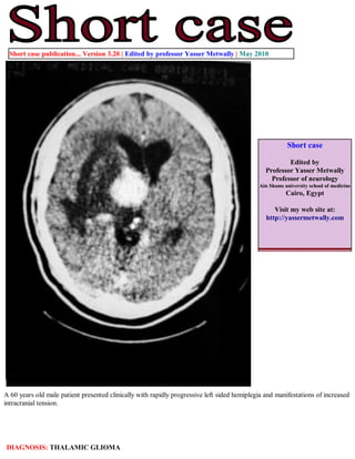
- 1. Short case publication... Version 3.20 | Edited by professor Yasser Metwally | May 2010 Short case Edited by Professor Yasser Metwally Professor of neurology Ain Shams university school of medicine Cairo, Egypt Visit my web site at: http://yassermetwally.com A 60 years old male patient presented clinically with rapidly progressive left sided hemiplegia and manifestations of increased intracranial tension. DIAGNOSIS: THALAMIC GLIOMA
- 2. Figure 1. Precontrast CT scan showing diffuse hypodensities involving the left thalamic area, left internal capsule and left basal ganglionic area. The hypodensities, most probably, represent tumor tissues, peritumoral edema and reactive astrogliosis. Figure 2. Postcontrast CT scan images showing a densely enhanced space occupying lesion involving the left thalamic area with positive mass effect and peritumoral edema involving the white matter and sparing the gray matter (vasogenic edema). Notice the peritumoral satellitosis (B)
- 3. Figure 3. A postcontrast CT scan image showing peritumoral satellitosis (Figure 2 B) References 1. Metwally, MYM: Textbook of neurimaging, A CD-ROM publication, (Metwally, MYM editor) WEB-CD agency for electronic publishing, version 11.2a April 2010 Addendum A new version of short case is uploaded in my web site every week (every Saturday and remains available till Friday.) To download the current version follow the link "http://pdf.yassermetwally.com/short.pdf". You can download the long case version of this short case during the same week from: http://pdf.yassermetwally.com/case.pdf or visit web site: http://pdf.yassermetwally.com To download the software version of the publication (crow.exe) follow the link: http://neurology.yassermetwally.com/crow.zip At the end of each year, all the publications are compiled on a single CD-ROM, please contact the author to know more details. Also to view a list of the previously published case records follow the following link: (http://wordpress.com/tag/case-record/) or click on it if it appears as a link in your PDF reader To inspect the patient's full radiological study, click on the attachment icon (the paper clip icon in the left pane) of the acrobat reader then double click on the attached file Click here to download the long case version of this short case in PDF format