Short case...Neurofibromatosis type 1
•
4 gefällt mir•891 views

A 16-year-old female patient presented with diminished vision, back pain, seizures, and poor school performance. Examination revealed optic nerve damage, cafe au lait spots on the skin, and skin growths. She was diagnosed with neurofibromatosis type 1 based on these findings. The document describes this genetic disorder and includes figures showing abnormalities in the patient's optic nerves, spine, and brain white matter seen on imaging. It also provides information on typical MRI findings in neurofibromatosis type 1 patients and references for further reading.

Empfohlen
Weitere ähnliche Inhalte
Was ist angesagt?
Was ist angesagt? (20)
Andere mochten auch
Andere mochten auch (20)
Ähnlich wie Short case...Neurofibromatosis type 1
Ähnlich wie Short case...Neurofibromatosis type 1 (20)
Mehr von Professor Yasser Metwally
Mehr von Professor Yasser Metwally (20)
Kürzlich hochgeladen
Kürzlich hochgeladen (20)
Short case...Neurofibromatosis type 1
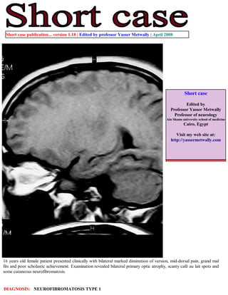
- 1. Short case publication... version 1.18 | Edited by professor Yasser Metwally | April 2008 Short case Edited by Professor Yasser Metwally Professor of neurology Ain Shams university school of medicine Cairo, Egypt Visit my web site at: http://yassermetwally.com 16 years old female patient presented clinically with bilateral marked diminution of version, mid-dorsal pain, grand mal fits and poor scholastic achievement. Examination revealed bilateral primary optic atrophy, scanty café au lait spots and some cutaneous neurofibromatosis. DIAGNOSIS: NEUROFIBROMATOSIS TYPE 1
- 2. Figure 1. Neurofibromatosis type 1. Optic pathway glioma demonstrated as barrel-shaped fusiform dilation of the optic nerves. (optic pathway gliomas in neurofibromatosis type 1 are commonly pilocytic tumors with very slow growth). Figure 2. Intramedullary cystic astrocytoma demonstrated in this patient. Surgical biopsy demonstrated a pilocytic astrocytoma.
- 3. Figure 3. Neurofibromatosis type 1. Scoliosis is demonstrated by plain X ray. Notice the heavily calcified dorsal disc herniation most probably secondary to the scoliotic process. Figure 4. Neurofibromatosis type 1 white matter changes
- 4. Table 1. MRI characteristic of neurofibromatosis type 1 MRI findings Comment Optic pathway gliomas Accounting for 2 to 5 percent of childhood brain tumors. However, up to 70 percent are associated with NF1. The vast majority of these are WHO grade I pilocytic astrocytomas. [1] Kyphoscoliosis with secondary Vertebral defects, including scalloping from dural ectasias, are not degenerative changes uncommon in NF1 [1]. Approximately 10 percent of affected individuals have scoliosis during late childhood and adolescence [1]. Sometimes this can be severe enough to warrant bracing or surgery and may be associated with the presence of an associated neurofibroma. Spinal pilocytic astrocytoma Uncommon in neurofibromatosis type 1, more common in neurofibromatosis type 2 Neurofibromatosis white matter Almost characteristic of neurofibromatosis type 1. [1] patches Addendum A new version of short case is uploaded in my web site every week (every Saturday and remains available till Friday.) To download the current version follow the link quot;http://pdf.yassermetwally.com/short.pdfquot;. You can download the long case version of this short case during the same week from: http://pdf.yassermetwally.com/case.pdf or visit web site: http://pdf.yassermetwally.com To download the software version of the publication (crow.exe) follow the link: http://neurology.yassermetwally.com/crow.zip At the end of each year, all the publications are compiled on a single CD-ROM, please contact the author to know more details. Screen resolution is better set at 1024*768 pixel screen area for optimum display For an archive of the previously reported cases go to www.yassermetwally.net, then under pages in the right panel, scroll down and click on the text entry quot;downloadable short cases in PDF formatquot; References 1. Metwally, MYM: Textbook of neurimaging, A CD-ROM publication, (Metwally, MYM editor) WEB-CD agency for electronic publishing, version 9.1a January 2008