Short case...Multiple sclerosis presented clinically with bilateral trigeminal neuralgia and a high cervicomedullary active demyelinating plaque
•
1 gefällt mir•524 views

Short case...Multiple sclerosis presented clinically with bilateral trigeminal neuralgia and a high cervicomedullary active demyelinating plaque

Empfohlen
Empfohlen
Weitere ähnliche Inhalte
Andere mochten auch
Andere mochten auch (18)
Mehr von Professor Yasser Metwally
Mehr von Professor Yasser Metwally (20)
Kürzlich hochgeladen
Kürzlich hochgeladen (20)
Short case...Multiple sclerosis presented clinically with bilateral trigeminal neuralgia and a high cervicomedullary active demyelinating plaque
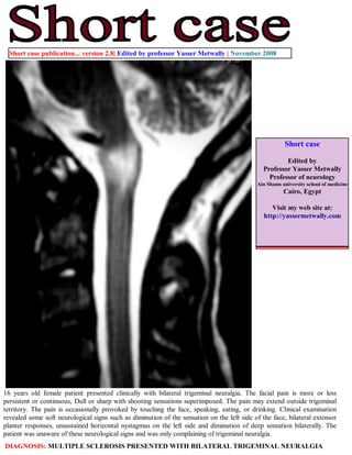
- 1. Short case publication... version 2.8| Edited by professor Yasser Metwally | November 2008 Short case Edited by Professor Yasser Metwally Professor of neurology Ain Shams university school of medicine Cairo, Egypt Visit my web site at: http://yassermetwally.com 16 years old female patient presented clinically with bilateral trigeminal neuralgia. The facial pain is more or less persistent or continuous, Dull or sharp with shooting sensations superimposed. The pain may extend outside trigeminal territory. The pain is occasionally provoked by touching the face, speaking, eating, or drinking. Clinical examination revealed some soft neurological signs such as diminution of the sensation on the left side of the face, bilateral extensor planter responses, unsustained horizontal nystagmus on the left side and diminution of deep sensation bilaterally. The patient was unaware of these neurological signs and was only complaining of trigeminal neuralgia. DIAGNOSIS: MULTIPLE SCLEROSIS PRESENTED WITH BILATERAL TRIGEMINAL NEURALGIA
- 2. Figure 1. Pre and postcontrast MRI T1 images showing a cervicomedullary enhanced two lesions involving mainly the anterior and posterior regions of cervicomedullary junction (the lesions are definitely separated from each other) with mild extension to the posterior parts of the medulla and probably involving also the inferior cerebellar peduncle. The contrast enhancement is an indication that the lesions were active during the time of examination . (The patient was in exacerbation during examination) Figure 2. Pre and postcontrast MRI T1 images showing a cervicomedullary enhanced two lesions involving mainly the anterior and posterior regions of cervicomedullary junction (the lesions are definitely separated from each other) with mild extension to the posterior parts of the medulla and probably involving also the inferior cerebellar peduncle. The contrast enhancement is an indication that the lesions were active during the time of examination . (The patient was in exacerbation during examination)
- 3. Figure 3. MRI T2 image (A) and postcontrast MRI T1 image (B). The cervicomedullary pathology appear hyperintense on the MRI T2 image and is composed almost of two lesions (an oval lesion anteriorly and a linear one posteriorly), the two lesions ate separated from each other by an isointense line of cleavage on the MRI T2 image. Only parts of the lesions as demonstrated on the MRI T2 image (A) are enhanced on the postcontrast MRI T1 image (B). Other parts of the lesions are not enhanced. The multiplicity of the lesions provides evidence for dissemination in place. However MRI can also provide evidence for quot; Dissemination in timequot;. When some lesions, or some parts of the same lesion - as seen on the T2 images- enhance on the T1 postcontrast images and others do not enhance, this simply mean that some lesions are old quot;burnt outquot; plaques (those which do not enhance) and other lesions are new and active plaques (those which enhance), and this provides evidence of quot; Dissemination in timequot;. Ring enhancement also provides evidence for quot; Dissemination in timequot;. Contrast enhancement (as demonstrated on the postcontrast T1 image) occurred at the periphery of the lesions demonstrated on the MRI T2 image (A) and this is the picture of reactivation of old plaques which takes place at the periphery of old plaques rather than at the center. Figure 4. MRI T2 images. The cervicomedullary pathology appear hyperintense on the MRI T2 images and is composed almost of two lesions (an oval lesion anteriorly and a linear one posteriorly), the two lesions ate separated from each other by an isointense line of cleavage on the MRI T2 images. The lesions are causing slight enlargement of the cervicomedullary junction.
- 4. Figure 5. MRI FLAIR image showing the typical picture of multiple sclerosis. References 1. Metwally, MYM: Textbook of neurimaging, A CD-ROM publication, (Metwally, MYM editor) WEB-CD agency for electronic publishing, version 9.4a October 2008 Addendum A new version of short case is uploaded in my web site every week (every Saturday and remains available till Friday.) To download the current version follow the link quot;http://pdf.yassermetwally.com/short.pdfquot;. You can download the long case version of this short case during the same week from: http://pdf.yassermetwally.com/case.pdf or visit web site: http://pdf.yassermetwally.com To download the software version of the publication (crow.exe) follow the link: http://neurology.yassermetwally.com/crow.zip At the end of each year, all the publications are compiled on a single CD-ROM, please contact the author to know more details. Screen resolution is better set at 1024*768 pixel screen area for optimum display For an archive of the previously reported cases go to www.yassermetwally.net, then under pages in the right panel, scroll down and click on the text entry quot;downloadable short cases in PDF formatquot; Also to view a list of the previously published case records follow the following link (http://wordpress.com/tag/case- record/) or click on it if it appears as a link in your PDF reader