Issues in brainmapping...Intermittent rhythmic delta activity and its brainmap counterpart
•
1 gefällt mir•1,122 views

Issues in brainmapping...Intermittent rhythmic delta activity and its brainmap counterpart
![INTERMITTENT RHYTHMIC SLOW WAVE ACTIVITY
Rhythmic often symmetric but cansinusoidal waveformsadults, the delta activity has aoccur intermittently in the EEG recording. It
is most
delta activity consists of
be lateralized. In
of approximately 2.5 Hz that
frontal predominance (frontal intermittent
rhythmic delta activity [FIRDA]). In children, it is maximal posteriorly (occipital intermittent rhythmic delta activity [OIRDA]).
Intermittent rhythmic delta activity is associated with structural lesions, most commonly diencephalic, infratentorial or
intraventricular tumors, or with diffuse encephalopathies. FIRDA occurring in patients with a normal EEG background suggests that
the pattern is due to a structural lesion; when associated with EEG background abnormalities, it is likely to be due to encephalopathy.
In cases of encephalopathy with FIRDA, the pathophysiologic processes are believed to involve cortical and subcortical gray matter.
OIRDA is associated with absence epilepsy in children aged 6-10 years.
Intermittentsynchronousdelta activityappearing usually occurs atThe ascending of 2-2.5is Hz withmore steeplysinusoidal,descent, and
bilaterally
rhythmic
waveforms
(IRDA)
in short bursts.
frequencies
phase sloped
relatively
than the
stereotypic,
waves are typically bilateral and widespread with peak amplitude frontally in older individuals (FIRDA) and occipitally in children
(OIRDA). These patterns attenuate with alerting or eye opening. Eye closure, drowsiness, and hyperventilation accentuate IRDA.
Although IRDA disappears in stage 2 and deeper non–rapid eye movement (REM) sleep, it may reappear in REM sleep.
Multiple etiologies can result in IRDA, including metabolic, toxic, hypoxic, or various diffuse or focal intracranial diseases. Even when
IRDA occurs unilaterally in association with a focal cerebral lesion, the lateralization of IRDA may be ipsilateral or contralateral to
the lesion. Thus IRDA is a nonspecific nonlocalizing EEG pattern, unless associated with other focal findings on the EEG. Although
the mechanisms for production of IRDA are understood incompletely, studies correlating with pathologic specimens suggest that IRDA
is associated primarily with diffuse gray matter disease.
The degree of encephalopathy manifested appears to correspond to the proportion of IRDA on the EEG. This pattern must be
distinguished from the frequently encountered frontally maximal intermittent delta that can be seen in drowsy elderly patients.
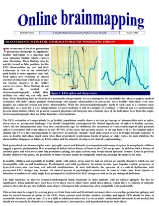
Figure 2. The intermittent
rhythmic delta activity [left
image] and the the polymorphic
slow wave activity [right image]
Consists of sinusoidal waveforms of approximately 2.5 Hz that occur intermittently in the EEG recording.
It is most often symmetric but can be lateralized.
In adults, the delta activity has a frontal predominance (frontal intermittent rhythmic delta activity
[FIRDA]). In children, it is maximal posteriorly (occipital intermittent rhythmic delta activity [OIRDA])
The intermittent rhythmic delta activity shows visual reactivity and is commonly suppressed in the eye
open state unless the patient is comatose.
Intermittent rhythmic delta activity is associated with structural lesions, most commonly diencephalic,
infratentorial or intraventricular tumors, or with diffuse encephalopathies.
FIRDA occurring in patients with a normal EEG background suggests that the pattern is due to a
structural lesion; when associated with EEG background abnormalities, it is likely to be due to
encephalopathy.
OIRDA is associated with absence epilepsy in children aged 6-10 years.](data:image/gif;base64,R0lGODlhAQABAIAAAAAAAP///yH5BAEAAAAALAAAAAABAAEAAAIBRAA7)
Empfohlen
Weitere ähnliche Inhalte
Andere mochten auch
Andere mochten auch (18)
Mehr von Professor Yasser Metwally
Mehr von Professor Yasser Metwally (20)
Kürzlich hochgeladen
Kürzlich hochgeladen (20)
Issues in brainmapping...Intermittent rhythmic delta activity and its brainmap counterpart
- 1. Beta III version A Monthly Publication presented by Professor Yasser Metwally March 2008 THE OCCURRENCE OF EPILEPTIC DISCHARGE IN HEALTHY NONEPILEPTIC PERSONS he occurrence of focal or generalized paroxysmal discharges in apparently healthy individuals is a puzzling and even annoying finding which requires some discussion. These findings may be quickly termed as false positives, but the EEG abnormalities are real and their irrelevance in view of the individual's good health is more apparent than real. Such spikes give testimony of certain cerebral dysfunctions which may or may not become manifest in the further course of events. These findings do not discredit the method of electroencephalography, which, after Figure 1. EEG spikes and sharp waves artifacts are ruled out, can only show facts. These disturbing facts are in need of a reasonable interpretation. Let us contemplate the indubitable fact that a complete medical evaluation will yield certain physical shortcomings and organic abnormalities in practically every healthy individual; even acne pimples are cutaneous lesions and hence abnormalities. What the electroencephalographer needs in such cases is a common sense philosophy as a basis for a wise interpretation. General medicine is full of examples of seemingly irrelevant and yet unmistakably present abnormalities which the prudent, seasoned physician will integrate into a holistic view of the individual. Seen from this angle, electroencephalography does not differ from the rest of medicine. The EEG evaluation of comparatively large healthy populations usually shows a certain percentage of abnormalities such as spike, sharp wave or paroxysmal discharge. One should thoughtfully contemplated the clinical significance of spikes in healthy persons. Above all, the interpretation must take into consideration age. In childhood, the occurrence of central-midtemporal (also parietal) spikes is associated with overt seizures in only 50-70% of the cases; this pertains mainly to the age from 3-12 yr. In occipital spikes (mainly age 3-5 yr), the epileptogenicity is even lower. In general, quot;benignquot; focal spikes (such as seen in benign Rolandic epilepsy) is seen, in healthy individual, much more often than generalized synchronous bursts of spikes or spike waves. In most children, the abnormalities disappear on follow up EGG studies and the minority develop overt clinical symptomatology. Both generalized synchronous (spike wave, polyspike wave) and Rolandic (centroparieto-midtemporal) spikes in nonepileptic children suggest a genetic predisposition if no neurological deficit and no history of insult to the CNS are present. In children with a history of cerebral palsy and with no seizures but prominent spiking, the spike activity may herald future epileptic seizures. Even in perfectly healthy children with spikes, the possibility of future seizures cannot be completely ruled out, although the chances are slim. In healthy children and especially in healthy adults with spikes, stress must be laid on certain personality disorders which are not incompatible with normal functioning. Psychological and mild psychiatric deviations include poor impulse control, proneness to hysterical conversion reactions, and schizoid manifestations. In such individuals, the presence of a cerebral dysfunction with paroxysmal EEG changes may hamper the natural process of psychological maturation. In some of these cases, head injuries or infections of moderate severity might have prompted or facilitated the EEG changes as well as the psychological deviations. The high incidence of anterior temporal-midtemporal sharp transients in older patients with no clinical epileptic fits has no significance. These patients may even have overt sharp waves; others show small sharp spikes. Unless there is evidence of epileptic seizures, these discharges only indicate some degree of temporal lobe dysfunction, often compatible with good health. This section must be capped by a strong plea to refrain from rash and ill-advised statements that a seizure-free person has epilepsy and must be treated because of spikes in the EEG. These persons need further medical attention and repeat EEG should be done at reasonable intervals, such as every 2 yr in a child or adolescent and every 5 yr in an adult. Anticonvulsive treatment is not needed, but should not necessarily be denied to extremely apprehensive, introspective, and hypochondria-prone individuals.
- 2. INTERMITTENT RHYTHMIC SLOW WAVE ACTIVITY Rhythmic often symmetric but cansinusoidal waveformsadults, the delta activity has aoccur intermittently in the EEG recording. It is most delta activity consists of be lateralized. In of approximately 2.5 Hz that frontal predominance (frontal intermittent rhythmic delta activity [FIRDA]). In children, it is maximal posteriorly (occipital intermittent rhythmic delta activity [OIRDA]). Intermittent rhythmic delta activity is associated with structural lesions, most commonly diencephalic, infratentorial or intraventricular tumors, or with diffuse encephalopathies. FIRDA occurring in patients with a normal EEG background suggests that the pattern is due to a structural lesion; when associated with EEG background abnormalities, it is likely to be due to encephalopathy. In cases of encephalopathy with FIRDA, the pathophysiologic processes are believed to involve cortical and subcortical gray matter. OIRDA is associated with absence epilepsy in children aged 6-10 years. Intermittentsynchronousdelta activityappearing usually occurs atThe ascending of 2-2.5is Hz withmore steeplysinusoidal,descent, and bilaterally rhythmic waveforms (IRDA) in short bursts. frequencies phase sloped relatively than the stereotypic, waves are typically bilateral and widespread with peak amplitude frontally in older individuals (FIRDA) and occipitally in children (OIRDA). These patterns attenuate with alerting or eye opening. Eye closure, drowsiness, and hyperventilation accentuate IRDA. Although IRDA disappears in stage 2 and deeper non–rapid eye movement (REM) sleep, it may reappear in REM sleep. Multiple etiologies can result in IRDA, including metabolic, toxic, hypoxic, or various diffuse or focal intracranial diseases. Even when IRDA occurs unilaterally in association with a focal cerebral lesion, the lateralization of IRDA may be ipsilateral or contralateral to the lesion. Thus IRDA is a nonspecific nonlocalizing EEG pattern, unless associated with other focal findings on the EEG. Although the mechanisms for production of IRDA are understood incompletely, studies correlating with pathologic specimens suggest that IRDA is associated primarily with diffuse gray matter disease. The degree of encephalopathy manifested appears to correspond to the proportion of IRDA on the EEG. This pattern must be distinguished from the frequently encountered frontally maximal intermittent delta that can be seen in drowsy elderly patients. Figure 2. The intermittent rhythmic delta activity [left image] and the the polymorphic slow wave activity [right image] Consists of sinusoidal waveforms of approximately 2.5 Hz that occur intermittently in the EEG recording. It is most often symmetric but can be lateralized. In adults, the delta activity has a frontal predominance (frontal intermittent rhythmic delta activity [FIRDA]). In children, it is maximal posteriorly (occipital intermittent rhythmic delta activity [OIRDA]) The intermittent rhythmic delta activity shows visual reactivity and is commonly suppressed in the eye open state unless the patient is comatose. Intermittent rhythmic delta activity is associated with structural lesions, most commonly diencephalic, infratentorial or intraventricular tumors, or with diffuse encephalopathies. FIRDA occurring in patients with a normal EEG background suggests that the pattern is due to a structural lesion; when associated with EEG background abnormalities, it is likely to be due to encephalopathy. OIRDA is associated with absence epilepsy in children aged 6-10 years.
- 3. does not appear to is pathognomonic of epilepsy, and it This occasionally encountered in encephalopathic patients. pattern In summary,OIRDA be found almost exclusively in children.may befinding is probably epileptiform in nature. However, thisPrevious studies have found that most children whose EEGs depicted OIRDA had primary generalized epilepsy. However, the author detected a higher proportion of cases with localization-related epilepsy. Furthermore, the frequency of the OIRDA discharge appears to be higher when it occurs in association with absence epilepsy, and these cases are more likely to depict epileptiform activity intermixed with the rhythmic delta pattern than are cases of focal epilepsy. Frontal intermittent rhythmic delta activity is rare in children, is not associated with acute encephalopathy or with deep midline or infratentorial lesions, and tends to occur during wakefulness. The electrographic characteristics of frontal intermittent rhythmic delta activity appear to differ between cognitively normal and mentally retarded children Encephalopathy and coma result from conditions thatincluding metabolic, hemispheres or the reticular activatingendocrinologic, midbrain. The differential diagnosis is broad, affect both cerebral toxic, anoxic/ischemic, infectious, system in the degenerative, and inflammatory processes. These processes affect the brain diffusely, and, consequently, changes in the EEG often appear generalized. While most EEG findings in encephalopathy and coma are nonspecific with regard to etiology, information relevant to the clinical course and prognosis can be obtained using the EEG. In cases of mild encephalopathy, theta and delta activity is intermixed with the background alpha rhythm. Occasional generalized delta transients are also seen. As the encephalopathy worsens, there is loss of background alpha-range frequencies and an increased amount of generalized theta and delta activity. Intermittent-rhythm delta activity (IRDA) may appear, which in adults generally is frontally predominant (FIRDA), and is consistent with moderate diffuse bihemispheric cerebral dysfunction. In severe encephalopathy, there is generalized delta activity. Loss of reactivity in anyone of these stages implies greater severity, and, in specific clinical settings, a worse prognosis. In the clinical setting of severe anoxia (e.g., after cardiac arrest) or severe closed head injury, invariant patterns of persistent, generalized alpha activity (alpha coma), generalized periodic epileptiform discharges, or the burst suppression pattern. are associated with very poor outcome. Figure 3. Frontal intermittent rhythmic delta activity (FIRDA) The brainmap counterpart of intermittent rhythmic slow wave activity 1- Intermittent rhythmic slow wave activity is seen in diffuse encephalopathic process (except hepatic encephalopathy which has a different brainmap spectral profile as will be explained in a different issue) and in middle line thalamic, or huge parasellar or infratentorial tumors. It was also seen by the author in vertebro-basilar insufficiency and migraine. 2- There is reduction of alpha percentage activity, and reduced or reversed alpha visual reactivity. The degree of reduction of alpha percentage activity correlates with prognosis. The alpha topography is normal and retains its middle line topography with posterior maximum and is more reduced anteriorly than posteriorly. There is a fairly good degree of symmetry, synchrony and coherence in alpha percentage activity maps. however it is not very infrequent to see some degree of asymmetry with more alpha percentage activity seen on one side than the other. 3- The alpha land (the middle line electrodes) is now occupied by theta and delta percentage activity, which is now maximum in the middle line electrodes at F3,F4,O2,O1. The maximum middle line percentage activity is now shifted down to the theta rage in most
- 4. patients and occasionally to the delta rage depending on the severity of the encephalopathic process and the state of the patient. Combined theta and delta percentage activity maps clearly demonstrates the midline maximum. The topography of theta percentage activity is identical to that of normal alpha percentage activity maps in the eye closed state. Slow wave activity commonly show visual reactivity exactly as alpha percentage activity behaves under normal physiological conditions. 4- There is a fairly good degree of symmetry, synchrony and coherence in theta percentage activity. However it is not very infrequent to see some degree of asymmetry with more theta percentage activity seen on one side than the other. 5- Beta percentage activity is reduced in the midline frontal, central/parietal and occipital electrodes. Because the intermittent rhythmic slow wave activity occupies the same electrode regions that are normally occupied by the alpha activity, so it looks like that both (the intermittent rhythmic slow wave activity and alpha spindles) share a common generator. Similarities between both activities are: Both are sinusoidal activity that waxes and wanes (intermittent) Both show visual reactivity Both are maximum in the middle line electrodes So it looks like that the generator that is responsible for the generation of the intermittent rhythmic slow wave activity is the same generator that is responsible for the generation of alpha spindles under normal conditions, however in the former condition the generator (the pace-maker) is diseased and firing at a lower rate. A dysfunctioned pacemaker (subcortical gray matter, diencephalon) is probably necessary for the intermittent slow wave activity to be seen on EEG. Figure 4. Two studies showing the brainmap counterpart of intermittent rhythmic slow wave activity.. Notice the bilateral symmetrical theta activity occupying the same topography of the alpha activity with middle maximum. Some degree of visual reactivity is observed in the lower study. Some degree of asymmetry is observed in the lower maps Arousal stimuli in the thalamo-cortical circuitry are responsible for the production of alpha spindles under normal physiological conditions. However when a diffuse encephalopathic process involves the diencephalic neurons bilaterally, the dysfunctioned diencephalic neurons fire at a slower rate and instead of producing alpha spindles they produce theta or delta spindles that retain the same characteristics of alpha spindles in spatial topography and visual reactivity. In intermittent rhythmic slow wave activity cortical gray matter is not necessarily abnormal as this phenomenon is also observed in middle line tumors (thalamic) and some extra-axial tumors in the parasellar and infratentorial regions. A dysfunctioned subcortical gray matter affecting the diencephalic pace-making neurons is necessary for the generation of intermittent rhythmic slow wave activity. The diencephalic pace-making neurons are necessary for the genesis of alpha spindles under normal physiological conditions. In intermittent rhythmic slow wave activity there is a down-shift of the predominant middle line activity (the alpha land) from the alpha range to lower frequencies in the theta and delta ranges. In the author experience the predominant middle line activity is down-shifted to the theta range more often than the delta range. The condition is better termed intermittent rhythmic theta activity rather than intermittent rhythmic delta activity, although the later is more commonly used. In intermittent Figure 5. The brainmap counterpart of intermittent rhythmic rhythmic slow wave activity brainmapping has apparently shown that slow wave activity. Notice the Bifrontal middle line theta predominant middle line activity is more often down- shifted to the theta maximum with reduced alpha and beta activity. Maximum range rather than the delta range. theta activity is seen at F3,F4,FZ electrodes
- 5. Figure 6. A, Normal alpha map, B, Theta map picked up during intermittent rhythmic slow wave activity and C, Normal theta map. Notice that alpha activity is predominately middle line under normal conditions (A) and theta activity is maximum in the bitemporal (lateral) regions under normal conditions (C). In intermittent rhythmic slow wave activity there is a down-shift of the predominant middle line activity (the alpha land) from the alpha range to lower frequencies in the theta and delta ranges. In the author experience the predominant middle line activity is down-shifted to the theta range more often than the delta range. The topography of theta percentage activity during intermittent rhythmic slow wave activity (B) is identical to that of alpha map under normal physiological conditions (Compare The normal alpha map (A) and the theta map generated during intermittent rhythmic slow wave activity (B). Because the intermittent rhythmic slow wave activity occupies the same electrode regions that are normally occupied by the alpha activity, so it looks like that both (the intermittent rhythmic slow wave activity and alpha spindles) share a common generator. The generator (The pace maker) that is responsible for the generation of the intermittent rhythmic slow wave activity is the same generator which is responsible for the generation of alpha spindles under normal conditions, however in the former condition the generator (the pace-maker) is diseased and firing at a lower rate. In summary, although the condition is frequently termed frontal intermittent rhythmic delta activity (FIRDA) or occipital intermittent rhythmic delta activity (ORDA) in conventional EEG, However by using the brainmap technology The following are shown: Intermittent rhythmic slow wave activity is simply a condition where the predominant middle line activity is down-shifted from the alpha range to the theta range with maximum activity at F3, F4, FZ, O1,O2 electrode regions. During intermittent rhythmic slow wave activity the the alpha spindle (as the dominant EEG activity in middle line electrodes) is replaced by slow wave spindles (mostly in the theta range rather than the delta range) which now dominate EEG activity in the middle line electrode regions. In frontal intermittent rhythmic delta activity (FIRDA) the maximum slow wave activity is recorded at F3, F4, FZ electrode regions by brainmapping. This pattern is more commonly seen in adult and it reflects a diffuse encephalopathic process, it is also seen in middle line tumors, parasellar and infratentorial tumors, vertebro-basilar insufficiency and migraine. In occipital intermittent rhythmic delta activity (ORDA) the maximum slow wave activity is recorded at O1,O2 electrode regions by brainmapping. OIRDA is found almost exclusively in children. This pattern is probably epileptiform in nature. However, this pattern does not appear to be pathognomonic of epilepsy. The 3 c/s spike/wave discharge of absence attacks is occasionally proceeded by OIRDA. Whether OIRDA or FIRDA, the discharge is maximum in the middle line electrodes at F3, F4, FZ, C3, C4, CZ, P3, P4, PZ, O1,O2 and it replaces the alpha activity that normally reigns in these territories. The alpha activity is longer the prevalent activity at the middle line electrodes regions during intermittent rhythmic slow wave activity. Alpha activity is normally prevalent at O1,O2 and F3,F4,FZ electrode regions as well as the intermittent rhythmic slow wave activity which takes the upper hand in middle line electrodes and becomes the prevalent activity when they occur, they also retain the same characteristics of alpha spindles in spatial topography and visual reactivity. There is a fairly good degree of symmetry, synchrony and coherence in theta or delta percentage activity maps (during intermittent rhythmic slow wave activity). However it is not very infrequent to see some degree of asymmetry and lateralization with more theta or delta percentage activity seen on one side than the other.
- 6. Figure 7. The brainmap counterpart of intermittent rhythmic slow wave activity. Notice the Bifrontal middle line theta maximum with reduced alpha and beta activity. Maximum theta activity is seen at F3,F4,FZ electrodes References 1. Metwally, MYM: Textbook of neurimaging, A CD-ROM publication, (Metwally, MYM editor) WEB-CD agency for electronic publishing, version 9.1a January 2008 The author, Professor Yasser Metwally Professor of clinical neurology, Ain Shams university, Cairo, Egypt. www.yassermetwally.com A new version of this publication is uploaded in my web site every month Follow the following link to download the current version: http://brainmapping.yassermetwally.com/map.pdf © Yasser Metwally, all rights reserved