SlideShare ist ein Scribd-Unternehmen logo
1 von 14
Downloaden Sie, um offline zu lesen
CASE OF THE WEEK
                        PROFESSOR YASSER METWALLY
CLINICAL PICTURE

CLINICAL PICTURE:

A 60 years old male patient presented clinically with rapidly progressive left sided hemiplegia and manifestations of increased
intracranial tension. (To inspect the patient's full radiological study, click on the attachment icon (The paper clip icon in the
left pane) of the acrobat reader then double click on the attached file) (Click here to download the attached file)

RADIOLOGICAL FINDINGS

RADIOLOGICAL FINDINGS:


Figure 1. Precontrast CT scan showing diffuse hypodensities involving the left thalamic area, left internal capsule and left
basal ganglionic area. The hypodensities, most probably, represent tumor tissues, peritumoral edema and reactive astrogliosis.
Figure 2. Postcontrast CT scan images showing a densely enhanced space occupying lesion involving the left thalamic area
with positive mass effect and peritumoral edema involving the white matter and sparing the gray matter (vasogenic edema).
Notice the peritumoral satellitosis (B)


                                          Figure 3. A postcontrast CT scan image showing peritumoral satellitosis (Figure 2
                                          B)


CT guided stereotactic biopsy revealed Mixed astrocytoma (Grade III-Grade IV diffuse astrocytoma). The patient was not
operated upon, he received radiotherapy only to die 4 month following clinical diagnosis.

Common pathological characteristics of diffuse astrocytomas

      Diffuse astrocytomas are tumors predominantly composed of astrocytes. Unless otherwise indicated, the term usually
       applies to diffusely infiltrating neoplasms (WHO grades II through IV).

      Diffuse astrocytoma is unusual in the first decade of life and most commonly presents in older children or young adults
       up to the age of 40 to 45.

      All diffuse astrocytomas, particularly the diffusely infiltrating variety, have a tendency toward progression to more
       malignant forms. Diffuse astrocytomas have a peculiar tendency to change its grade over time into the next higher
       grade of malignancy and the condition is age dependant. A change in the grade of diffuse astrocytoma is more likely to
       occur in the older age group.
   Diffuse astrocytomas commonly start as grade II at a younger age group then gradually change its grade over time into
       the next higher grade until they ultimately dedifferentiate into glioblastomas (secondary glioblastoma multiforme), on
       the other hand, glioblastoma multiforme in older patients are usually primary-that is, they occur as glioblastoma
       multiforme from their inception, without progression from a lower- grade tumor.

      Diffuse astrocytomas appear to form a continuum of both biological and histological aggression. They vary from lesions
       with almost normal cytology (grade I and grade II astrocytomas) through intermediate stages (grade III, anaplastic
       astrocytomas) and up to the most aggressive of all human brain tumours (grade IV astrocytomas or glioblastoma
       multiforme).

      Diffuse astrocytoma often spreads widely through the brain but without destruction and also without interruption of
       normal function. Microscopically, tumor cells infiltrate between myelinated fibers in a nondestructive manner
       (perineuronal satellitosis). The local spread of diffuse astrocytomas (forming gliomatosis cerebri and butterfly gliomas)
       does not mean that the tumour grade is grade IV (glioblastoma multiforme), local spread can occur in grade II and
       grade III and in the author experience gliomatosis cerebri and butterfly gliomas are much more commonly seen in
       grade II astrocytomas and has not been encountered in grade III (anaplastic astrocytomas) and grade IV (glioblastoma
       multiforme). It takes a long time for a diffuse astrocytoma to cross the corpus callosum to the opposite hemisphere to
       form a butterfly glioma. Patients harbouring glioblastomas have a much shorter life span for their tumours to form
       butterfly gliomas, however cases were reported for glioblastomas forming butterfly tumours.

      These glioma cells migrate through the normal parenchyma, collect just below the pial margin (subpial spread),
       surround neurons and vessels (perineuronal and perivascular satellitosis), and migrate through the white matter tracks
       (intrafacicular spread). This invasive behavior of the individual cells may correspond to the neoplastic cell's
       reacquisition of primitive migratory behavior during central nervous system development. The ultimate result of this
       behavior is the spread of individual tumor cells diffusely over long distances and into regions of brain essential for
       survival of the patient. The extreme example of this behavior is a condition referred to as gliomatosis cerebri, in which
       the entire brain is diffusely infiltrated by neoplastic cells with minimal or no central focal area of tumor per se.
       Furthermore, 25% of patients with GBM have multiple or multicentric GBMs at autopsy. Although GBMs can be
       visualized on MRI scans as mass lesions that enhance with contrast, the neoplastic cells extend far beyond the area of
       enhancement.

      In practice considerable histological heterogeneity in astrocytic tumours is found ( i.e., low grade areas with Rosenthal
       fibers and calcification can be intermixed with with frankly malignant ones).

      The differences in histologic features, potential for invasiveness, and extent of progression likely reflect genetic
       differences acquired during astrocytoma growth.

      Grade IV astrocytomas (glioblastoma multiforme) differ from diffuse astrocytoma grade II and grade III (anaplastic
       astrocytomas) in the presence of gross necrosis, and microscopically in the presence of vascular endothelial hyperplasia
       and tumour hemorrhage.


Primary tumors of the thalamus account for only 1-1.5% of all intracranial tumors and approximately 25% of them arise in
children aged 15 years or under. Thalamic gliomias might be unilateral or bilateral.

Diffuse and involvement of thalamic nuclei by these tumors makes surgical therapy very difficult and no case of radical
removal has been described in the literature. Consequently, the main role of surgery is limited and usually performed for a
histological diagnosis. Generally, these gliomas are low-grade astrocytomas (grade II of WHO classification), but limited
anaplastic areas may be encountered.

Radiotherapy and chemotherapy are sometimes utilized as adjuvant therapy, but their role is questionable. Outcome is
generally poor, independently of the therapy that is utilized. Rapid fatal evolution after diagnosis and the almost complete
unresponsiveness of these tumors to radiotherapy make these rare tumors difficult to treat. Anaplastic gliomas usually show
enhancement after contrast administration.

Severe dementia and personality modification observed in adults affected by bilateral thalamic glioma is attributed to the
involvement of dorsomedial nuclei of thalami and their connections with temporal and frontal lobes [9]. Bilateral thalamic
glial tumors are rare and less than 50 cases have been published in the literature [1-8].

DIAGNOSIS:

DIAGNOSIS: THALAMIC GLIOMA
DISCUSSION

DISCUSSION:

Bailey and Cushing observed that gliomas become more aggressive with time, writing – all of these lesions, so far as our
records permit us to judge, show an increasing degree of malignancy, the recurrent tumors giving evidence of more active cell
division than the original lesion. 2 It is now understood that the accumulation of molecular abnormalities1 underlies the
increasingly aggressive clinical behavior of an individual patient s glioma over time. Moreover, while GBMs may be
histologically identical, they are molecularly distinct, [51] and each tumor may require individually tailored treatment with
the combination of agents predicted to impact multiple molecular abnormalities. Successful therapy of a molecularly complex
disease such as GBM may also require simultaneous administration of multiple agents, including both traditional
chemotherapies and several pathway inhibitors.

      The past of glioblastoma multiforme

The most common cancer arising from the brain is the glioblastoma multiforme (GBM). It is also the most deadly, [1]
representing the most aggressive subtype among the gliomas, a collection of tumors including astrocytomas and
oligodendrogliomas (see Table 1). In 1926, Bailey and Cushing, in describing spongioblastoma multiforme , the label then used
for GBM, noted that:

- It is from this group doubtless that the generally unfavorable impression regarding gliomas as a whole has been gained. It is
not only the largest single group in the series but at the same time is one of the most malignant. In the five unoperated cases,
the average duration of life from the onset of symptoms was only three months, which speaks well on the whole for the
average survival period of twelve months for those surgically treated. [2]

Since their seminal work, the median survival of 12 months has not changed markedly. Both data from the 1960s3 and
current data [4] confirm that the extent of surgical resection is an important prognostic factor. However, as Bailey and
Cushing observed, GBMs have – infiltrating propensities, and when enucleation is attempted, the growth is found at the depth
to spread into and merge with the normal cerebral tissue without recognizable demarcation. [2] In prior eras, radical surgical
excisions, including removal of the entire cerebral hemisphere containing the tumor, [5] were occasionally attempted, yet
patients who survived the hemispherectomy died of recurrent tumor, [6] clinically proving the importance of the histologic
observation that tumor cells invade throughout the brain. In the modern age, brain imaging may disclose macroscopic tumor
in the opposite hemisphere (see Figure 1) or even gliomatosis cerebri literally a brain full of tumor. In the years leading to up
to World War II, the German pathologist Scherer, whose scientific discoveries were tainted by his Nazi activities, [7]
described secondary structures [8,9] that further characterized invasive tumor cells. These structures are secondary because
they are dependent for their formation on underlying normal brain structures, as opposed to primary structures of the tumor
such as pseudopalisading necrosis and microvascular proliferation. Examples include perineuronal and perivascular
satellitosis (accumulation of tumor cells around neurons and blood vessels), subpial spread, and intrafascicular tracking such
as infiltration along corpus callosum and other white matter tracks (see Figure 2).

Advances in surgical technique, imaging, and targeting of radiotherapy (RT) are important contributions to local control.
However, changing GBM from a disease that kills quickly to one that can be managed as a chronic illness, such as
hypertension or diabetes mellitus, will require systemic therapies targeting tumor cells infiltrating throughout the brain, such
as chemotherapy, immunotherapy, and small molecule pathway inhibitors.

      The current status of glioblastoma multiforme

Currently, treatment for GBM involves both local and systemic therapy. Surgery and partial brain RT are the standard
locally directed therapies. Some physicians also advise intra-operative placement of chemotherapy containing polymers (i.e.
Gliadel wafers ) directly into the surgical bed in an attempt to prolong local control. [10] While there is a modest survival
benefit, the use of these polymers remains controversial because of the potential for toxicity. Other treatment modalities that
target disease localized to the surgical bed or the surrounding area have included brachytherapy and stereotactic
radiosurgery (with either a linear accelerator or gammaknife), neither of which are commonly advised. Convection-based
chemotherapies delivered by catheter infusion, such as local delivery of pseudomonas exotoxin linked to either interleukin [13]
(IL-13) or transforming growth factor-a (TGFa), [11] are available in clinical trials for some patients. These trials take
advantage of differences in the expression of proteins (such as growth factor receptors) on the surface of residual tumor cells
in the periphery of the operative bed to deliver the toxin to tumor cells, but spare normal brain.

By contrast, systemic chemotherapy targets tumor cells beyond the reach of local therapies. The most commonly prescribed
systemic chemotherapy for GBM is temozolomide (Temodar®), an alkylator that became available during the last decade.
The effectiveness of temozolomide in the management of GBM at diagnosis was recently demonstrated by a large
multinational study.4 A modest survival benefit of 2.5 months for concurrent temozolomide with RT (14.6 months median
survival) was observed relative to RT alone (12.1 months median survival).4 In addition, while the survival benefit was still
present two years after diagnosis, only 10.7% of patients were progression-free and only 26.5% of patients were alive at that
point. [4] While systemic chemotherapy improves the outcome for some patients, long-term disease control therefore remains
elusive.

Table 1. Common Gliomas. The gliomas are assigned a grade by the World Health Organization (WHO) depending on
histologic features that predict behavior.This classification scheme is derived from the clinicopathologic studies of Bailey and
Cushing.2 Grade I tumors, such as juvenile pilocytic astrocytomas, are generally focal rather than diffuse and are potentially
curable by surgical excision.WHO grade II–IV tumors are diffusely infiltrative.WHO grade III–IV tumors are termed ‘high
grade’ or malignant. GBMs, or grade IV astrocytomas, are the most aggressive subtype.


 Discoveries during the last several years have improved the understanding of glioma and general cancer biology markedly. 
Generally, a cancer comprises cells that either divide or survive when they should instead undergo either cell cycle arrest or
die. These abnormalities are also not mutually exclusive, and most cancers, including GBMs, are driven by several molecular
abnormalities. The signal to divide is typically provided by a growth factor (ligand). Examples include TGF?, epidermal
growth factor (EGF), platelet-derived growth factor (PDGF), and vascular endothelial growth factor (VEGF). Such ligands
interact with cells through receptors including EGF receptors (EGFRs), PDGF receptors (PDGFRs), and VEGF receptors
(VEGFRs). Receptor activity is linked with cellular processes such as mitosis or invasion by signal transduction cascades.
Examples of signal transduction cascades important in human GBMs include those activated by the oncogenes Ras, Akt, and
Src. [12] In cancer cells, these pathways are disrupted through several mechanisms. For example, EGFR is overexpressed in
up to 92% of astrocytomas, [13] and up to 62% of GBMs express EGFRvIII, [14] a mutant receptor that is active
independently of ligand. Co-expression of EGF and EGFR15,16 in GBMs leads to a potential autocrine loop.An analogous
loop is created by PDGF and PDGFR co-expression in up to 94% of high-grade oligodendrogliomas. [17–19] Regardless of
ligand or receptor status, close to 100% of GBMs exhibit activation of Ras,20,21 and approximately 70% exhibit activated
Akt, [21,22] the latter typically through loss of the tumor suppressor gene phosphatase tensin homolog on chromosome ten
(PTEN), [23–25] which normally represses Akt activation. Src is detected in 67% of GBMs.26 Finally, control over cell
division is normally maintained by tumor suppressors, such as an inhibitor of CDK4A (INK4A) and its alternative reading
frame (ARF), as well as p53, which also contributes to DNA repair and apoptosis, and other enzymes. Disruptions of normal
cell cycle control of one form or another have been observed in almost all GBMs. [12,27]


                              Figure 1. Magnetic Resonance Imaging Findings in Glioblastoma Multiforme. Contrast
                              enhanced magnetic resonance image (MRI) of the brain demonstrating a large GBM with a
                              smaller site in the contralateral hemisphere. This infiltrative nature of GBMs, essentially
                              effecting the entire brain, underscores the failure of even radical surgery, such as
                              hemispherectomy, to effect cure.


Moreover, the modeling of gliomas in mice has demonstrated that abnormalities of ligands, receptors, signal transducers, and
proliferation cause gliomas. For example, combined activation of Ras with Akt in glial progenitors is sufficient to induce
GBMs in mice, [22] and transgenic expression of activated forms of Ras [28] or Src [29] in glia leads to GBMs following
spontaneous development of cooperative oncogenic abnormalities. Modeling has also demonstrated that PTEN loss is
functionally equivalent to Akt activation, [30] when combined with activated Ras. PDGF overexpression in glia causes high-
grade oligodendrogliomas [31,32] that also exhibit pathologic features of GBMs, including pseudopalisading necrosis and
microvascular proliferation. The threshold to tumor formation is lowered by disruption of Ink4a-Arf or p53 expression.
[31,33,34]

While more is learned about glioma biology, small molecule inhibitors are being developed that target the causal pathways.
[35] For example, several inhibitors of EGFRs are under investigation in clinical trials. These include the EGFR inhibitors
erlotinib (OSI- 774/Tarceva), gefitinib (ZD-1839/Iressa), and lapatinib (GW572016). The PDGFR inhibitors imatinib (STI-
571/Gleevec) and PTK787, both of which have other targets, are also in use. Signal transduction cascade blockers are also
being studied. One example is R11577, which targets the enzyme that activates Ras. Rapamycin (sirolimus), CCI-779
(temsirolimus), and Rad-001 (everolimus) target mTOR, one of the key enzymes activated by Akt.


Figure 2. Histology of Gliomas. (A) Pseudopalisading (arrow) necrosis (arrow head) and (B) microvascular proliferation
(arrow) are the classic histologic findings in glioblastoma multiforme (GBM). Secondary Scherer structures (C) involve tumor
cells (arrow heads) accumulating around blood vessels (BV, long arrow), and neurons (N, long arrow) in a low grade (WHO
grade II) oligodendroglioma. Such perivascular and perineuronal satellitosis, along with intrafascicular growth and subpial
accumulation (not shown), contribute to the diffusely infiltrative nature of gliomas throughout normal brain structures..
(Click to enlarge figure)

Unfortunately, despite initial enthusiasm, treatment of GBMs as well as systemic malignancies with these small molecule
inhibitors as single agents has generally been disappointing. For example, published interim and final reports of trials
involving gefitinib, [36,37] erlotinib, [38–40] imatinib, [41,42] PTK787, [43,44] and CCI-77945 monotherapy for recurrent
high-grade gliomas have not shown response or survival rates that are markedly superior to those observed with traditional
chemotherapies, such as temozolomide4,46–48 or carmustine (BCNU). [49] However, there are individual patients treated
with these agents who experience durable objective responses or sustained stable disease.Therefore, these agents are likely to
have a role in GBM management.


                                                                 Figure 3. Pathways Important in GBM Biology. An
                                                                 extracellular ligand such as EGF,TGFa, or PDGF induces
                                                                 dimerization of receptors such as EGFR or PDGFR.
                                                                 Receptor stimulation activates intrinsic tyrosine kinase (TK)
                                                                 activity and EGFR and PDGFR are therefore called
                                                                 receptor tyrosine kinases (RTKs). RTKs then activate the
                                                                 Akt, Src, and Ras signal transduction cascades.Tumor cell
                                                                 growth is driven by ligand or receptor overexpression,
                                                                 constitutively    activating    receptor    mutations     (e.g.
                                                                 EGFRvIII), or signal transduction activity. Pointed (green)
                                                                 and block (red) arrows indicate pathway activation and
                                                                 inhibition, respectively.The inhibitors shown and others are
                                                                 under investigation in the treatment of GBMs.


      Future perceptive of glioblastoma multiforme

In addition to surgical resection and RT, the future of GBM therapy is likely to involve both additional measures to improve
local control (such as convection or catheter delivery of antitumor agents into the operative cavity) and systemic treatment to
address infiltrative disease distant from the main tumor bed. However, a major thrust of research will be tissue analyses
looking for molecular features that predict sensitivity of GBMs to either traditional chemotherapies or small molecule
inhibitors. Tailoring therapy with specific drugs to those patients is most likely to improve response rates and spare patients
who are unlikely to benefit the expense and potential toxicity of these agents. Determination of a molecularly effective dose
(MED) (inhibits a pathway), may also be more useful than the traditionally used maximally tolerated dose (MTD).

An example of a molecular prognostic factor is loss of heterozygosity for chromosomes 1p and 19q in anaplastic
oligodendrogliomas, which predicts both sensitivity to chemotherapy and radiation, as well as longer overall survival. [50]
Consequently, some neurooncologists are currently using results of 1p/19q analysis to guide therapy, [51] although this
remains an area of controversy. Other genomic alterations are also predictive—PTEN loss is associated with poor survival for
patients with anaplastic oligodendrogliomas [52] and is likely to predict poor outcome from GBM. [23,53,54] More recently it
was reported that GBMs, in which O6-methylguanine- DNA methyltransferase (MGMT) expression was silenced by gene
methylation, were more sensitive to temozolomide than tumors with unmethylated MGMT. The likely explanation is that
MGMT may counteract temozolomide activity by removing alkyl groups on DNA. [55] It is unclear whether MGMT
methylation impacts sensitivity of other glioma subtypes to temozolomide, yet MGMT methylation status may be used in the
near future to guide therapy.

Individualized medicine determined by molecular rather than simply histologic phenotype may also guide therapy with small
molecule inhibitors. Somatic mutations in exons 18 21 of EGFR are associated with sensitivity of lung cancer to gefitinib [56
58] or erlotinib. [58] However, the authors and others have not found these mutations in gliomas. [34,57,59,60] Efforts are
under way to identify the molecular features that predict sensitivity of GBMs to EGFR and other receptor tyrosine kinase
(RTK) inhibitors.

Response and survival rates may also be improved through combination therapy. For example, preliminary data suggest that
concurrent therapy with imatinib (PDGFR/VEGFR inhibitor) and hydroxyurea (a more traditional chemotherapy) is more
effective than imatinib monotherapy. A small series with 14 evaluable patients with recurrent GBMs demonstrated a disease
control rate (complete response or partial response or stable disease) of 64% for patients treated with this combination. [61]
By contrast, imatinib monotherapy led to a disease control rate of 29%. [42] Larger trials of this and other combinations, such
as temozolomide with PTK787, are under way. (Click here to download master degree thesis " Non surgical management of
brain tumors" in PDF format)


SUMMARY


SUMMARY

The last five years have seen an evolution in the management of highgrade astrocytic tumors comparable in scope yet greater
in magnitude to that of the prior 40 years. This is thanks to the convergence of three factors: the introduction of an oral agent
with antitumor activity beyond the blood–brain barrier and modest systemic toxicity (temozolomide); the demonstration
through a well-conducted randomized trial of the superiority of multimodality therapy; and the fact that we now stand on the
threshold of additional progress through key advances in translational biology, which, in many cancers, is providing new
targets for therapeutic intervention.

Astrocytic tumors have long been the bane of neurosurgeons, radiation therapists, and neuro-oncologists. Although they
account for only 2.3% of all cancer-related deaths in the US, [63] little if any substantial progress in brain imaging and
treatment had been made until the first years of this millennium. Characteristics of high-grade glial tumors compared with
other cancers are its unique location, robust invasive and angiogenic capabilities without a significant propensity to
metastasize outside of the central nervous system (CNS), and the profound histological and molecular heterogeneity within
tumor specimens.

      Advances in the Management of Glioblastomas— Multimodality Strategies

In 2003, a phase III study in 240 newly diagnosed patients with surgically resectable malignant gliomas—including 207 with
glioblastoma multiforme (GBM)—compared surgery plus radiotherapy and placebo wafers with surgery plus radiotherapy
and the addition of 3.8% bischloroethyl nitrosourea (BCNU, carmustine) wafers (Gliadel wafers) into the tumor bed. The
study demonstrated a modest, albeit significant, prolongation of survival in the latter group (13.9 versus 11.6 months). [64]
Long-term follow-up of this study showed that the survival advantage with BCNU wafers was maintained at one, two, and
three years, and was statistically significant (p=0.01) at three years compared with placebo, although the absolute number of
patients evaluated at this latter timepoint was quite small. [65]

Perhaps the most significant advance in the management of glioblastomas emanates from the work of Stupp et al. [65] who, in
a safety and efficacy study, randomized 573 patients with newly diagnosed glioblastoma from 85 centers, primarily in Europe,
to radiotherapy alone or to radiotherapy plus concomitant temozolomide followed by monthly temozolomide for six cycles. At
a median follow-up of 28 months the median survival in the radiotherapy group alone was 12.1 months compared with 14.6
months in the group receiving both treatment modalities (p<0.001). The two-year survival rate was 10.4% with radiotherapy
alone versus 26.5% with radiotherapy and temozolomide. The results of this study produced level 1A evidence for the benefit
of this combined modality treatment in initially diagnosed patients, and was incorporated into the new National
Comprehensive Cancer Network (NCCN) guidelines for CNS tumors in 2005.

Thus, as a next logical step, a fusion of these two prior studies was evaluated in a phase II setting in patients with newly
diagnosed, highgrade GBM undergoing resection with BCNU wafer insertion followed by the combination of radiotherapy
plus temozolomide. Early interim data have been presented in abstract form. [67] The study end-points include survival and
progression-free survival (PFS). Of 35 patients enrolled so far, 34 were diagnosed with GBM. At median follow-up of 10.4
months, [87] patients had documented recurrence and 19 patients had died. Six patients remain on active treatment. The one-
year survival rate is 64%, and median survival is 18.6 months. These early data suggest that combination therapy with BCNU
wafers followed by therapy plus temozolomide may be an effective regimen in patients with initial highgrade resectable
malignant gliomas, although randomized trials will ultimately be needed to assess the efficacy of this treatment modality.
Other treatment modalities that have been investigated for the treatment of high-grade astrocytic tumors—especially in terms
of targeting disease localized to the surgical bed or the surrounding area—have included stereotactic radiosurgery and
brachytherapy. A randomized trial conducted by the Radiation Therapy Oncology Group (RTOG) compared post-operative
conventional radiotherapy plus systemic BCNU alone or preceded by stereotactic radiosurgery—including both linear
accelerator or gamma-knife—in patients with GBM (>4cm tumor size). The results of the trial were disappointing with no
improvement in local control or survival with stereotactic radiosurgery. [68]

The US Food and Drug Administration (FDA) has recently approved GliaSite, a novel brachytherapy device, to provide local
post-operative irradiation to high- grade gliomas. However, to date, no efficacy trials have been conducted with the system. In
a retrospective, multiinstitutional analysis, median survival— measured from the date of GliaSite placement—was 35.9 weeks
for patients with an initial diagnosis of GBM. The patient population consisted of patients with recurrent high-grade gliomas
who had previously undergone resection and had received external beam radiotherapy as part of their initial treatment.
Following surgical debulking of the recurrent lesion, an expandable balloon catheter (GliaSite) was placed in the tumor cavity.
Although reirradiation of malignant gliomas with the GliaSite system appeared to provide a modest survival benefit, it is
difficult to assess the value of any survival without the benefit of a control group. [69]

Convection-enhanced delivery (CED) of toxins to the tumor site is a new treatment modality under investigation for malignant
gliomas. It was developed as a method to treat brain tumors by circumventing the normal limitations imposed by the blood–
brain barrier. CED involves the stereotactically guided implantation of delivery catheters directly into the residual tumor or
around the resection cavity to facilitate the local delivery by high-flow micro-infusion of the targeted toxin to tumor cells. A
combined summary of three phase I clinical trials investigating the use of cintredekin besudotox—a recombinant protein
consisting of interleukin-13 (IL-13) and a truncated form of Pseudomonas exotoxin— delivered via CED in the treatment of
recurrent malignant glioma following tumor resection, demonstrated an overall median survival after treatment of 45.9
weeks. [70] The Phase III Randomized Evaluation of Convection Enhanced Delivery of IL13-Pe38qqr with Survival Endpoint
(PRECISE) Trial was designed to compare CED of cintredekin besudotox to treatment with the BCNU wafers in 294 patients
with first recurrence or progression of GBM. Unfortunately, the study was stopped in December 2006 after the efficacy end-
point of a statistically significant difference in overall survival was not met. Indeed, the median survival in the CED arm was
36.4 weeks, while that of the BCNU wafer arm was 35.3 weeks. An NCI-sponsored phase I trial is currently evaluating CED of
131I-chTNT-1/B, a chimeric tumor necrosis therapy antibody attached to the radioisotope iodine 131 in malignant glioma.
Although CED is a promising alternative for targeted delivery, it remains a complex, interdisciplinary technique that needs
further investigation to optimize catheter positioning and drug distribution.

      Molecular Targets and Prognostic Factors

Turning to recent advances in the genomic analysis of glioblastoma, four molecular markers are currently being explored.
First is the identification of loss of the chromosome 1p/19q in anaplastic oligodendroglioma as a predictor of response to
chemotherapy—particularly PCV (procarbazine, CCNU [chloroethylnitrosourea, lomustine], and vincristine). The initial
results, published by Cairncross et al. in 1998, [9] led to two randomized clinical trials. The first—European Organization for
Research and Treatment of Cancer (EORTC) 26951—evaluated radiotherapy versus radiotherapy followed by PCV in
patients with newly diagnosed anaplastic oligodendroglioma or anaplastic oligo-astrocytomas. [10] The second (RTOG 94-02)
evaluated PCV given prior to radiotherapy. [73] Both studies demonstrated that the addition of PCV improved PFS without
impacting on overall survival (OS). Although chromosome-1p/19q loss does predict chemosensitivity, it did not identify
patients who have a better outcome after adjuvant chemotherapy. Moreover, it became apparent that patients with the
combined chromosomal 1p/19q loss have a better outcome after radiotherapy compared with patients whose tumor does not
contain this chromosomal aberration. At a molecular level, up to 50% of glioblastoma specimens express dysregulated
epidermal growth factor receptor (HER1/EGFR). [74] This observation has spurred interest in the use of the small molecule
HER1/EGFR-targeted therapy agents such as erlotinib and gefitinib. Initial phase II studies evaluating gefitinib failed to
demonstrate significant objective tumor regressions, with a six-month PFS of only 13% in 53 patients with recurrent
glioblastoma. [75]

In contrast, the data relating to erlotinib initially appeared somewhat more promising in one study of 31 patients with
recurrent glioblastomas, in which six patients achieved a partial response and the six-month PFS was 26%. [76,77] Of note is
that these authors could not determine any correlation between response and EGFR expression or amplification within the
tumor specimens. Also, a second phase II study of 30 patients treated with erlotinib failed to result in any objective responses
or six-month PFS. [78] Unfortunately, a recent EORTC trial comparing erlotinib with either temozolomide or BCNU in 110
patients with recurrent glioblastoma failed to demonstrate a benefit of the oral targeted therapy with respect to sixmonth PFS
or 12-month survival. [79] Optimal dosing of these oral agents, especially while patients are taking enzyme-inducing anti-
epileptics or drugs with similar pharmacological effects, may be a significant confounding variable in determining their true
clinical efficacy. [80]

Another molecular target of some promise in the management of patients with GBM is transforming growth factor beta
(TGF-B). Not only does it stimulate cell migration, invasion, and angiogenesis, but it also appears to play an important role in
the disruption of afferent and efferent immune responses. [81] Several in vitro systems, as well as rodent glioma models,
delineate the potential therapeutic impact of TGF-B antagonism, employing not only antisense strategies, but also specific
TGF-B receptor kinase antagonists. In particular, the use of such agents in conjunction with vaccines, or perhaps novel
approaches of cellular immunotherapy, bears further study. Another molecular marker of interest is the O6-methylguanine-
DNA methyltransferase (MGMT) promoter gene, also known as O6-alkylguanine-DNA alkyltransferase or AGT. The gene
itself expresses alkyltransferase, which plays a role in resistance to alkylating and methylating agents. Methylation of this gene
disrupts the expression of alkyltransferase and thus renders the cell more susceptible to alkylating and methylating
chemotherapy agents such as temozolomide. Hegi et al. analyzed tissue from newly diagnosed patients with glioblastoma
enrolled into the EORTC 26981 trial, and documented a significant correlation between MGMT methylation and outcome
from treatment. [82] Methylation of the MGMT promoter was demonstrated in 45% of 206 tumors analyzed, and this was
associated with a 46% survival rate at two years compared with only 13.8% in those patients with non-methylated promoter
status. [82] Although the preliminary conclusion from this translational study is that MGMT promoter methylation may be
predictive of outcome to multimodality treatment in glioblastoma, validation from additional prospective studies is required.

A novel oral protein kinase C inhibitor that initially appeared to have activity in recurrent glioblastoma was enzastaurin. This
agent, an oral inhibitor of PKCß and PI3K/AKT pathways, is well tolerated, possesses antiangiogenic properties in pre-
clinical models, and induces tumor cell apoptosis. [83] Enzastaurin is currently being evaluated in phase II trials for the
treatment of patients with recurrent high-grade gliomas. Initial results in 87 evaluable patients with recurrent high-grade
gliomas showed that enzastaurin treatment was well tolerated and objective radiographic responses were seen in 22% of
patients with GBM. The exposure to enzastaurin was significantly lower in patients treated with enzyme-inducing
antiepileptic drugs (EIADs). [84] Enzastaurin also appears to be safe in conjunction with radiation therapy and temozolomide
in patients with newly diagnosed GBM. [85] GBM is highly angiogenic, and vascular endothelial growth factor (VEGF) is
amplified in most GBM tumors. [86] Over the last three years, there has also been an evolution in the understanding of the
‘brain tumor stem cell.’ If the concept of a brain tumor stem cell proves to be a real entity, identifiable perhaps by CD-133
expression, and correlated with a significant angiogenic effect associated with VEGF expression and production, this could
confirm an important role for antiangiogenic therapy in this cancer. [87] This prompted the evaluation of the recombinant
humanized anti-VEGF monoclonal antibody bevacizumab in patients with malignant gliomas. A recent phase II trial studied
the effect of bevacizumab in combination with the cytotoxic agent irinotecan in patients with recurrent high-grade astrocytic
neoplasms. [26] The investigators demonstrated a radiographic response rate of 63% with the combination therapy and six-
month overall survival was estimated at 72%. A randomized phase II study in patients with recurrent glioblastomas
evaluating bevacizumab alone versus bevacizumab with irinotecan was recently completed and the results are anxiously
awaited . Additional agents with antiangiogenic properties such as the multitargeted agents sorafenib and sunitinib are also
being investigated in malignant gliomas. [89,90,91]

      Conclusion

Significant advances are being made in the understanding of the biology of high-grade gliomas, which are contributing to the
development of promising targeted therapies and treatment modalities. Over the last couple of years, there has been an
evolution in the understanding of the ‘brain tumor stem cell.’ If the concept of a brain tumor stem cell proves to be a real
entity identifiable by CD-133 expression, and if this correlates with a significant angiogenic effect associated with VEGF
expression and production, it opens new possibilities for targeted therapy. [89] Multitargeted therapy is a necessity to manage
high-grade brain tumors optimally. The potential of quadruple multimodality therapy for the management of brain tumors,
which includes surgery, radiotherapy, systemic therapy, and localized chemotherapy, needs to be further investigated.
Furthermore, with the promising results seen with bevacizumab, there is the possibility of a fifth modality—an
antiangiogenesis inhibitor.


      Addendum

             A new version of this PDF file (with a new case) is uploaded in my web site every week (every Saturday and remains
              available till Friday.)

             To download the current version follow the link "http://pdf.yassermetwally.com/case.pdf".
             You can also download the current version from my web site at "http://yassermetwally.com".
             To download the software version of the publication (crow.exe) follow the link:
              http://neurology.yassermetwally.com/crow.zip
             The case is also presented as a short case in PDF format, to download the short case follow the link:
              http://pdf.yassermetwally.com/short.pdf
   At the end of each year, all the publications are compiled on a single CD-ROM, please contact the author to know more
             details.
            Screen resolution is better set at 1024*768 pixel screen area for optimum display.
            Also to view a list of the previously published case records follow the following link (http://wordpress.com/tag/case-
             record/) or click on it if it appears as a link in your PDF reader
            To inspect the patient's full radiological study, click on the attachment icon (The paper clip icon in the left pane) of the
             acrobat reader then double click on the attached file.
            Click here to download the short case version of this case record in PDF format


REFERENCES

References

1. Kleihues P, Cavanee W K, Pathology and Genetics of Tumours of the Nervous System, International Agency for Research
on Cancer (IARC) Press, Lyon:WHO/IARC (2000).

2.Bailey P, Cushing H, A classification of the tumors of the glioma group on a histo-genetic basis with a correlated study of
prognosis, Philadelphia: J B Lippincott Co. (1926).

3.Jelsma R K, Bucy P C, “The treatment of glioblastoma multiforme”, Trans.Am. Neurol. Assoc. (1967);92: pp. 90-93.

4.Stupp R et al., “Radiotherapy plus Concomitant and Adjuvant Temozolomide for Glioblastoma”, N. Engl. J. Med.
(2005);352(10): pp. 987-996.

5.Dandy W E,”Removal of right cerebral hemisphere for certain tumors with hemiplegia”, JAMA (1928);90: pp. 823-825.

6.Gardner W J et al., “Residual functioning following hemispherectomy for tumouir and for infantile hemiplegia”, Brain
(1955);78(4): pp. 487-502.

7.Peiffer J, Kleihues P,”Hans-Joachim Scherer (1906-1945), Pioneer in Glioma Research, Brain Pathol. (1999);9(2): pp. 241-
245.

8.Scherer H J, “Structural development in gliomas”, Am. J. Cancer (1938);34(3): pp. 333-351.

9.Scherer H J, “The forms of growth in gliomas and their practical significance”, Brain (1940);63(part 1): pp. 1-35.

10.Kleinberg L R et al., “Clinical course and pathologic findings after Gliadel and radiotherapy for newly diagnosed
malignant gliama: implications for patient management”, Cancer Invest. (2004);22(1): pp. 1-9.

11.Sampson J H et al.,”Sustained radiographic and clinical response in patient with bifrontal recurrent glioblastoma
multiforme with intracerebral infusion of the recombinant targeted toxin TP-38: case study”, Neuro-oncol. (2005);7(1): pp.
90-96.

12.Lassman A B, Holland E C, “Molecular Biology and Genetic Models of Gliomas and Medulloblastomas”, in Russell and
Rubinstein s Pathology of Tumors of the Nervous System 7th Edition, McLendon et al. (eds) (2005), Arnold Health Sciences:
London (in press).

13.Schlegel J et al.,”Amplification and differential expression of members of the erbB-gene family in human glioblastoma”, J.
Neurooncol. (1994);22(3): pp. 201-207.

14.Wikstrand C J et al., “Monoclonal antibodies against EGFRvIII are tumor specific and react with breast and lung
carcinomas and malignant gliomas”, Cancer Res. (1995);55(14): pp. 3,140-3,148.

15.Mishima K et al., “Heparin-binding epidermal growth factor-like growth factor stimulates mitogenic signaling and is
highly expressed in human malignant gliomas”, Acta. Neuropathol. (Berl) (1998);96: pp. 322-328.

16.Ekstrand A J et al.,”Genes for epidermal growth factor receptor, transforming growth factor and their expression in
human gliomas in vivo”, Cancer Res. (1991);51(8): pp. 2,164-2,172.

17.Di Rocco F et al., “Platelet-derived growth factor and its receptor expression in human oligodendrogliomas”, Neurosurgery
(1998);42(2): pp. 341-346.
18.Robinson S et al., “Constitutive Expression of Growth-related Oncogene and Its Receptor in Oligodendrogliomas”,
Neurosurgery (2001);48(4): pp. 864-873; discussion pp. 873-874.

19.Shoshan Y et al., “Expression of oligodendrocyte progenitor cell antigens by gliomas: implications for the histogenesis of
brain tumors”, Proc. Natl. Acad. Sci. USA (1999);96(18): pp. 10,361-10,366.

20.Guha et al., “Proliferation of human malignant astrocytomas is dependent on Ras activation”, Oncogene (1997);15(23): pp.
2,755-2,765.

21.Rajasekhar V K et al., “Oncogenic Ras and Akt signaling contribute to glioblastoma formation by differential recruitment
of existing mRNAs to polysomes”, Mol. Cell. (2003);12(4): pp. 889-901.

22.22. Holland E C et al., Combined activation of Ras and Akt in neural progenitors induces glioblastoma formation in mice”,
Nat. Genet. (2000);25(1): pp. 55-57.

23.Sano T et al., “Differential Expression of MMAC/PTEN in Glioblastoma Multiforme: relationship to localization and
prognosis”, Cancer Res. (1999);59(8): pp. 1,820-1,824.

24.Choe G et al.,”Analysis of the phosphatidylinositol 3 -kinase signaling pathway in glioblastoma patients in vivo”, Cancer
Res. (2003);63(11): pp. 2,742-2,746.

25.Ermoian R P et al., “Dysregulation of PTEN and protein kinase B is associated with glioma histology and patient survival”,
Clin. Cancer Res. (2002);8(5): pp. 1,100-1,106.

26.Takenaka N et al.,”Immunohistochemical detection of the gene product of Rous sarcoma virus in human brain tumors”,
Brain Res. (1985);337(2): pp. 201-207.

27.Fulci G et al.,”p53 gene mutation and ink4a-p14 deletion appear to be two mutually exclusive events in human
glioblastoma”, Oncogene (2000);19(33): pp. 3,816-3,822.

28.Ding H et al.,”Astrocyte-specific expression of activated p21-ras results in malignant astrocytoma formation in a transgenic
mouse model of human gliomas”, Cancer Res. (2001);61(9): pp. 3,826-3,836.

29.Weissenberger J et al., “Development and malignant progression of astrocytomas in GFAF-v-src transgenic mice”,
Oncogene (1997);14(17): pp. 2,005-2,013.

30.Hu X et al.,”PTEN loss is equivalent to Akt and cooperates with Ras in the formation of GBMs”, Neoplasia (2005); in press.

31.Dai C et al., “PDGF autocrine stimulation dedifferentiates cultured astrocytes and induces oligodendrogliomas and
oligoastrocytomas from neural progenitors and astrocytes in vivo”, Genes Dev. (2001);15(15): pp. 1,913-1,925.

32.Shih A H et al., “Dose-dependent effects of platelet-derived growth factor-B on glial tumorigenesis”, Cancer Res. (2004);64
(14): pp. 4,783-4,789.

33.Uhrbom L et al., “Ink4a-Arf loss cooperates with KRas activation in astrocytes and neural progenitors to generate
glioblastomas of various morphologies depending on activated Akt”, Cancer Res. (2002);62(19): pp. 5,551-5,558.

34.Holland laboratory, unpublished results.

35.Mischel P S, Cloughesy T F,”Targeted molecular therapy of GBM”, Brain Pathol. (2003);13(1): pp. 52-61.

36.Lieberman F S et al.,”Phase I-II study of ZD-1839 for recurrent malignant gliomas and meningiomas progressing after
radiation therapy”, J. Clin. Oncol. (2003);22: p. 105 (abstract 421).

37.Rich J N et al., “Phase II Trial of Gefitinib in Recurrent Glioblastoma”, J. Clin. Oncol. (2004);22(1): pp. 133-142.

38.Vogelbaum M A et al., “Phase II trial of the EGFR tyrosine kinase inhibitor erlotinib for single agent therapy of recurrent
Glioblastoma Multiforme: Interim results”, J. Clin. Oncol. (2004);22(14S): p. 1,558 (abstract).

39.Raizer J J et al., “A phase II trial of erlotinib (OSI-774) in patients (pts) with recurrent malignant gliomas (MG) not on
EIAEDs.”, J. Clin. Oncol. (2004);22(14S): p. 1,502 (abstract).

40.Yung A et al., “Erlotinib HCL for glioblastoma multiforme in first relapse, a phase II trial”, J. Clin. Oncol. (2004);22(14S):
p. 1,555 (abstract).

41.van den Bent M et al., “Multicentre phase II study of imatinib mesylkate (Gleevec) in patients with recurrent
glioblastoma”, Neuro-oncol. (2004);6(4): p. 383 (abstract).

42.Wen P Y et al., “Phase I/II study of imatinib mesylate (STI571) for patients with recurrent malignant gliomas (NABTC 99-
08)”, Neuro-oncol. (2004);6(4): p. 385 (abstract).

43.Yung W K A et al., “A phase I trial of PTK787/ZK 222584 (PTK/ZK), a novel oral VEGFR TK inhibitor in recurrent
glioblastoma”, Proc.Am. Soc. Clin. Oncol. (2004), abstract no. 315.

44.Conrad C et al.,”A phase I/II trial of single-agent PTK 787/ZK222584 (PTK/ZK), a novel, oral angiogenesis inhibitor, in
patients with recurrent glioblastoma multiforme (GBM)”, J. Clin. Oncol. (2004);22(14S): p. 1,512 (abstract).

45.Chang S et al.,”Phase II/pharmacokinetic study of CCI-779 in recurrent GBM”, Neuro-oncol. (2003);5(4): p. 349
(abstract).

46.Yung W K et al., “A phase II study of temozolomide vs. procarbazine in patients with glioblastoma multiforme at first
relapse”, Br. J. Cancer (2000);83(5): p. 588-593.

47.Brada M, Hoang-Xuan K, Rampling R, “Multicenter phase II trial of temozolomide in patients with GBM at first relapse”,
Ann. Oncol. (2001);12(2): pp. 259-266.

48.Chang S M,Theodosopoulos P, Lamborn K, Malec M, Rabbitt J, Page M et al., “Temozolomide in the treatment of
recurrent malignant glioma”, Cancer (2004);100(3): p. 605-611.

49.Brandes A A,Tosoni A, Amista P, Nicolardi L et al., “How effective is BCNU in recurrent glioblastoma in the modern era?
A phase II trial”, Neurology (2004);63(7): pp. 1,281-1,284.

50.Cairncross J G, Ueki K, Zlatescu M C et al., “Specific genetic predictors of chemotherapeutic response and survival in
patients with anaplastic oligodendrogliomas”, J. Natl. Cancer Inst. (1998);90(19): p. 1,473-1,479.

51.Louis D N, Holland E C, Cairncross J G et al.,”Glioma classification: a molecular reappraisal”, Am. J. Pathol. (2001);159
(3): pp. 779-786.

52.Sasaki H et al.,”PTEN is a target of chromosome 10q loss in anaplastic oligodendrogliomas and PTEN alterations are
associated with poor prognosis”, Am. J. Pathol. (2001);159(1): p. 359-367.

53.Zhou Y H et al., “The expression of PAX6, PTEN, vascular endothelial growth factor, and epidermal growth factor
receptor in gliomas: relationship to tumor grade and survival”, Clin. Cancer Res. (2003);9(9): pp. 3,369-3,375.

54.Hill C, Hunter S B, Brat D J,”Genetic markers in glioblastoma: prognostic significance and future therapeutic
implications”, Adv. Anat. Pathol. (2003);10(4): pp. 212-217.

55.Hegi M E, Diserens A C, Gorlia T et al.,”MGMT gene silencing and benefit from temozolomide in glioblastoma”, N. Engl.
J. Med. (2005);352(10): p. 997-1,003.

56.Paez J G et al., “EGFR mutations in lung cancer: correlation with clinical response to gefitinib therapy”, Science
(2004);304(5676): pp. 1,497-1,500.

57.Lynch T J, Bell D W, Sordella R et al.,”Activating mutations in the epidermal growth factor receptor underlying
responsiveness of non-small-cell lung cancer to gefitinib”, N. Engl. J. Med. (2004);350(21): p. 2,129-2,139.

58.Pao W, Miller V, Zakowski M et al., “EGF receptor gene mutations are common in lung cancers from never smokers and
are associated with sensitivity of tumors to gefitinib and erlotinib”, Proc. Natl.Acad. Sci.USA (2004);101(36): p. 13,306-13,311.

59.Barber T D et al.,”Somatic mutations of EGFR in colorectal cancers and glioblastomas”,N.Engl. J.Med. (2004);351(27): p.
2,883.

60.Rich J N, Rasheed B K,Yan H et al., “EGFR mutations and sensitivity to gefitinib”, N. Engl. J. Med. (16 September
2004);351(12): p. 1,260.

61.Dresemann G, “Imatinib (STI571) plus hydroxyurea: Safety and efficacy in pre-treated, progressive glioblastoma
multiforme (GBM) patients (pts)”, J. Clin. Oncol. (2004);22(14S): p. 1,550

62. Metwally, MYM: Textbook of neuroimaging, A CD-ROM publication, (Metwally, MYM editor) WEB-CD agency for
electronic publication, version 11.2a. April 2010

63.American Cancer Society, Cancer Facts and Figures 2006, Atlanta, American Cancer Society, 2006. Available at:
http://www.cancer.org/downloads/STT/CAFF2006PWSecured.pdf

64.Westphal M, Ram Z, Riddle V, et al., on behalf of the Executive Committee of the Gliadel Study Group, Gliadel wafer in
initial surgery for malignant glioma: long-term follow-up of a multicenter controlled trial, Acta Neurochir (Wien),
2006;148:269–75.

65.Stupp R, Mason WP, van den Bent MJ, et al., Radiotherapy plus concomitant and adjuvant temozolomide for
glioblastoma, N Engl J Med, 2005;352:987–96.

66.Westphal M, Hilt DC, Bortey E, et al., A phase III trial of local chemotherapy with biodegradable carmustine (BCNU)
wafers (Gliadel wafers) in patients with primary malignant glioma, Neuro-oncol, 2003;5:79–88.

67.LaRocca RV, Hodes J, Villaneuva WG, et al., A phase II study of radiation with concomitant and then sequential
temozolomide (TMZ) in patients (pts) with newly diagnosed supratentorial high grade malignant glioma (MG) who have
undergone surgery with carmustine (BCNU) wafer insertion, Annual Meeting of the Society of Neuro-Oncology, Neuro-Oncol,
2006;8:445, Abstract TA-28.

68.Souhami L, Seiferheld W, Brachman D, et al., Randomized comparison of stereotactic radiosurgery followed by
conventional radiotherapy with carmustine to conventional radiotherapy with carmustine for patients with glioblastoma
multiforme: report of Radiation Therapy Oncology Group 93-05 protocol, Int J Radiat Oncol Biol Phys, 2004;60:853–60.

69.Gabayan AJ, Green SB, Sanan A, et al., GliaSite brachytherapy for treatment of recurrent malignant gliomas: a
retrospective multiinstitutional analysis, Neurosurgery, 2006;58(4):701–9.

70.Kunwar S, Prados MD, Chang SM, et al., Direct intracerebral delivery of cintredekin besudotox (IL13-PE38QQR) in
recurrent malignant glioma: a report by the Cintredekin Besudotox Intraparenchymal Study Group, J Clin Oncol, 2007;25
(7):837–44.

71.Cairncross JG, Ueki K, Zlatescu MC, et al., Specific genetic predictors of chemotherapeutic response and survival in
patients with anaplastic oligodendrogliomas, J Natl Cancer Inst, 1998;90:1473–9.

72.van den Bent MJ, Carpentier AF, Brandes AA, et al., Adjuvant procarbazine, lomustine, and vincristine improves
progressionfree survival but not overall survival in newly diagnosed anaplastic oligodendrogliomas and oligoastrocytomas: a
randomized European Organisation for Research and Treatment of Cancer phase III trial, J Clin Oncol, 2006;24:2715–22.

73. Intergroup Radiation Therapy Oncology Group Trial 9402, Cairncross G, Berkey B, Shaw E, et al., Phase III trial of
chemotherapy plus radiotherapy compared with radiotherapy alone for pure and mixed anaplastic oligodendroglioma:
Intergroup Radiation Therapy Oncology Group Trial 9402, J Clin Oncol, 2006;24:2707–14.

74.Halatsch ME, Schmidt U, Behnke-Mursch J, et al., Epidermal growth factor receptor inhibition for the treatment of
glioblastoma multiforme and other malignant brain tumors, Cancer Treat Rev, 2006;32:72–89.

75.Rich JN, Reardon DA, Peery T, et al., Phase II trial of gefitinib in recureent glioblastoma, J Clin Oncol, 2004;22:133–42.

76.Vogelbaum MA, Peereboom D, Stevens G, et al., Response rate to single agent therapy with the EGFR tyrosine kinase
inhibitor erlotinib in recurrent glioblastoma multiforme: results of a phase II trial, Neuro-Oncol, 2004;6(4):384, Abstract TA-
59.

77.Vogelbaum MA, Peereboom D, Stevens G, et al., Phase II trial of the EGFR tyrosine kinase inhibitor erlotinib for single
agent therapy of recurrent Glioblastoma Multiforme: Interim results, J Clin Oncol, 2004 ASCO Annual Meeting Proceedings,
2004;22(14S):Abstract 1558.

78.Raizer JJ, Abrey LE,Wen P, et al., A phase II trial of erlotinib (OSI- 774) in patients (pts) with recurrent malignant
gliomas (MG), not on EIAEDS, J Clin Oncol, 2004 ASCO Annual Meeting Proceedings, 2004;22(14S):Abstract 1502.

79.Van Den Bent MJ, Brandes A, Rampling R, et al., Randomized phase II trial of erlotinib versus temozolomide or BCNU in
recurrent glioblastoma multiforme (GBM): EORTC 26034, J Clin Oncol, 2007 ASCO Annual Meeting Proceedings Part I,
2007;25:Abstract 2005.

80.Mellinghoff IK,Wang MY, Vivanco I, et al., Molecular determinants of the response of glioblastomas to EGFR kinaase
inhibitors, N Engl J Med, 2005;353:2012–24.

81.Wick W, Naumann U and Weller M, Transforming growth factorbeta: a molecular target for the future therapy of
glioblastoma, Curr Pharm Des, 2006;12:341–9.

82.Hegi ME, Diserens AC, Gorlia T, et al., MGMT gene silencing and benefit from temozolomide in glioblastoma, N Engl J
Med, 2005;352:997–1003.

83.Graff JR, McNulty AM, Hanna KR, et al., The protein kinase Cbeta-selective inhibitor, Enzastaurin (LY317615.HCl),
suppresses signaling through the AKT pathway, induces apoptosis, and suppresses growth of human colon cancer and
glioblastoma xenografts, Cancer Res, 2005;65:7462–9.

84.Fine HA, Kim L, Royce C, et al., Results from phase II trial of enzastaurin (LY317615) in patients with recurrent high
grade gliomas, J Clin Oncol, 2005 ASCO Annual Meeting Proceedings, 2005;23(16S Pt I of II):Abstract 1504.

85.Butowski NA, Lamborn K, Chang S, et al., Study of enzastaurin plus temozolomide during and following radiation therapy
in patients with newly diagnosed glioblastoma multiforme (GBM) or gliosarcoma, J Clin Oncology, 2007 ASCO Annual
Meeting Proceedings, 2007;25(18S):Abstract 12511.

86.Kaur B, Tan C, Brat DJ, et al., Genetic and hypoxic regulation of angiogenesis in gliomas, J Neurooncol, 2004;70:229–43.

87.Bao S,Wu Q, Sathornsumetee S, et al., Stem cell-like glioma cells promote tumor angiogenesis through vascular endothelial
growth factor, Cancer Res, 2006;66:7843–8.

88.Vredenburgh JJ, Desjardins A, Herndon JE 2nd, et al., Phase II trial of bevacizumab and irinotecan in recurrent
malignant glioma, Clin Cancer Res, 2007;13(4):1253–9.

90.Farhadi MR, Capelle HH, Erber R, et al., Combined inhibition of vascular endothelial growth factor and platelet-derived
growth factor signaling: effects on the angiogenesis, microcirculation, and growth of orthotopic malignant gliomas, J
Neurosurg, 2005;102:363–70.

91. Kesari S, Ramakrishna N, Sauvageot C, et al., Targeted molecular therapy of malignant gliomas, Curr Neurol Neurosci
Rep, 2005;5:186–97.

Weitere ähnliche Inhalte

Was ist angesagt?

Neuro imaging in epilepsy
Neuro imaging in                                               epilepsyNeuro imaging in                                               epilepsy
Neuro imaging in epilepsyEhab Elftouh
 
Imaging of spinal dysraphism
Imaging of spinal dysraphismImaging of spinal dysraphism
Imaging of spinal dysraphismMuthu Magesh
 
Diagnostic Imaging of Degenerative & White Matter Diseases
Diagnostic Imaging of Degenerative & White Matter DiseasesDiagnostic Imaging of Degenerative & White Matter Diseases
Diagnostic Imaging of Degenerative & White Matter DiseasesMohamed M.A. Zaitoun
 
Dandy-Walker Malformation: Classification and Management
Dandy-Walker Malformation: Classification and ManagementDandy-Walker Malformation: Classification and Management
Dandy-Walker Malformation: Classification and ManagementDr. Shahnawaz Alam
 
Anatomy of the middle cerebral artery (MCA)
Anatomy of the middle cerebral artery (MCA)Anatomy of the middle cerebral artery (MCA)
Anatomy of the middle cerebral artery (MCA)Mohamed M.A. Zaitoun
 
Mri in white matter diseases
Mri in white matter diseasesMri in white matter diseases
Mri in white matter diseasesSindhu Gowdar
 
Imaging in stroke
Imaging in stroke Imaging in stroke
Imaging in stroke Deepak Garg
 
Anatomy of cerebral veins
Anatomy of cerebral veinsAnatomy of cerebral veins
Anatomy of cerebral veinsAmar Patil
 
Vascular malformations of the brain
Vascular malformations of the brainVascular malformations of the brain
Vascular malformations of the brainMohamed M.A. Zaitoun
 
Chiari malformation
Chiari malformationChiari malformation
Chiari malformationSourabh Jain
 
Frontotemporal FTOZ craniotomy
Frontotemporal FTOZ craniotomyFrontotemporal FTOZ craniotomy
Frontotemporal FTOZ craniotomyDr. Shahnawaz Alam
 
Third ventricular surgical approaches
Third ventricular surgical approachesThird ventricular surgical approaches
Third ventricular surgical approachessuresh Bishokarma
 
Ich imaging mbs kota
Ich imaging mbs kotaIch imaging mbs kota
Ich imaging mbs kotaNeurologyKota
 
Neuronal migration disorders
Neuronal migration disordersNeuronal migration disorders
Neuronal migration disordersAmr Hassan
 
Presentation1.pptx sellar and para sellar masses
Presentation1.pptx sellar and para sellar massesPresentation1.pptx sellar and para sellar masses
Presentation1.pptx sellar and para sellar massesAbdellah Nazeer
 

Was ist angesagt? (20)

Neuro imaging in epilepsy
Neuro imaging in                                               epilepsyNeuro imaging in                                               epilepsy
Neuro imaging in epilepsy
 
Imaging of spinal dysraphism
Imaging of spinal dysraphismImaging of spinal dysraphism
Imaging of spinal dysraphism
 
Diagnostic Imaging of Degenerative & White Matter Diseases
Diagnostic Imaging of Degenerative & White Matter DiseasesDiagnostic Imaging of Degenerative & White Matter Diseases
Diagnostic Imaging of Degenerative & White Matter Diseases
 
Foramen Magnum Meningioma
Foramen Magnum MeningiomaForamen Magnum Meningioma
Foramen Magnum Meningioma
 
Dandy-Walker Malformation: Classification and Management
Dandy-Walker Malformation: Classification and ManagementDandy-Walker Malformation: Classification and Management
Dandy-Walker Malformation: Classification and Management
 
APPROACH TO PINEAL TUMOR
APPROACH TO PINEAL TUMORAPPROACH TO PINEAL TUMOR
APPROACH TO PINEAL TUMOR
 
Compressive Myelopathy
Compressive MyelopathyCompressive Myelopathy
Compressive Myelopathy
 
Meningioma falcine and parasagittal
Meningioma falcine and parasagittalMeningioma falcine and parasagittal
Meningioma falcine and parasagittal
 
Anatomy of the middle cerebral artery (MCA)
Anatomy of the middle cerebral artery (MCA)Anatomy of the middle cerebral artery (MCA)
Anatomy of the middle cerebral artery (MCA)
 
Mri in white matter diseases
Mri in white matter diseasesMri in white matter diseases
Mri in white matter diseases
 
Imaging in stroke
Imaging in stroke Imaging in stroke
Imaging in stroke
 
Anatomy of cerebral veins
Anatomy of cerebral veinsAnatomy of cerebral veins
Anatomy of cerebral veins
 
Vascular malformations of the brain
Vascular malformations of the brainVascular malformations of the brain
Vascular malformations of the brain
 
Chiari malformation
Chiari malformationChiari malformation
Chiari malformation
 
Cisterns of brain
Cisterns of brainCisterns of brain
Cisterns of brain
 
Frontotemporal FTOZ craniotomy
Frontotemporal FTOZ craniotomyFrontotemporal FTOZ craniotomy
Frontotemporal FTOZ craniotomy
 
Third ventricular surgical approaches
Third ventricular surgical approachesThird ventricular surgical approaches
Third ventricular surgical approaches
 
Ich imaging mbs kota
Ich imaging mbs kotaIch imaging mbs kota
Ich imaging mbs kota
 
Neuronal migration disorders
Neuronal migration disordersNeuronal migration disorders
Neuronal migration disorders
 
Presentation1.pptx sellar and para sellar masses
Presentation1.pptx sellar and para sellar massesPresentation1.pptx sellar and para sellar masses
Presentation1.pptx sellar and para sellar masses
 

Andere mochten auch

case record...Heavily calcified dorsal disc herniation at D11,D2 disc level
case record...Heavily calcified dorsal disc herniation at D11,D2 disc levelcase record...Heavily calcified dorsal disc herniation at D11,D2 disc level
case record...Heavily calcified dorsal disc herniation at D11,D2 disc levelProfessor Yasser Metwally
 
Topic of the month: Radiological pathology of intracranial aneurysms
Topic of the month: Radiological pathology of intracranial aneurysmsTopic of the month: Radiological pathology of intracranial aneurysms
Topic of the month: Radiological pathology of intracranial aneurysmsProfessor Yasser Metwally
 
Case record...Congenital craniocervical anomalies
Case record...Congenital craniocervical anomaliesCase record...Congenital craniocervical anomalies
Case record...Congenital craniocervical anomaliesProfessor Yasser Metwally
 
Topic of the month.... Pathology of primary CNS lymphoma
Topic of the month.... Pathology of primary CNS lymphomaTopic of the month.... Pathology of primary CNS lymphoma
Topic of the month.... Pathology of primary CNS lymphomaProfessor Yasser Metwally
 
Risk factors for idiopathic Parkinson disease
Risk factors for idiopathic Parkinson diseaseRisk factors for idiopathic Parkinson disease
Risk factors for idiopathic Parkinson diseaseProfessor Yasser Metwally
 

Andere mochten auch (20)

Topic of the month.... Back pain
Topic of the month.... Back painTopic of the month.... Back pain
Topic of the month.... Back pain
 
Case record...Wilson disease
Case record...Wilson diseaseCase record...Wilson disease
Case record...Wilson disease
 
Short case...Neuromyelitis optica
Short case...Neuromyelitis opticaShort case...Neuromyelitis optica
Short case...Neuromyelitis optica
 
Issues in brainmapping...Neonatal EEG
Issues in brainmapping...Neonatal EEGIssues in brainmapping...Neonatal EEG
Issues in brainmapping...Neonatal EEG
 
Short case...Cerebral palsy
Short case...Cerebral palsyShort case...Cerebral palsy
Short case...Cerebral palsy
 
Short case...Spinal metastasis
Short case...Spinal metastasisShort case...Spinal metastasis
Short case...Spinal metastasis
 
Short case...Myxopapillary ependymoma
Short case...Myxopapillary ependymomaShort case...Myxopapillary ependymoma
Short case...Myxopapillary ependymoma
 
Short case...Neuro-Behcet
Short case...Neuro-BehcetShort case...Neuro-Behcet
Short case...Neuro-Behcet
 
case record...Heavily calcified dorsal disc herniation at D11,D2 disc level
case record...Heavily calcified dorsal disc herniation at D11,D2 disc levelcase record...Heavily calcified dorsal disc herniation at D11,D2 disc level
case record...Heavily calcified dorsal disc herniation at D11,D2 disc level
 
Short case...Septo-optic dysplasia
Short case...Septo-optic dysplasiaShort case...Septo-optic dysplasia
Short case...Septo-optic dysplasia
 
Short case...Glomus jugulare tumor
Short case...Glomus jugulare tumorShort case...Glomus jugulare tumor
Short case...Glomus jugulare tumor
 
Case record...Neurofibromatosis type 2
Case record...Neurofibromatosis type 2Case record...Neurofibromatosis type 2
Case record...Neurofibromatosis type 2
 
Restorative neurology
Restorative neurologyRestorative neurology
Restorative neurology
 
Topic of the month: Radiological pathology of intracranial aneurysms
Topic of the month: Radiological pathology of intracranial aneurysmsTopic of the month: Radiological pathology of intracranial aneurysms
Topic of the month: Radiological pathology of intracranial aneurysms
 
Case record...Cortical/cerebellar dysplasia
Case record...Cortical/cerebellar dysplasiaCase record...Cortical/cerebellar dysplasia
Case record...Cortical/cerebellar dysplasia
 
Case record...Congenital craniocervical anomalies
Case record...Congenital craniocervical anomaliesCase record...Congenital craniocervical anomalies
Case record...Congenital craniocervical anomalies
 
Case record...Herpes simplex encephalitis
Case record...Herpes simplex encephalitisCase record...Herpes simplex encephalitis
Case record...Herpes simplex encephalitis
 
Topic of the month.... Pathology of primary CNS lymphoma
Topic of the month.... Pathology of primary CNS lymphomaTopic of the month.... Pathology of primary CNS lymphoma
Topic of the month.... Pathology of primary CNS lymphoma
 
Risk factors for idiopathic Parkinson disease
Risk factors for idiopathic Parkinson diseaseRisk factors for idiopathic Parkinson disease
Risk factors for idiopathic Parkinson disease
 
Raselbar city, Egypt at automn
Raselbar city, Egypt at automnRaselbar city, Egypt at automn
Raselbar city, Egypt at automn
 

Ähnlich wie Case record...Thalamic glioma

Case record... Grade II,III mixed astrocytoma
Case record... Grade II,III mixed astrocytomaCase record... Grade II,III mixed astrocytoma
Case record... Grade II,III mixed astrocytomaProfessor Yasser Metwally
 
Low Grade Gliomas
Low  Grade  GliomasLow  Grade  Gliomas
Low Grade GliomasArnab Bose
 
Cellular pathology of cns.pptx
Cellular pathology of cns.pptxCellular pathology of cns.pptx
Cellular pathology of cns.pptxSafuraIjaz
 
Brain tumors Bs Nursing and sign and symptoms
Brain tumors Bs Nursing and sign and symptomsBrain tumors Bs Nursing and sign and symptoms
Brain tumors Bs Nursing and sign and symptomswajidullah9551
 
Radiological pathology of diffuse astrocytomas
Radiological pathology of diffuse astrocytomasRadiological pathology of diffuse astrocytomas
Radiological pathology of diffuse astrocytomasProfessor Yasser Metwally
 
Pediatric Supratentorial Tumors / Tumores Supratentoriais Pediátrico
Pediatric Supratentorial Tumors / Tumores Supratentoriais PediátricoPediatric Supratentorial Tumors / Tumores Supratentoriais Pediátrico
Pediatric Supratentorial Tumors / Tumores Supratentoriais PediátricoErion Junior de Andrade
 
Liposarcoma: A Pictorial and Literature Review
Liposarcoma: A Pictorial and Literature ReviewLiposarcoma: A Pictorial and Literature Review
Liposarcoma: A Pictorial and Literature Reviewasclepiuspdfs
 
Glioblastoma - Diffuse guerilla war by Dr Paloma Jimenez Arribas
Glioblastoma - Diffuse guerilla war by Dr Paloma Jimenez Arribas Glioblastoma - Diffuse guerilla war by Dr Paloma Jimenez Arribas
Glioblastoma - Diffuse guerilla war by Dr Paloma Jimenez Arribas Jonathan McFarland
 
Comprehensive Notes on the Molecular Basis of Cancer
Comprehensive Notes on the Molecular Basis of CancerComprehensive Notes on the Molecular Basis of Cancer
Comprehensive Notes on the Molecular Basis of Cancermeducationdotnet
 
Cancer of the CNS
Cancer of the CNSCancer of the CNS
Cancer of the CNSBen Alcini
 

Ähnlich wie Case record...Thalamic glioma (20)

Case record...Brain stem glioma
Case record...Brain stem gliomaCase record...Brain stem glioma
Case record...Brain stem glioma
 
Case record... Grade II,III mixed astrocytoma
Case record... Grade II,III mixed astrocytomaCase record... Grade II,III mixed astrocytoma
Case record... Grade II,III mixed astrocytoma
 
Case record...brain stem glioma
Case record...brain stem gliomaCase record...brain stem glioma
Case record...brain stem glioma
 
Low Grade Gliomas
Low  Grade  GliomasLow  Grade  Gliomas
Low Grade Gliomas
 
Short case...brain stem glioma
Short case...brain stem gliomaShort case...brain stem glioma
Short case...brain stem glioma
 
Cellular pathology of cns.pptx
Cellular pathology of cns.pptxCellular pathology of cns.pptx
Cellular pathology of cns.pptx
 
Brain tumors Bs Nursing and sign and symptoms
Brain tumors Bs Nursing and sign and symptomsBrain tumors Bs Nursing and sign and symptoms
Brain tumors Bs Nursing and sign and symptoms
 
high grade glioma.pptx
high grade glioma.pptxhigh grade glioma.pptx
high grade glioma.pptx
 
Harbor UCLA Neuro-Radiology Case 6
Harbor UCLA Neuro-Radiology Case 6Harbor UCLA Neuro-Radiology Case 6
Harbor UCLA Neuro-Radiology Case 6
 
Radiological pathology of diffuse astrocytomas
Radiological pathology of diffuse astrocytomasRadiological pathology of diffuse astrocytomas
Radiological pathology of diffuse astrocytomas
 
Pediatric Supratentorial Tumors / Tumores Supratentoriais Pediátrico
Pediatric Supratentorial Tumors / Tumores Supratentoriais PediátricoPediatric Supratentorial Tumors / Tumores Supratentoriais Pediátrico
Pediatric Supratentorial Tumors / Tumores Supratentoriais Pediátrico
 
Liposarcoma: A Pictorial and Literature Review
Liposarcoma: A Pictorial and Literature ReviewLiposarcoma: A Pictorial and Literature Review
Liposarcoma: A Pictorial and Literature Review
 
Glioblastoma - Diffuse guerilla war by Dr Paloma Jimenez Arribas
Glioblastoma - Diffuse guerilla war by Dr Paloma Jimenez Arribas Glioblastoma - Diffuse guerilla war by Dr Paloma Jimenez Arribas
Glioblastoma - Diffuse guerilla war by Dr Paloma Jimenez Arribas
 
Comprehensive Notes on the Molecular Basis of Cancer
Comprehensive Notes on the Molecular Basis of CancerComprehensive Notes on the Molecular Basis of Cancer
Comprehensive Notes on the Molecular Basis of Cancer
 
Cancer of the CNS
Cancer of the CNSCancer of the CNS
Cancer of the CNS
 
Harbor UCLA Neuro-Radiology Case 6
Harbor UCLA Neuro-Radiology Case 6Harbor UCLA Neuro-Radiology Case 6
Harbor UCLA Neuro-Radiology Case 6
 
Mesothelioma Help - Mesothelioma Cell Types
Mesothelioma Help - Mesothelioma Cell TypesMesothelioma Help - Mesothelioma Cell Types
Mesothelioma Help - Mesothelioma Cell Types
 
Mesothelioma Cell Types
Mesothelioma Cell TypesMesothelioma Cell Types
Mesothelioma Cell Types
 
Brain tumors advanced
Brain tumors advancedBrain tumors advanced
Brain tumors advanced
 
Advances in thymoma_imaging
Advances in thymoma_imagingAdvances in thymoma_imaging
Advances in thymoma_imaging
 

Mehr von Professor Yasser Metwally

The Snake, the Scorpion, the turtle in Egypt
The Snake, the Scorpion, the turtle in EgyptThe Snake, the Scorpion, the turtle in Egypt
The Snake, the Scorpion, the turtle in EgyptProfessor Yasser Metwally
 
Radiological pathology of epileptic disorders
Radiological pathology of epileptic disordersRadiological pathology of epileptic disorders
Radiological pathology of epileptic disordersProfessor Yasser Metwally
 
Radiological pathology of cerebrovascular disorders
Radiological pathology of cerebrovascular disordersRadiological pathology of cerebrovascular disorders
Radiological pathology of cerebrovascular disordersProfessor Yasser Metwally
 
Radiological pathology of spontaneous cerebral hemorrhage
Radiological pathology of spontaneous cerebral hemorrhageRadiological pathology of spontaneous cerebral hemorrhage
Radiological pathology of spontaneous cerebral hemorrhageProfessor Yasser Metwally
 
Radiological pathology of cerebral amyloid angiography
Radiological pathology of cerebral amyloid angiographyRadiological pathology of cerebral amyloid angiography
Radiological pathology of cerebral amyloid angiographyProfessor Yasser Metwally
 
Radiological pathology of cerebral microbleeds
Radiological pathology of cerebral microbleedsRadiological pathology of cerebral microbleeds
Radiological pathology of cerebral microbleedsProfessor Yasser Metwally
 
Issues in radiological pathology: Radiological pathology of watershed infarct...
Issues in radiological pathology: Radiological pathology of watershed infarct...Issues in radiological pathology: Radiological pathology of watershed infarct...
Issues in radiological pathology: Radiological pathology of watershed infarct...Professor Yasser Metwally
 
Radiological pathology of cortical laminar necrosis
Radiological pathology of cortical laminar necrosisRadiological pathology of cortical laminar necrosis
Radiological pathology of cortical laminar necrosisProfessor Yasser Metwally
 

Mehr von Professor Yasser Metwally (20)

The Egyptian Zoo in Cairo 2015
The Egyptian Zoo in Cairo 2015The Egyptian Zoo in Cairo 2015
The Egyptian Zoo in Cairo 2015
 
End of the great nile river in Ras Elbar
End of the great nile river in Ras ElbarEnd of the great nile river in Ras Elbar
End of the great nile river in Ras Elbar
 
The Lion and The tiger in Egypt
The Lion and The tiger in EgyptThe Lion and The tiger in Egypt
The Lion and The tiger in Egypt
 
The monkeys in Egypt
The monkeys in EgyptThe monkeys in Egypt
The monkeys in Egypt
 
The Snake, the Scorpion, the turtle in Egypt
The Snake, the Scorpion, the turtle in EgyptThe Snake, the Scorpion, the turtle in Egypt
The Snake, the Scorpion, the turtle in Egypt
 
The Egyptian Parrot
The Egyptian ParrotThe Egyptian Parrot
The Egyptian Parrot
 
The Egyptian Deer
The Egyptian DeerThe Egyptian Deer
The Egyptian Deer
 
The Egyptian Pelican
The Egyptian PelicanThe Egyptian Pelican
The Egyptian Pelican
 
The Flamingo bird in Egypt
The Flamingo bird in EgyptThe Flamingo bird in Egypt
The Flamingo bird in Egypt
 
Egyptian Cats
Egyptian CatsEgyptian Cats
Egyptian Cats
 
Radiological pathology of epileptic disorders
Radiological pathology of epileptic disordersRadiological pathology of epileptic disorders
Radiological pathology of epileptic disorders
 
Radiological pathology of cerebrovascular disorders
Radiological pathology of cerebrovascular disordersRadiological pathology of cerebrovascular disorders
Radiological pathology of cerebrovascular disorders
 
Radiological pathology of spontaneous cerebral hemorrhage
Radiological pathology of spontaneous cerebral hemorrhageRadiological pathology of spontaneous cerebral hemorrhage
Radiological pathology of spontaneous cerebral hemorrhage
 
Radiological pathology of cerebral amyloid angiography
Radiological pathology of cerebral amyloid angiographyRadiological pathology of cerebral amyloid angiography
Radiological pathology of cerebral amyloid angiography
 
Radiological pathology of cerebral microbleeds
Radiological pathology of cerebral microbleedsRadiological pathology of cerebral microbleeds
Radiological pathology of cerebral microbleeds
 
The Egyptian Zoo in Cairo
The Egyptian Zoo in CairoThe Egyptian Zoo in Cairo
The Egyptian Zoo in Cairo
 
Progressive multifocal leukoencephalopathy
Progressive multifocal leukoencephalopathyProgressive multifocal leukoencephalopathy
Progressive multifocal leukoencephalopathy
 
Progressive multifocal leukoencephalopathy
Progressive multifocal leukoencephalopathyProgressive multifocal leukoencephalopathy
Progressive multifocal leukoencephalopathy
 
Issues in radiological pathology: Radiological pathology of watershed infarct...
Issues in radiological pathology: Radiological pathology of watershed infarct...Issues in radiological pathology: Radiological pathology of watershed infarct...
Issues in radiological pathology: Radiological pathology of watershed infarct...
 
Radiological pathology of cortical laminar necrosis
Radiological pathology of cortical laminar necrosisRadiological pathology of cortical laminar necrosis
Radiological pathology of cortical laminar necrosis
 

Kürzlich hochgeladen

VIP Mumbai Call Girls Hiranandani Gardens Just Call 9920874524 with A/C Room ...
VIP Mumbai Call Girls Hiranandani Gardens Just Call 9920874524 with A/C Room ...VIP Mumbai Call Girls Hiranandani Gardens Just Call 9920874524 with A/C Room ...
VIP Mumbai Call Girls Hiranandani Gardens Just Call 9920874524 with A/C Room ...Garima Khatri
 
Call Girls Varanasi Just Call 9907093804 Top Class Call Girl Service Available
Call Girls Varanasi Just Call 9907093804 Top Class Call Girl Service AvailableCall Girls Varanasi Just Call 9907093804 Top Class Call Girl Service Available
Call Girls Varanasi Just Call 9907093804 Top Class Call Girl Service AvailableDipal Arora
 
Lucknow Call girls - 8800925952 - 24x7 service with hotel room
Lucknow Call girls - 8800925952 - 24x7 service with hotel roomLucknow Call girls - 8800925952 - 24x7 service with hotel room
Lucknow Call girls - 8800925952 - 24x7 service with hotel roomdiscovermytutordmt
 
Call Girls Faridabad Just Call 9907093804 Top Class Call Girl Service Available
Call Girls Faridabad Just Call 9907093804 Top Class Call Girl Service AvailableCall Girls Faridabad Just Call 9907093804 Top Class Call Girl Service Available
Call Girls Faridabad Just Call 9907093804 Top Class Call Girl Service AvailableDipal Arora
 
Night 7k to 12k Chennai City Center Call Girls 👉👉 7427069034⭐⭐ 100% Genuine E...
Night 7k to 12k Chennai City Center Call Girls 👉👉 7427069034⭐⭐ 100% Genuine E...Night 7k to 12k Chennai City Center Call Girls 👉👉 7427069034⭐⭐ 100% Genuine E...
Night 7k to 12k Chennai City Center Call Girls 👉👉 7427069034⭐⭐ 100% Genuine E...hotbabesbook
 
Call Girls Dehradun Just Call 9907093804 Top Class Call Girl Service Available
Call Girls Dehradun Just Call 9907093804 Top Class Call Girl Service AvailableCall Girls Dehradun Just Call 9907093804 Top Class Call Girl Service Available
Call Girls Dehradun Just Call 9907093804 Top Class Call Girl Service AvailableDipal Arora
 
Call Girls Bangalore Just Call 9907093804 Top Class Call Girl Service Available
Call Girls Bangalore Just Call 9907093804 Top Class Call Girl Service AvailableCall Girls Bangalore Just Call 9907093804 Top Class Call Girl Service Available
Call Girls Bangalore Just Call 9907093804 Top Class Call Girl Service AvailableDipal Arora
 
Best Rate (Hyderabad) Call Girls Jahanuma ⟟ 8250192130 ⟟ High Class Call Girl...
Best Rate (Hyderabad) Call Girls Jahanuma ⟟ 8250192130 ⟟ High Class Call Girl...Best Rate (Hyderabad) Call Girls Jahanuma ⟟ 8250192130 ⟟ High Class Call Girl...
Best Rate (Hyderabad) Call Girls Jahanuma ⟟ 8250192130 ⟟ High Class Call Girl...astropune
 
Call Girls Jabalpur Just Call 9907093804 Top Class Call Girl Service Available
Call Girls Jabalpur Just Call 9907093804 Top Class Call Girl Service AvailableCall Girls Jabalpur Just Call 9907093804 Top Class Call Girl Service Available
Call Girls Jabalpur Just Call 9907093804 Top Class Call Girl Service AvailableDipal Arora
 
Bangalore Call Girls Nelamangala Number 7001035870 Meetin With Bangalore Esc...
Bangalore Call Girls Nelamangala Number 7001035870  Meetin With Bangalore Esc...Bangalore Call Girls Nelamangala Number 7001035870  Meetin With Bangalore Esc...
Bangalore Call Girls Nelamangala Number 7001035870 Meetin With Bangalore Esc...narwatsonia7
 
Top Quality Call Girl Service Kalyanpur 6378878445 Available Call Girls Any Time
Top Quality Call Girl Service Kalyanpur 6378878445 Available Call Girls Any TimeTop Quality Call Girl Service Kalyanpur 6378878445 Available Call Girls Any Time
Top Quality Call Girl Service Kalyanpur 6378878445 Available Call Girls Any TimeCall Girls Delhi
 
Call Girls Horamavu WhatsApp Number 7001035870 Meeting With Bangalore Escorts
Call Girls Horamavu WhatsApp Number 7001035870 Meeting With Bangalore EscortsCall Girls Horamavu WhatsApp Number 7001035870 Meeting With Bangalore Escorts
Call Girls Horamavu WhatsApp Number 7001035870 Meeting With Bangalore Escortsvidya singh
 
Book Paid Powai Call Girls Mumbai 𖠋 9930245274 𖠋Low Budget Full Independent H...
Book Paid Powai Call Girls Mumbai 𖠋 9930245274 𖠋Low Budget Full Independent H...Book Paid Powai Call Girls Mumbai 𖠋 9930245274 𖠋Low Budget Full Independent H...
Book Paid Powai Call Girls Mumbai 𖠋 9930245274 𖠋Low Budget Full Independent H...Call Girls in Nagpur High Profile
 
Manyata Tech Park ( Call Girls ) Bangalore ✔ 6297143586 ✔ Hot Model With Sexy...
Manyata Tech Park ( Call Girls ) Bangalore ✔ 6297143586 ✔ Hot Model With Sexy...Manyata Tech Park ( Call Girls ) Bangalore ✔ 6297143586 ✔ Hot Model With Sexy...
Manyata Tech Park ( Call Girls ) Bangalore ✔ 6297143586 ✔ Hot Model With Sexy...vidya singh
 
Night 7k to 12k Navi Mumbai Call Girl Photo 👉 BOOK NOW 9833363713 👈 ♀️ night ...
Night 7k to 12k Navi Mumbai Call Girl Photo 👉 BOOK NOW 9833363713 👈 ♀️ night ...Night 7k to 12k Navi Mumbai Call Girl Photo 👉 BOOK NOW 9833363713 👈 ♀️ night ...
Night 7k to 12k Navi Mumbai Call Girl Photo 👉 BOOK NOW 9833363713 👈 ♀️ night ...aartirawatdelhi
 
Call Girls Ooty Just Call 9907093804 Top Class Call Girl Service Available
Call Girls Ooty Just Call 9907093804 Top Class Call Girl Service AvailableCall Girls Ooty Just Call 9907093804 Top Class Call Girl Service Available
Call Girls Ooty Just Call 9907093804 Top Class Call Girl Service AvailableDipal Arora
 
♛VVIP Hyderabad Call Girls Chintalkunta🖕7001035870🖕Riya Kappor Top Call Girl ...
♛VVIP Hyderabad Call Girls Chintalkunta🖕7001035870🖕Riya Kappor Top Call Girl ...♛VVIP Hyderabad Call Girls Chintalkunta🖕7001035870🖕Riya Kappor Top Call Girl ...
♛VVIP Hyderabad Call Girls Chintalkunta🖕7001035870🖕Riya Kappor Top Call Girl ...astropune
 
VIP Service Call Girls Sindhi Colony 📳 7877925207 For 18+ VIP Call Girl At Th...
VIP Service Call Girls Sindhi Colony 📳 7877925207 For 18+ VIP Call Girl At Th...VIP Service Call Girls Sindhi Colony 📳 7877925207 For 18+ VIP Call Girl At Th...
VIP Service Call Girls Sindhi Colony 📳 7877925207 For 18+ VIP Call Girl At Th...jageshsingh5554
 
Call Girls Siliguri Just Call 9907093804 Top Class Call Girl Service Available
Call Girls Siliguri Just Call 9907093804 Top Class Call Girl Service AvailableCall Girls Siliguri Just Call 9907093804 Top Class Call Girl Service Available
Call Girls Siliguri Just Call 9907093804 Top Class Call Girl Service AvailableDipal Arora
 
VIP Call Girls Indore Kirti 💚😋 9256729539 🚀 Indore Escorts
VIP Call Girls Indore Kirti 💚😋  9256729539 🚀 Indore EscortsVIP Call Girls Indore Kirti 💚😋  9256729539 🚀 Indore Escorts
VIP Call Girls Indore Kirti 💚😋 9256729539 🚀 Indore Escortsaditipandeya
 

Kürzlich hochgeladen (20)

VIP Mumbai Call Girls Hiranandani Gardens Just Call 9920874524 with A/C Room ...
VIP Mumbai Call Girls Hiranandani Gardens Just Call 9920874524 with A/C Room ...VIP Mumbai Call Girls Hiranandani Gardens Just Call 9920874524 with A/C Room ...
VIP Mumbai Call Girls Hiranandani Gardens Just Call 9920874524 with A/C Room ...
 
Call Girls Varanasi Just Call 9907093804 Top Class Call Girl Service Available
Call Girls Varanasi Just Call 9907093804 Top Class Call Girl Service AvailableCall Girls Varanasi Just Call 9907093804 Top Class Call Girl Service Available
Call Girls Varanasi Just Call 9907093804 Top Class Call Girl Service Available
 
Lucknow Call girls - 8800925952 - 24x7 service with hotel room
Lucknow Call girls - 8800925952 - 24x7 service with hotel roomLucknow Call girls - 8800925952 - 24x7 service with hotel room
Lucknow Call girls - 8800925952 - 24x7 service with hotel room
 
Call Girls Faridabad Just Call 9907093804 Top Class Call Girl Service Available
Call Girls Faridabad Just Call 9907093804 Top Class Call Girl Service AvailableCall Girls Faridabad Just Call 9907093804 Top Class Call Girl Service Available
Call Girls Faridabad Just Call 9907093804 Top Class Call Girl Service Available
 
Night 7k to 12k Chennai City Center Call Girls 👉👉 7427069034⭐⭐ 100% Genuine E...
Night 7k to 12k Chennai City Center Call Girls 👉👉 7427069034⭐⭐ 100% Genuine E...Night 7k to 12k Chennai City Center Call Girls 👉👉 7427069034⭐⭐ 100% Genuine E...
Night 7k to 12k Chennai City Center Call Girls 👉👉 7427069034⭐⭐ 100% Genuine E...
 
Call Girls Dehradun Just Call 9907093804 Top Class Call Girl Service Available
Call Girls Dehradun Just Call 9907093804 Top Class Call Girl Service AvailableCall Girls Dehradun Just Call 9907093804 Top Class Call Girl Service Available
Call Girls Dehradun Just Call 9907093804 Top Class Call Girl Service Available
 
Call Girls Bangalore Just Call 9907093804 Top Class Call Girl Service Available
Call Girls Bangalore Just Call 9907093804 Top Class Call Girl Service AvailableCall Girls Bangalore Just Call 9907093804 Top Class Call Girl Service Available
Call Girls Bangalore Just Call 9907093804 Top Class Call Girl Service Available
 
Best Rate (Hyderabad) Call Girls Jahanuma ⟟ 8250192130 ⟟ High Class Call Girl...
Best Rate (Hyderabad) Call Girls Jahanuma ⟟ 8250192130 ⟟ High Class Call Girl...Best Rate (Hyderabad) Call Girls Jahanuma ⟟ 8250192130 ⟟ High Class Call Girl...
Best Rate (Hyderabad) Call Girls Jahanuma ⟟ 8250192130 ⟟ High Class Call Girl...
 
Call Girls Jabalpur Just Call 9907093804 Top Class Call Girl Service Available
Call Girls Jabalpur Just Call 9907093804 Top Class Call Girl Service AvailableCall Girls Jabalpur Just Call 9907093804 Top Class Call Girl Service Available
Call Girls Jabalpur Just Call 9907093804 Top Class Call Girl Service Available
 
Bangalore Call Girls Nelamangala Number 7001035870 Meetin With Bangalore Esc...
Bangalore Call Girls Nelamangala Number 7001035870  Meetin With Bangalore Esc...Bangalore Call Girls Nelamangala Number 7001035870  Meetin With Bangalore Esc...
Bangalore Call Girls Nelamangala Number 7001035870 Meetin With Bangalore Esc...
 
Top Quality Call Girl Service Kalyanpur 6378878445 Available Call Girls Any Time
Top Quality Call Girl Service Kalyanpur 6378878445 Available Call Girls Any TimeTop Quality Call Girl Service Kalyanpur 6378878445 Available Call Girls Any Time
Top Quality Call Girl Service Kalyanpur 6378878445 Available Call Girls Any Time
 
Call Girls Horamavu WhatsApp Number 7001035870 Meeting With Bangalore Escorts
Call Girls Horamavu WhatsApp Number 7001035870 Meeting With Bangalore EscortsCall Girls Horamavu WhatsApp Number 7001035870 Meeting With Bangalore Escorts
Call Girls Horamavu WhatsApp Number 7001035870 Meeting With Bangalore Escorts
 
Book Paid Powai Call Girls Mumbai 𖠋 9930245274 𖠋Low Budget Full Independent H...
Book Paid Powai Call Girls Mumbai 𖠋 9930245274 𖠋Low Budget Full Independent H...Book Paid Powai Call Girls Mumbai 𖠋 9930245274 𖠋Low Budget Full Independent H...
Book Paid Powai Call Girls Mumbai 𖠋 9930245274 𖠋Low Budget Full Independent H...
 
Manyata Tech Park ( Call Girls ) Bangalore ✔ 6297143586 ✔ Hot Model With Sexy...
Manyata Tech Park ( Call Girls ) Bangalore ✔ 6297143586 ✔ Hot Model With Sexy...Manyata Tech Park ( Call Girls ) Bangalore ✔ 6297143586 ✔ Hot Model With Sexy...
Manyata Tech Park ( Call Girls ) Bangalore ✔ 6297143586 ✔ Hot Model With Sexy...
 
Night 7k to 12k Navi Mumbai Call Girl Photo 👉 BOOK NOW 9833363713 👈 ♀️ night ...
Night 7k to 12k Navi Mumbai Call Girl Photo 👉 BOOK NOW 9833363713 👈 ♀️ night ...Night 7k to 12k Navi Mumbai Call Girl Photo 👉 BOOK NOW 9833363713 👈 ♀️ night ...
Night 7k to 12k Navi Mumbai Call Girl Photo 👉 BOOK NOW 9833363713 👈 ♀️ night ...
 
Call Girls Ooty Just Call 9907093804 Top Class Call Girl Service Available
Call Girls Ooty Just Call 9907093804 Top Class Call Girl Service AvailableCall Girls Ooty Just Call 9907093804 Top Class Call Girl Service Available
Call Girls Ooty Just Call 9907093804 Top Class Call Girl Service Available
 
♛VVIP Hyderabad Call Girls Chintalkunta🖕7001035870🖕Riya Kappor Top Call Girl ...
♛VVIP Hyderabad Call Girls Chintalkunta🖕7001035870🖕Riya Kappor Top Call Girl ...♛VVIP Hyderabad Call Girls Chintalkunta🖕7001035870🖕Riya Kappor Top Call Girl ...
♛VVIP Hyderabad Call Girls Chintalkunta🖕7001035870🖕Riya Kappor Top Call Girl ...
 
VIP Service Call Girls Sindhi Colony 📳 7877925207 For 18+ VIP Call Girl At Th...
VIP Service Call Girls Sindhi Colony 📳 7877925207 For 18+ VIP Call Girl At Th...VIP Service Call Girls Sindhi Colony 📳 7877925207 For 18+ VIP Call Girl At Th...
VIP Service Call Girls Sindhi Colony 📳 7877925207 For 18+ VIP Call Girl At Th...
 
Call Girls Siliguri Just Call 9907093804 Top Class Call Girl Service Available
Call Girls Siliguri Just Call 9907093804 Top Class Call Girl Service AvailableCall Girls Siliguri Just Call 9907093804 Top Class Call Girl Service Available
Call Girls Siliguri Just Call 9907093804 Top Class Call Girl Service Available
 
VIP Call Girls Indore Kirti 💚😋 9256729539 🚀 Indore Escorts
VIP Call Girls Indore Kirti 💚😋  9256729539 🚀 Indore EscortsVIP Call Girls Indore Kirti 💚😋  9256729539 🚀 Indore Escorts
VIP Call Girls Indore Kirti 💚😋 9256729539 🚀 Indore Escorts
 

Case record...Thalamic glioma

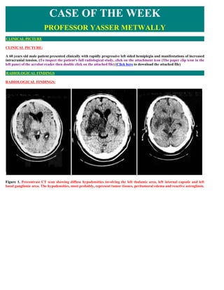
  • 1. CASE OF THE WEEK PROFESSOR YASSER METWALLY CLINICAL PICTURE CLINICAL PICTURE: A 60 years old male patient presented clinically with rapidly progressive left sided hemiplegia and manifestations of increased intracranial tension. (To inspect the patient's full radiological study, click on the attachment icon (The paper clip icon in the left pane) of the acrobat reader then double click on the attached file) (Click here to download the attached file) RADIOLOGICAL FINDINGS RADIOLOGICAL FINDINGS: Figure 1. Precontrast CT scan showing diffuse hypodensities involving the left thalamic area, left internal capsule and left basal ganglionic area. The hypodensities, most probably, represent tumor tissues, peritumoral edema and reactive astrogliosis.
  • 2. Figure 2. Postcontrast CT scan images showing a densely enhanced space occupying lesion involving the left thalamic area with positive mass effect and peritumoral edema involving the white matter and sparing the gray matter (vasogenic edema). Notice the peritumoral satellitosis (B) Figure 3. A postcontrast CT scan image showing peritumoral satellitosis (Figure 2 B) CT guided stereotactic biopsy revealed Mixed astrocytoma (Grade III-Grade IV diffuse astrocytoma). The patient was not operated upon, he received radiotherapy only to die 4 month following clinical diagnosis. Common pathological characteristics of diffuse astrocytomas  Diffuse astrocytomas are tumors predominantly composed of astrocytes. Unless otherwise indicated, the term usually applies to diffusely infiltrating neoplasms (WHO grades II through IV).  Diffuse astrocytoma is unusual in the first decade of life and most commonly presents in older children or young adults up to the age of 40 to 45.  All diffuse astrocytomas, particularly the diffusely infiltrating variety, have a tendency toward progression to more malignant forms. Diffuse astrocytomas have a peculiar tendency to change its grade over time into the next higher grade of malignancy and the condition is age dependant. A change in the grade of diffuse astrocytoma is more likely to occur in the older age group.
  • 3. Diffuse astrocytomas commonly start as grade II at a younger age group then gradually change its grade over time into the next higher grade until they ultimately dedifferentiate into glioblastomas (secondary glioblastoma multiforme), on the other hand, glioblastoma multiforme in older patients are usually primary-that is, they occur as glioblastoma multiforme from their inception, without progression from a lower- grade tumor.  Diffuse astrocytomas appear to form a continuum of both biological and histological aggression. They vary from lesions with almost normal cytology (grade I and grade II astrocytomas) through intermediate stages (grade III, anaplastic astrocytomas) and up to the most aggressive of all human brain tumours (grade IV astrocytomas or glioblastoma multiforme).  Diffuse astrocytoma often spreads widely through the brain but without destruction and also without interruption of normal function. Microscopically, tumor cells infiltrate between myelinated fibers in a nondestructive manner (perineuronal satellitosis). The local spread of diffuse astrocytomas (forming gliomatosis cerebri and butterfly gliomas) does not mean that the tumour grade is grade IV (glioblastoma multiforme), local spread can occur in grade II and grade III and in the author experience gliomatosis cerebri and butterfly gliomas are much more commonly seen in grade II astrocytomas and has not been encountered in grade III (anaplastic astrocytomas) and grade IV (glioblastoma multiforme). It takes a long time for a diffuse astrocytoma to cross the corpus callosum to the opposite hemisphere to form a butterfly glioma. Patients harbouring glioblastomas have a much shorter life span for their tumours to form butterfly gliomas, however cases were reported for glioblastomas forming butterfly tumours.  These glioma cells migrate through the normal parenchyma, collect just below the pial margin (subpial spread), surround neurons and vessels (perineuronal and perivascular satellitosis), and migrate through the white matter tracks (intrafacicular spread). This invasive behavior of the individual cells may correspond to the neoplastic cell's reacquisition of primitive migratory behavior during central nervous system development. The ultimate result of this behavior is the spread of individual tumor cells diffusely over long distances and into regions of brain essential for survival of the patient. The extreme example of this behavior is a condition referred to as gliomatosis cerebri, in which the entire brain is diffusely infiltrated by neoplastic cells with minimal or no central focal area of tumor per se. Furthermore, 25% of patients with GBM have multiple or multicentric GBMs at autopsy. Although GBMs can be visualized on MRI scans as mass lesions that enhance with contrast, the neoplastic cells extend far beyond the area of enhancement.  In practice considerable histological heterogeneity in astrocytic tumours is found ( i.e., low grade areas with Rosenthal fibers and calcification can be intermixed with with frankly malignant ones).  The differences in histologic features, potential for invasiveness, and extent of progression likely reflect genetic differences acquired during astrocytoma growth.  Grade IV astrocytomas (glioblastoma multiforme) differ from diffuse astrocytoma grade II and grade III (anaplastic astrocytomas) in the presence of gross necrosis, and microscopically in the presence of vascular endothelial hyperplasia and tumour hemorrhage. Primary tumors of the thalamus account for only 1-1.5% of all intracranial tumors and approximately 25% of them arise in children aged 15 years or under. Thalamic gliomias might be unilateral or bilateral. Diffuse and involvement of thalamic nuclei by these tumors makes surgical therapy very difficult and no case of radical removal has been described in the literature. Consequently, the main role of surgery is limited and usually performed for a histological diagnosis. Generally, these gliomas are low-grade astrocytomas (grade II of WHO classification), but limited anaplastic areas may be encountered. Radiotherapy and chemotherapy are sometimes utilized as adjuvant therapy, but their role is questionable. Outcome is generally poor, independently of the therapy that is utilized. Rapid fatal evolution after diagnosis and the almost complete unresponsiveness of these tumors to radiotherapy make these rare tumors difficult to treat. Anaplastic gliomas usually show enhancement after contrast administration. Severe dementia and personality modification observed in adults affected by bilateral thalamic glioma is attributed to the involvement of dorsomedial nuclei of thalami and their connections with temporal and frontal lobes [9]. Bilateral thalamic glial tumors are rare and less than 50 cases have been published in the literature [1-8]. DIAGNOSIS: DIAGNOSIS: THALAMIC GLIOMA
  • 4. DISCUSSION DISCUSSION: Bailey and Cushing observed that gliomas become more aggressive with time, writing – all of these lesions, so far as our records permit us to judge, show an increasing degree of malignancy, the recurrent tumors giving evidence of more active cell division than the original lesion. 2 It is now understood that the accumulation of molecular abnormalities1 underlies the increasingly aggressive clinical behavior of an individual patient s glioma over time. Moreover, while GBMs may be histologically identical, they are molecularly distinct, [51] and each tumor may require individually tailored treatment with the combination of agents predicted to impact multiple molecular abnormalities. Successful therapy of a molecularly complex disease such as GBM may also require simultaneous administration of multiple agents, including both traditional chemotherapies and several pathway inhibitors.  The past of glioblastoma multiforme The most common cancer arising from the brain is the glioblastoma multiforme (GBM). It is also the most deadly, [1] representing the most aggressive subtype among the gliomas, a collection of tumors including astrocytomas and oligodendrogliomas (see Table 1). In 1926, Bailey and Cushing, in describing spongioblastoma multiforme , the label then used for GBM, noted that: - It is from this group doubtless that the generally unfavorable impression regarding gliomas as a whole has been gained. It is not only the largest single group in the series but at the same time is one of the most malignant. In the five unoperated cases, the average duration of life from the onset of symptoms was only three months, which speaks well on the whole for the average survival period of twelve months for those surgically treated. [2] Since their seminal work, the median survival of 12 months has not changed markedly. Both data from the 1960s3 and current data [4] confirm that the extent of surgical resection is an important prognostic factor. However, as Bailey and Cushing observed, GBMs have – infiltrating propensities, and when enucleation is attempted, the growth is found at the depth to spread into and merge with the normal cerebral tissue without recognizable demarcation. [2] In prior eras, radical surgical excisions, including removal of the entire cerebral hemisphere containing the tumor, [5] were occasionally attempted, yet patients who survived the hemispherectomy died of recurrent tumor, [6] clinically proving the importance of the histologic observation that tumor cells invade throughout the brain. In the modern age, brain imaging may disclose macroscopic tumor in the opposite hemisphere (see Figure 1) or even gliomatosis cerebri literally a brain full of tumor. In the years leading to up to World War II, the German pathologist Scherer, whose scientific discoveries were tainted by his Nazi activities, [7] described secondary structures [8,9] that further characterized invasive tumor cells. These structures are secondary because they are dependent for their formation on underlying normal brain structures, as opposed to primary structures of the tumor such as pseudopalisading necrosis and microvascular proliferation. Examples include perineuronal and perivascular satellitosis (accumulation of tumor cells around neurons and blood vessels), subpial spread, and intrafascicular tracking such as infiltration along corpus callosum and other white matter tracks (see Figure 2). Advances in surgical technique, imaging, and targeting of radiotherapy (RT) are important contributions to local control. However, changing GBM from a disease that kills quickly to one that can be managed as a chronic illness, such as hypertension or diabetes mellitus, will require systemic therapies targeting tumor cells infiltrating throughout the brain, such as chemotherapy, immunotherapy, and small molecule pathway inhibitors.  The current status of glioblastoma multiforme Currently, treatment for GBM involves both local and systemic therapy. Surgery and partial brain RT are the standard locally directed therapies. Some physicians also advise intra-operative placement of chemotherapy containing polymers (i.e. Gliadel wafers ) directly into the surgical bed in an attempt to prolong local control. [10] While there is a modest survival benefit, the use of these polymers remains controversial because of the potential for toxicity. Other treatment modalities that target disease localized to the surgical bed or the surrounding area have included brachytherapy and stereotactic radiosurgery (with either a linear accelerator or gammaknife), neither of which are commonly advised. Convection-based chemotherapies delivered by catheter infusion, such as local delivery of pseudomonas exotoxin linked to either interleukin [13] (IL-13) or transforming growth factor-a (TGFa), [11] are available in clinical trials for some patients. These trials take advantage of differences in the expression of proteins (such as growth factor receptors) on the surface of residual tumor cells in the periphery of the operative bed to deliver the toxin to tumor cells, but spare normal brain. By contrast, systemic chemotherapy targets tumor cells beyond the reach of local therapies. The most commonly prescribed systemic chemotherapy for GBM is temozolomide (Temodar®), an alkylator that became available during the last decade. The effectiveness of temozolomide in the management of GBM at diagnosis was recently demonstrated by a large multinational study.4 A modest survival benefit of 2.5 months for concurrent temozolomide with RT (14.6 months median survival) was observed relative to RT alone (12.1 months median survival).4 In addition, while the survival benefit was still
  • 5. present two years after diagnosis, only 10.7% of patients were progression-free and only 26.5% of patients were alive at that point. [4] While systemic chemotherapy improves the outcome for some patients, long-term disease control therefore remains elusive. Table 1. Common Gliomas. The gliomas are assigned a grade by the World Health Organization (WHO) depending on histologic features that predict behavior.This classification scheme is derived from the clinicopathologic studies of Bailey and Cushing.2 Grade I tumors, such as juvenile pilocytic astrocytomas, are generally focal rather than diffuse and are potentially curable by surgical excision.WHO grade II–IV tumors are diffusely infiltrative.WHO grade III–IV tumors are termed ‘high grade’ or malignant. GBMs, or grade IV astrocytomas, are the most aggressive subtype.  Discoveries during the last several years have improved the understanding of glioma and general cancer biology markedly.  Generally, a cancer comprises cells that either divide or survive when they should instead undergo either cell cycle arrest or die. These abnormalities are also not mutually exclusive, and most cancers, including GBMs, are driven by several molecular abnormalities. The signal to divide is typically provided by a growth factor (ligand). Examples include TGF?, epidermal growth factor (EGF), platelet-derived growth factor (PDGF), and vascular endothelial growth factor (VEGF). Such ligands interact with cells through receptors including EGF receptors (EGFRs), PDGF receptors (PDGFRs), and VEGF receptors (VEGFRs). Receptor activity is linked with cellular processes such as mitosis or invasion by signal transduction cascades. Examples of signal transduction cascades important in human GBMs include those activated by the oncogenes Ras, Akt, and Src. [12] In cancer cells, these pathways are disrupted through several mechanisms. For example, EGFR is overexpressed in up to 92% of astrocytomas, [13] and up to 62% of GBMs express EGFRvIII, [14] a mutant receptor that is active independently of ligand. Co-expression of EGF and EGFR15,16 in GBMs leads to a potential autocrine loop.An analogous loop is created by PDGF and PDGFR co-expression in up to 94% of high-grade oligodendrogliomas. [17–19] Regardless of ligand or receptor status, close to 100% of GBMs exhibit activation of Ras,20,21 and approximately 70% exhibit activated Akt, [21,22] the latter typically through loss of the tumor suppressor gene phosphatase tensin homolog on chromosome ten (PTEN), [23–25] which normally represses Akt activation. Src is detected in 67% of GBMs.26 Finally, control over cell division is normally maintained by tumor suppressors, such as an inhibitor of CDK4A (INK4A) and its alternative reading frame (ARF), as well as p53, which also contributes to DNA repair and apoptosis, and other enzymes. Disruptions of normal cell cycle control of one form or another have been observed in almost all GBMs. [12,27] Figure 1. Magnetic Resonance Imaging Findings in Glioblastoma Multiforme. Contrast enhanced magnetic resonance image (MRI) of the brain demonstrating a large GBM with a smaller site in the contralateral hemisphere. This infiltrative nature of GBMs, essentially effecting the entire brain, underscores the failure of even radical surgery, such as hemispherectomy, to effect cure. Moreover, the modeling of gliomas in mice has demonstrated that abnormalities of ligands, receptors, signal transducers, and proliferation cause gliomas. For example, combined activation of Ras with Akt in glial progenitors is sufficient to induce GBMs in mice, [22] and transgenic expression of activated forms of Ras [28] or Src [29] in glia leads to GBMs following spontaneous development of cooperative oncogenic abnormalities. Modeling has also demonstrated that PTEN loss is functionally equivalent to Akt activation, [30] when combined with activated Ras. PDGF overexpression in glia causes high- grade oligodendrogliomas [31,32] that also exhibit pathologic features of GBMs, including pseudopalisading necrosis and microvascular proliferation. The threshold to tumor formation is lowered by disruption of Ink4a-Arf or p53 expression. [31,33,34] While more is learned about glioma biology, small molecule inhibitors are being developed that target the causal pathways. [35] For example, several inhibitors of EGFRs are under investigation in clinical trials. These include the EGFR inhibitors erlotinib (OSI- 774/Tarceva), gefitinib (ZD-1839/Iressa), and lapatinib (GW572016). The PDGFR inhibitors imatinib (STI- 571/Gleevec) and PTK787, both of which have other targets, are also in use. Signal transduction cascade blockers are also
  • 6. being studied. One example is R11577, which targets the enzyme that activates Ras. Rapamycin (sirolimus), CCI-779 (temsirolimus), and Rad-001 (everolimus) target mTOR, one of the key enzymes activated by Akt. Figure 2. Histology of Gliomas. (A) Pseudopalisading (arrow) necrosis (arrow head) and (B) microvascular proliferation (arrow) are the classic histologic findings in glioblastoma multiforme (GBM). Secondary Scherer structures (C) involve tumor cells (arrow heads) accumulating around blood vessels (BV, long arrow), and neurons (N, long arrow) in a low grade (WHO grade II) oligodendroglioma. Such perivascular and perineuronal satellitosis, along with intrafascicular growth and subpial accumulation (not shown), contribute to the diffusely infiltrative nature of gliomas throughout normal brain structures.. (Click to enlarge figure) Unfortunately, despite initial enthusiasm, treatment of GBMs as well as systemic malignancies with these small molecule inhibitors as single agents has generally been disappointing. For example, published interim and final reports of trials involving gefitinib, [36,37] erlotinib, [38–40] imatinib, [41,42] PTK787, [43,44] and CCI-77945 monotherapy for recurrent high-grade gliomas have not shown response or survival rates that are markedly superior to those observed with traditional chemotherapies, such as temozolomide4,46–48 or carmustine (BCNU). [49] However, there are individual patients treated with these agents who experience durable objective responses or sustained stable disease.Therefore, these agents are likely to have a role in GBM management. Figure 3. Pathways Important in GBM Biology. An extracellular ligand such as EGF,TGFa, or PDGF induces dimerization of receptors such as EGFR or PDGFR. Receptor stimulation activates intrinsic tyrosine kinase (TK) activity and EGFR and PDGFR are therefore called receptor tyrosine kinases (RTKs). RTKs then activate the Akt, Src, and Ras signal transduction cascades.Tumor cell growth is driven by ligand or receptor overexpression, constitutively activating receptor mutations (e.g. EGFRvIII), or signal transduction activity. Pointed (green) and block (red) arrows indicate pathway activation and inhibition, respectively.The inhibitors shown and others are under investigation in the treatment of GBMs.  Future perceptive of glioblastoma multiforme In addition to surgical resection and RT, the future of GBM therapy is likely to involve both additional measures to improve local control (such as convection or catheter delivery of antitumor agents into the operative cavity) and systemic treatment to address infiltrative disease distant from the main tumor bed. However, a major thrust of research will be tissue analyses looking for molecular features that predict sensitivity of GBMs to either traditional chemotherapies or small molecule inhibitors. Tailoring therapy with specific drugs to those patients is most likely to improve response rates and spare patients who are unlikely to benefit the expense and potential toxicity of these agents. Determination of a molecularly effective dose (MED) (inhibits a pathway), may also be more useful than the traditionally used maximally tolerated dose (MTD). An example of a molecular prognostic factor is loss of heterozygosity for chromosomes 1p and 19q in anaplastic oligodendrogliomas, which predicts both sensitivity to chemotherapy and radiation, as well as longer overall survival. [50] Consequently, some neurooncologists are currently using results of 1p/19q analysis to guide therapy, [51] although this remains an area of controversy. Other genomic alterations are also predictive—PTEN loss is associated with poor survival for patients with anaplastic oligodendrogliomas [52] and is likely to predict poor outcome from GBM. [23,53,54] More recently it was reported that GBMs, in which O6-methylguanine- DNA methyltransferase (MGMT) expression was silenced by gene methylation, were more sensitive to temozolomide than tumors with unmethylated MGMT. The likely explanation is that MGMT may counteract temozolomide activity by removing alkyl groups on DNA. [55] It is unclear whether MGMT
  • 7. methylation impacts sensitivity of other glioma subtypes to temozolomide, yet MGMT methylation status may be used in the near future to guide therapy. Individualized medicine determined by molecular rather than simply histologic phenotype may also guide therapy with small molecule inhibitors. Somatic mutations in exons 18 21 of EGFR are associated with sensitivity of lung cancer to gefitinib [56 58] or erlotinib. [58] However, the authors and others have not found these mutations in gliomas. [34,57,59,60] Efforts are under way to identify the molecular features that predict sensitivity of GBMs to EGFR and other receptor tyrosine kinase (RTK) inhibitors. Response and survival rates may also be improved through combination therapy. For example, preliminary data suggest that concurrent therapy with imatinib (PDGFR/VEGFR inhibitor) and hydroxyurea (a more traditional chemotherapy) is more effective than imatinib monotherapy. A small series with 14 evaluable patients with recurrent GBMs demonstrated a disease control rate (complete response or partial response or stable disease) of 64% for patients treated with this combination. [61] By contrast, imatinib monotherapy led to a disease control rate of 29%. [42] Larger trials of this and other combinations, such as temozolomide with PTK787, are under way. (Click here to download master degree thesis " Non surgical management of brain tumors" in PDF format) SUMMARY SUMMARY The last five years have seen an evolution in the management of highgrade astrocytic tumors comparable in scope yet greater in magnitude to that of the prior 40 years. This is thanks to the convergence of three factors: the introduction of an oral agent with antitumor activity beyond the blood–brain barrier and modest systemic toxicity (temozolomide); the demonstration through a well-conducted randomized trial of the superiority of multimodality therapy; and the fact that we now stand on the threshold of additional progress through key advances in translational biology, which, in many cancers, is providing new targets for therapeutic intervention. Astrocytic tumors have long been the bane of neurosurgeons, radiation therapists, and neuro-oncologists. Although they account for only 2.3% of all cancer-related deaths in the US, [63] little if any substantial progress in brain imaging and treatment had been made until the first years of this millennium. Characteristics of high-grade glial tumors compared with other cancers are its unique location, robust invasive and angiogenic capabilities without a significant propensity to metastasize outside of the central nervous system (CNS), and the profound histological and molecular heterogeneity within tumor specimens.  Advances in the Management of Glioblastomas— Multimodality Strategies In 2003, a phase III study in 240 newly diagnosed patients with surgically resectable malignant gliomas—including 207 with glioblastoma multiforme (GBM)—compared surgery plus radiotherapy and placebo wafers with surgery plus radiotherapy and the addition of 3.8% bischloroethyl nitrosourea (BCNU, carmustine) wafers (Gliadel wafers) into the tumor bed. The study demonstrated a modest, albeit significant, prolongation of survival in the latter group (13.9 versus 11.6 months). [64] Long-term follow-up of this study showed that the survival advantage with BCNU wafers was maintained at one, two, and three years, and was statistically significant (p=0.01) at three years compared with placebo, although the absolute number of patients evaluated at this latter timepoint was quite small. [65] Perhaps the most significant advance in the management of glioblastomas emanates from the work of Stupp et al. [65] who, in a safety and efficacy study, randomized 573 patients with newly diagnosed glioblastoma from 85 centers, primarily in Europe, to radiotherapy alone or to radiotherapy plus concomitant temozolomide followed by monthly temozolomide for six cycles. At a median follow-up of 28 months the median survival in the radiotherapy group alone was 12.1 months compared with 14.6 months in the group receiving both treatment modalities (p<0.001). The two-year survival rate was 10.4% with radiotherapy alone versus 26.5% with radiotherapy and temozolomide. The results of this study produced level 1A evidence for the benefit of this combined modality treatment in initially diagnosed patients, and was incorporated into the new National Comprehensive Cancer Network (NCCN) guidelines for CNS tumors in 2005. Thus, as a next logical step, a fusion of these two prior studies was evaluated in a phase II setting in patients with newly
  • 8. diagnosed, highgrade GBM undergoing resection with BCNU wafer insertion followed by the combination of radiotherapy plus temozolomide. Early interim data have been presented in abstract form. [67] The study end-points include survival and progression-free survival (PFS). Of 35 patients enrolled so far, 34 were diagnosed with GBM. At median follow-up of 10.4 months, [87] patients had documented recurrence and 19 patients had died. Six patients remain on active treatment. The one- year survival rate is 64%, and median survival is 18.6 months. These early data suggest that combination therapy with BCNU wafers followed by therapy plus temozolomide may be an effective regimen in patients with initial highgrade resectable malignant gliomas, although randomized trials will ultimately be needed to assess the efficacy of this treatment modality. Other treatment modalities that have been investigated for the treatment of high-grade astrocytic tumors—especially in terms of targeting disease localized to the surgical bed or the surrounding area—have included stereotactic radiosurgery and brachytherapy. A randomized trial conducted by the Radiation Therapy Oncology Group (RTOG) compared post-operative conventional radiotherapy plus systemic BCNU alone or preceded by stereotactic radiosurgery—including both linear accelerator or gamma-knife—in patients with GBM (>4cm tumor size). The results of the trial were disappointing with no improvement in local control or survival with stereotactic radiosurgery. [68] The US Food and Drug Administration (FDA) has recently approved GliaSite, a novel brachytherapy device, to provide local post-operative irradiation to high- grade gliomas. However, to date, no efficacy trials have been conducted with the system. In a retrospective, multiinstitutional analysis, median survival— measured from the date of GliaSite placement—was 35.9 weeks for patients with an initial diagnosis of GBM. The patient population consisted of patients with recurrent high-grade gliomas who had previously undergone resection and had received external beam radiotherapy as part of their initial treatment. Following surgical debulking of the recurrent lesion, an expandable balloon catheter (GliaSite) was placed in the tumor cavity. Although reirradiation of malignant gliomas with the GliaSite system appeared to provide a modest survival benefit, it is difficult to assess the value of any survival without the benefit of a control group. [69] Convection-enhanced delivery (CED) of toxins to the tumor site is a new treatment modality under investigation for malignant gliomas. It was developed as a method to treat brain tumors by circumventing the normal limitations imposed by the blood– brain barrier. CED involves the stereotactically guided implantation of delivery catheters directly into the residual tumor or around the resection cavity to facilitate the local delivery by high-flow micro-infusion of the targeted toxin to tumor cells. A combined summary of three phase I clinical trials investigating the use of cintredekin besudotox—a recombinant protein consisting of interleukin-13 (IL-13) and a truncated form of Pseudomonas exotoxin— delivered via CED in the treatment of recurrent malignant glioma following tumor resection, demonstrated an overall median survival after treatment of 45.9 weeks. [70] The Phase III Randomized Evaluation of Convection Enhanced Delivery of IL13-Pe38qqr with Survival Endpoint (PRECISE) Trial was designed to compare CED of cintredekin besudotox to treatment with the BCNU wafers in 294 patients with first recurrence or progression of GBM. Unfortunately, the study was stopped in December 2006 after the efficacy end- point of a statistically significant difference in overall survival was not met. Indeed, the median survival in the CED arm was 36.4 weeks, while that of the BCNU wafer arm was 35.3 weeks. An NCI-sponsored phase I trial is currently evaluating CED of 131I-chTNT-1/B, a chimeric tumor necrosis therapy antibody attached to the radioisotope iodine 131 in malignant glioma. Although CED is a promising alternative for targeted delivery, it remains a complex, interdisciplinary technique that needs further investigation to optimize catheter positioning and drug distribution.  Molecular Targets and Prognostic Factors Turning to recent advances in the genomic analysis of glioblastoma, four molecular markers are currently being explored. First is the identification of loss of the chromosome 1p/19q in anaplastic oligodendroglioma as a predictor of response to chemotherapy—particularly PCV (procarbazine, CCNU [chloroethylnitrosourea, lomustine], and vincristine). The initial results, published by Cairncross et al. in 1998, [9] led to two randomized clinical trials. The first—European Organization for Research and Treatment of Cancer (EORTC) 26951—evaluated radiotherapy versus radiotherapy followed by PCV in patients with newly diagnosed anaplastic oligodendroglioma or anaplastic oligo-astrocytomas. [10] The second (RTOG 94-02) evaluated PCV given prior to radiotherapy. [73] Both studies demonstrated that the addition of PCV improved PFS without impacting on overall survival (OS). Although chromosome-1p/19q loss does predict chemosensitivity, it did not identify patients who have a better outcome after adjuvant chemotherapy. Moreover, it became apparent that patients with the combined chromosomal 1p/19q loss have a better outcome after radiotherapy compared with patients whose tumor does not contain this chromosomal aberration. At a molecular level, up to 50% of glioblastoma specimens express dysregulated epidermal growth factor receptor (HER1/EGFR). [74] This observation has spurred interest in the use of the small molecule HER1/EGFR-targeted therapy agents such as erlotinib and gefitinib. Initial phase II studies evaluating gefitinib failed to demonstrate significant objective tumor regressions, with a six-month PFS of only 13% in 53 patients with recurrent glioblastoma. [75] In contrast, the data relating to erlotinib initially appeared somewhat more promising in one study of 31 patients with recurrent glioblastomas, in which six patients achieved a partial response and the six-month PFS was 26%. [76,77] Of note is that these authors could not determine any correlation between response and EGFR expression or amplification within the tumor specimens. Also, a second phase II study of 30 patients treated with erlotinib failed to result in any objective responses or six-month PFS. [78] Unfortunately, a recent EORTC trial comparing erlotinib with either temozolomide or BCNU in 110 patients with recurrent glioblastoma failed to demonstrate a benefit of the oral targeted therapy with respect to sixmonth PFS or 12-month survival. [79] Optimal dosing of these oral agents, especially while patients are taking enzyme-inducing anti-
  • 9. epileptics or drugs with similar pharmacological effects, may be a significant confounding variable in determining their true clinical efficacy. [80] Another molecular target of some promise in the management of patients with GBM is transforming growth factor beta (TGF-B). Not only does it stimulate cell migration, invasion, and angiogenesis, but it also appears to play an important role in the disruption of afferent and efferent immune responses. [81] Several in vitro systems, as well as rodent glioma models, delineate the potential therapeutic impact of TGF-B antagonism, employing not only antisense strategies, but also specific TGF-B receptor kinase antagonists. In particular, the use of such agents in conjunction with vaccines, or perhaps novel approaches of cellular immunotherapy, bears further study. Another molecular marker of interest is the O6-methylguanine- DNA methyltransferase (MGMT) promoter gene, also known as O6-alkylguanine-DNA alkyltransferase or AGT. The gene itself expresses alkyltransferase, which plays a role in resistance to alkylating and methylating agents. Methylation of this gene disrupts the expression of alkyltransferase and thus renders the cell more susceptible to alkylating and methylating chemotherapy agents such as temozolomide. Hegi et al. analyzed tissue from newly diagnosed patients with glioblastoma enrolled into the EORTC 26981 trial, and documented a significant correlation between MGMT methylation and outcome from treatment. [82] Methylation of the MGMT promoter was demonstrated in 45% of 206 tumors analyzed, and this was associated with a 46% survival rate at two years compared with only 13.8% in those patients with non-methylated promoter status. [82] Although the preliminary conclusion from this translational study is that MGMT promoter methylation may be predictive of outcome to multimodality treatment in glioblastoma, validation from additional prospective studies is required. A novel oral protein kinase C inhibitor that initially appeared to have activity in recurrent glioblastoma was enzastaurin. This agent, an oral inhibitor of PKCß and PI3K/AKT pathways, is well tolerated, possesses antiangiogenic properties in pre- clinical models, and induces tumor cell apoptosis. [83] Enzastaurin is currently being evaluated in phase II trials for the treatment of patients with recurrent high-grade gliomas. Initial results in 87 evaluable patients with recurrent high-grade gliomas showed that enzastaurin treatment was well tolerated and objective radiographic responses were seen in 22% of patients with GBM. The exposure to enzastaurin was significantly lower in patients treated with enzyme-inducing antiepileptic drugs (EIADs). [84] Enzastaurin also appears to be safe in conjunction with radiation therapy and temozolomide in patients with newly diagnosed GBM. [85] GBM is highly angiogenic, and vascular endothelial growth factor (VEGF) is amplified in most GBM tumors. [86] Over the last three years, there has also been an evolution in the understanding of the ‘brain tumor stem cell.’ If the concept of a brain tumor stem cell proves to be a real entity, identifiable perhaps by CD-133 expression, and correlated with a significant angiogenic effect associated with VEGF expression and production, this could confirm an important role for antiangiogenic therapy in this cancer. [87] This prompted the evaluation of the recombinant humanized anti-VEGF monoclonal antibody bevacizumab in patients with malignant gliomas. A recent phase II trial studied the effect of bevacizumab in combination with the cytotoxic agent irinotecan in patients with recurrent high-grade astrocytic neoplasms. [26] The investigators demonstrated a radiographic response rate of 63% with the combination therapy and six- month overall survival was estimated at 72%. A randomized phase II study in patients with recurrent glioblastomas evaluating bevacizumab alone versus bevacizumab with irinotecan was recently completed and the results are anxiously awaited . Additional agents with antiangiogenic properties such as the multitargeted agents sorafenib and sunitinib are also being investigated in malignant gliomas. [89,90,91]  Conclusion Significant advances are being made in the understanding of the biology of high-grade gliomas, which are contributing to the development of promising targeted therapies and treatment modalities. Over the last couple of years, there has been an evolution in the understanding of the ‘brain tumor stem cell.’ If the concept of a brain tumor stem cell proves to be a real entity identifiable by CD-133 expression, and if this correlates with a significant angiogenic effect associated with VEGF expression and production, it opens new possibilities for targeted therapy. [89] Multitargeted therapy is a necessity to manage high-grade brain tumors optimally. The potential of quadruple multimodality therapy for the management of brain tumors, which includes surgery, radiotherapy, systemic therapy, and localized chemotherapy, needs to be further investigated. Furthermore, with the promising results seen with bevacizumab, there is the possibility of a fifth modality—an antiangiogenesis inhibitor.  Addendum  A new version of this PDF file (with a new case) is uploaded in my web site every week (every Saturday and remains available till Friday.)  To download the current version follow the link "http://pdf.yassermetwally.com/case.pdf".  You can also download the current version from my web site at "http://yassermetwally.com".  To download the software version of the publication (crow.exe) follow the link: http://neurology.yassermetwally.com/crow.zip  The case is also presented as a short case in PDF format, to download the short case follow the link: http://pdf.yassermetwally.com/short.pdf
  • 10. At the end of each year, all the publications are compiled on a single CD-ROM, please contact the author to know more details.  Screen resolution is better set at 1024*768 pixel screen area for optimum display.  Also to view a list of the previously published case records follow the following link (http://wordpress.com/tag/case- record/) or click on it if it appears as a link in your PDF reader  To inspect the patient's full radiological study, click on the attachment icon (The paper clip icon in the left pane) of the acrobat reader then double click on the attached file.  Click here to download the short case version of this case record in PDF format REFERENCES References 1. Kleihues P, Cavanee W K, Pathology and Genetics of Tumours of the Nervous System, International Agency for Research on Cancer (IARC) Press, Lyon:WHO/IARC (2000). 2.Bailey P, Cushing H, A classification of the tumors of the glioma group on a histo-genetic basis with a correlated study of prognosis, Philadelphia: J B Lippincott Co. (1926). 3.Jelsma R K, Bucy P C, “The treatment of glioblastoma multiforme”, Trans.Am. Neurol. Assoc. (1967);92: pp. 90-93. 4.Stupp R et al., “Radiotherapy plus Concomitant and Adjuvant Temozolomide for Glioblastoma”, N. Engl. J. Med. (2005);352(10): pp. 987-996. 5.Dandy W E,”Removal of right cerebral hemisphere for certain tumors with hemiplegia”, JAMA (1928);90: pp. 823-825. 6.Gardner W J et al., “Residual functioning following hemispherectomy for tumouir and for infantile hemiplegia”, Brain (1955);78(4): pp. 487-502. 7.Peiffer J, Kleihues P,”Hans-Joachim Scherer (1906-1945), Pioneer in Glioma Research, Brain Pathol. (1999);9(2): pp. 241- 245. 8.Scherer H J, “Structural development in gliomas”, Am. J. Cancer (1938);34(3): pp. 333-351. 9.Scherer H J, “The forms of growth in gliomas and their practical significance”, Brain (1940);63(part 1): pp. 1-35. 10.Kleinberg L R et al., “Clinical course and pathologic findings after Gliadel and radiotherapy for newly diagnosed malignant gliama: implications for patient management”, Cancer Invest. (2004);22(1): pp. 1-9. 11.Sampson J H et al.,”Sustained radiographic and clinical response in patient with bifrontal recurrent glioblastoma multiforme with intracerebral infusion of the recombinant targeted toxin TP-38: case study”, Neuro-oncol. (2005);7(1): pp. 90-96. 12.Lassman A B, Holland E C, “Molecular Biology and Genetic Models of Gliomas and Medulloblastomas”, in Russell and Rubinstein s Pathology of Tumors of the Nervous System 7th Edition, McLendon et al. (eds) (2005), Arnold Health Sciences: London (in press). 13.Schlegel J et al.,”Amplification and differential expression of members of the erbB-gene family in human glioblastoma”, J. Neurooncol. (1994);22(3): pp. 201-207. 14.Wikstrand C J et al., “Monoclonal antibodies against EGFRvIII are tumor specific and react with breast and lung carcinomas and malignant gliomas”, Cancer Res. (1995);55(14): pp. 3,140-3,148. 15.Mishima K et al., “Heparin-binding epidermal growth factor-like growth factor stimulates mitogenic signaling and is highly expressed in human malignant gliomas”, Acta. Neuropathol. (Berl) (1998);96: pp. 322-328. 16.Ekstrand A J et al.,”Genes for epidermal growth factor receptor, transforming growth factor and their expression in human gliomas in vivo”, Cancer Res. (1991);51(8): pp. 2,164-2,172. 17.Di Rocco F et al., “Platelet-derived growth factor and its receptor expression in human oligodendrogliomas”, Neurosurgery (1998);42(2): pp. 341-346.
  • 11. 18.Robinson S et al., “Constitutive Expression of Growth-related Oncogene and Its Receptor in Oligodendrogliomas”, Neurosurgery (2001);48(4): pp. 864-873; discussion pp. 873-874. 19.Shoshan Y et al., “Expression of oligodendrocyte progenitor cell antigens by gliomas: implications for the histogenesis of brain tumors”, Proc. Natl. Acad. Sci. USA (1999);96(18): pp. 10,361-10,366. 20.Guha et al., “Proliferation of human malignant astrocytomas is dependent on Ras activation”, Oncogene (1997);15(23): pp. 2,755-2,765. 21.Rajasekhar V K et al., “Oncogenic Ras and Akt signaling contribute to glioblastoma formation by differential recruitment of existing mRNAs to polysomes”, Mol. Cell. (2003);12(4): pp. 889-901. 22.22. Holland E C et al., Combined activation of Ras and Akt in neural progenitors induces glioblastoma formation in mice”, Nat. Genet. (2000);25(1): pp. 55-57. 23.Sano T et al., “Differential Expression of MMAC/PTEN in Glioblastoma Multiforme: relationship to localization and prognosis”, Cancer Res. (1999);59(8): pp. 1,820-1,824. 24.Choe G et al.,”Analysis of the phosphatidylinositol 3 -kinase signaling pathway in glioblastoma patients in vivo”, Cancer Res. (2003);63(11): pp. 2,742-2,746. 25.Ermoian R P et al., “Dysregulation of PTEN and protein kinase B is associated with glioma histology and patient survival”, Clin. Cancer Res. (2002);8(5): pp. 1,100-1,106. 26.Takenaka N et al.,”Immunohistochemical detection of the gene product of Rous sarcoma virus in human brain tumors”, Brain Res. (1985);337(2): pp. 201-207. 27.Fulci G et al.,”p53 gene mutation and ink4a-p14 deletion appear to be two mutually exclusive events in human glioblastoma”, Oncogene (2000);19(33): pp. 3,816-3,822. 28.Ding H et al.,”Astrocyte-specific expression of activated p21-ras results in malignant astrocytoma formation in a transgenic mouse model of human gliomas”, Cancer Res. (2001);61(9): pp. 3,826-3,836. 29.Weissenberger J et al., “Development and malignant progression of astrocytomas in GFAF-v-src transgenic mice”, Oncogene (1997);14(17): pp. 2,005-2,013. 30.Hu X et al.,”PTEN loss is equivalent to Akt and cooperates with Ras in the formation of GBMs”, Neoplasia (2005); in press. 31.Dai C et al., “PDGF autocrine stimulation dedifferentiates cultured astrocytes and induces oligodendrogliomas and oligoastrocytomas from neural progenitors and astrocytes in vivo”, Genes Dev. (2001);15(15): pp. 1,913-1,925. 32.Shih A H et al., “Dose-dependent effects of platelet-derived growth factor-B on glial tumorigenesis”, Cancer Res. (2004);64 (14): pp. 4,783-4,789. 33.Uhrbom L et al., “Ink4a-Arf loss cooperates with KRas activation in astrocytes and neural progenitors to generate glioblastomas of various morphologies depending on activated Akt”, Cancer Res. (2002);62(19): pp. 5,551-5,558. 34.Holland laboratory, unpublished results. 35.Mischel P S, Cloughesy T F,”Targeted molecular therapy of GBM”, Brain Pathol. (2003);13(1): pp. 52-61. 36.Lieberman F S et al.,”Phase I-II study of ZD-1839 for recurrent malignant gliomas and meningiomas progressing after radiation therapy”, J. Clin. Oncol. (2003);22: p. 105 (abstract 421). 37.Rich J N et al., “Phase II Trial of Gefitinib in Recurrent Glioblastoma”, J. Clin. Oncol. (2004);22(1): pp. 133-142. 38.Vogelbaum M A et al., “Phase II trial of the EGFR tyrosine kinase inhibitor erlotinib for single agent therapy of recurrent Glioblastoma Multiforme: Interim results”, J. Clin. Oncol. (2004);22(14S): p. 1,558 (abstract). 39.Raizer J J et al., “A phase II trial of erlotinib (OSI-774) in patients (pts) with recurrent malignant gliomas (MG) not on EIAEDs.”, J. Clin. Oncol. (2004);22(14S): p. 1,502 (abstract). 40.Yung A et al., “Erlotinib HCL for glioblastoma multiforme in first relapse, a phase II trial”, J. Clin. Oncol. (2004);22(14S):
  • 12. p. 1,555 (abstract). 41.van den Bent M et al., “Multicentre phase II study of imatinib mesylkate (Gleevec) in patients with recurrent glioblastoma”, Neuro-oncol. (2004);6(4): p. 383 (abstract). 42.Wen P Y et al., “Phase I/II study of imatinib mesylate (STI571) for patients with recurrent malignant gliomas (NABTC 99- 08)”, Neuro-oncol. (2004);6(4): p. 385 (abstract). 43.Yung W K A et al., “A phase I trial of PTK787/ZK 222584 (PTK/ZK), a novel oral VEGFR TK inhibitor in recurrent glioblastoma”, Proc.Am. Soc. Clin. Oncol. (2004), abstract no. 315. 44.Conrad C et al.,”A phase I/II trial of single-agent PTK 787/ZK222584 (PTK/ZK), a novel, oral angiogenesis inhibitor, in patients with recurrent glioblastoma multiforme (GBM)”, J. Clin. Oncol. (2004);22(14S): p. 1,512 (abstract). 45.Chang S et al.,”Phase II/pharmacokinetic study of CCI-779 in recurrent GBM”, Neuro-oncol. (2003);5(4): p. 349 (abstract). 46.Yung W K et al., “A phase II study of temozolomide vs. procarbazine in patients with glioblastoma multiforme at first relapse”, Br. J. Cancer (2000);83(5): p. 588-593. 47.Brada M, Hoang-Xuan K, Rampling R, “Multicenter phase II trial of temozolomide in patients with GBM at first relapse”, Ann. Oncol. (2001);12(2): pp. 259-266. 48.Chang S M,Theodosopoulos P, Lamborn K, Malec M, Rabbitt J, Page M et al., “Temozolomide in the treatment of recurrent malignant glioma”, Cancer (2004);100(3): p. 605-611. 49.Brandes A A,Tosoni A, Amista P, Nicolardi L et al., “How effective is BCNU in recurrent glioblastoma in the modern era? A phase II trial”, Neurology (2004);63(7): pp. 1,281-1,284. 50.Cairncross J G, Ueki K, Zlatescu M C et al., “Specific genetic predictors of chemotherapeutic response and survival in patients with anaplastic oligodendrogliomas”, J. Natl. Cancer Inst. (1998);90(19): p. 1,473-1,479. 51.Louis D N, Holland E C, Cairncross J G et al.,”Glioma classification: a molecular reappraisal”, Am. J. Pathol. (2001);159 (3): pp. 779-786. 52.Sasaki H et al.,”PTEN is a target of chromosome 10q loss in anaplastic oligodendrogliomas and PTEN alterations are associated with poor prognosis”, Am. J. Pathol. (2001);159(1): p. 359-367. 53.Zhou Y H et al., “The expression of PAX6, PTEN, vascular endothelial growth factor, and epidermal growth factor receptor in gliomas: relationship to tumor grade and survival”, Clin. Cancer Res. (2003);9(9): pp. 3,369-3,375. 54.Hill C, Hunter S B, Brat D J,”Genetic markers in glioblastoma: prognostic significance and future therapeutic implications”, Adv. Anat. Pathol. (2003);10(4): pp. 212-217. 55.Hegi M E, Diserens A C, Gorlia T et al.,”MGMT gene silencing and benefit from temozolomide in glioblastoma”, N. Engl. J. Med. (2005);352(10): p. 997-1,003. 56.Paez J G et al., “EGFR mutations in lung cancer: correlation with clinical response to gefitinib therapy”, Science (2004);304(5676): pp. 1,497-1,500. 57.Lynch T J, Bell D W, Sordella R et al.,”Activating mutations in the epidermal growth factor receptor underlying responsiveness of non-small-cell lung cancer to gefitinib”, N. Engl. J. Med. (2004);350(21): p. 2,129-2,139. 58.Pao W, Miller V, Zakowski M et al., “EGF receptor gene mutations are common in lung cancers from never smokers and are associated with sensitivity of tumors to gefitinib and erlotinib”, Proc. Natl.Acad. Sci.USA (2004);101(36): p. 13,306-13,311. 59.Barber T D et al.,”Somatic mutations of EGFR in colorectal cancers and glioblastomas”,N.Engl. J.Med. (2004);351(27): p. 2,883. 60.Rich J N, Rasheed B K,Yan H et al., “EGFR mutations and sensitivity to gefitinib”, N. Engl. J. Med. (16 September 2004);351(12): p. 1,260. 61.Dresemann G, “Imatinib (STI571) plus hydroxyurea: Safety and efficacy in pre-treated, progressive glioblastoma
  • 13. multiforme (GBM) patients (pts)”, J. Clin. Oncol. (2004);22(14S): p. 1,550 62. Metwally, MYM: Textbook of neuroimaging, A CD-ROM publication, (Metwally, MYM editor) WEB-CD agency for electronic publication, version 11.2a. April 2010 63.American Cancer Society, Cancer Facts and Figures 2006, Atlanta, American Cancer Society, 2006. Available at: http://www.cancer.org/downloads/STT/CAFF2006PWSecured.pdf 64.Westphal M, Ram Z, Riddle V, et al., on behalf of the Executive Committee of the Gliadel Study Group, Gliadel wafer in initial surgery for malignant glioma: long-term follow-up of a multicenter controlled trial, Acta Neurochir (Wien), 2006;148:269–75. 65.Stupp R, Mason WP, van den Bent MJ, et al., Radiotherapy plus concomitant and adjuvant temozolomide for glioblastoma, N Engl J Med, 2005;352:987–96. 66.Westphal M, Hilt DC, Bortey E, et al., A phase III trial of local chemotherapy with biodegradable carmustine (BCNU) wafers (Gliadel wafers) in patients with primary malignant glioma, Neuro-oncol, 2003;5:79–88. 67.LaRocca RV, Hodes J, Villaneuva WG, et al., A phase II study of radiation with concomitant and then sequential temozolomide (TMZ) in patients (pts) with newly diagnosed supratentorial high grade malignant glioma (MG) who have undergone surgery with carmustine (BCNU) wafer insertion, Annual Meeting of the Society of Neuro-Oncology, Neuro-Oncol, 2006;8:445, Abstract TA-28. 68.Souhami L, Seiferheld W, Brachman D, et al., Randomized comparison of stereotactic radiosurgery followed by conventional radiotherapy with carmustine to conventional radiotherapy with carmustine for patients with glioblastoma multiforme: report of Radiation Therapy Oncology Group 93-05 protocol, Int J Radiat Oncol Biol Phys, 2004;60:853–60. 69.Gabayan AJ, Green SB, Sanan A, et al., GliaSite brachytherapy for treatment of recurrent malignant gliomas: a retrospective multiinstitutional analysis, Neurosurgery, 2006;58(4):701–9. 70.Kunwar S, Prados MD, Chang SM, et al., Direct intracerebral delivery of cintredekin besudotox (IL13-PE38QQR) in recurrent malignant glioma: a report by the Cintredekin Besudotox Intraparenchymal Study Group, J Clin Oncol, 2007;25 (7):837–44. 71.Cairncross JG, Ueki K, Zlatescu MC, et al., Specific genetic predictors of chemotherapeutic response and survival in patients with anaplastic oligodendrogliomas, J Natl Cancer Inst, 1998;90:1473–9. 72.van den Bent MJ, Carpentier AF, Brandes AA, et al., Adjuvant procarbazine, lomustine, and vincristine improves progressionfree survival but not overall survival in newly diagnosed anaplastic oligodendrogliomas and oligoastrocytomas: a randomized European Organisation for Research and Treatment of Cancer phase III trial, J Clin Oncol, 2006;24:2715–22. 73. Intergroup Radiation Therapy Oncology Group Trial 9402, Cairncross G, Berkey B, Shaw E, et al., Phase III trial of chemotherapy plus radiotherapy compared with radiotherapy alone for pure and mixed anaplastic oligodendroglioma: Intergroup Radiation Therapy Oncology Group Trial 9402, J Clin Oncol, 2006;24:2707–14. 74.Halatsch ME, Schmidt U, Behnke-Mursch J, et al., Epidermal growth factor receptor inhibition for the treatment of glioblastoma multiforme and other malignant brain tumors, Cancer Treat Rev, 2006;32:72–89. 75.Rich JN, Reardon DA, Peery T, et al., Phase II trial of gefitinib in recureent glioblastoma, J Clin Oncol, 2004;22:133–42. 76.Vogelbaum MA, Peereboom D, Stevens G, et al., Response rate to single agent therapy with the EGFR tyrosine kinase inhibitor erlotinib in recurrent glioblastoma multiforme: results of a phase II trial, Neuro-Oncol, 2004;6(4):384, Abstract TA- 59. 77.Vogelbaum MA, Peereboom D, Stevens G, et al., Phase II trial of the EGFR tyrosine kinase inhibitor erlotinib for single agent therapy of recurrent Glioblastoma Multiforme: Interim results, J Clin Oncol, 2004 ASCO Annual Meeting Proceedings, 2004;22(14S):Abstract 1558. 78.Raizer JJ, Abrey LE,Wen P, et al., A phase II trial of erlotinib (OSI- 774) in patients (pts) with recurrent malignant gliomas (MG), not on EIAEDS, J Clin Oncol, 2004 ASCO Annual Meeting Proceedings, 2004;22(14S):Abstract 1502. 79.Van Den Bent MJ, Brandes A, Rampling R, et al., Randomized phase II trial of erlotinib versus temozolomide or BCNU in recurrent glioblastoma multiforme (GBM): EORTC 26034, J Clin Oncol, 2007 ASCO Annual Meeting Proceedings Part I,
  • 14. 2007;25:Abstract 2005. 80.Mellinghoff IK,Wang MY, Vivanco I, et al., Molecular determinants of the response of glioblastomas to EGFR kinaase inhibitors, N Engl J Med, 2005;353:2012–24. 81.Wick W, Naumann U and Weller M, Transforming growth factorbeta: a molecular target for the future therapy of glioblastoma, Curr Pharm Des, 2006;12:341–9. 82.Hegi ME, Diserens AC, Gorlia T, et al., MGMT gene silencing and benefit from temozolomide in glioblastoma, N Engl J Med, 2005;352:997–1003. 83.Graff JR, McNulty AM, Hanna KR, et al., The protein kinase Cbeta-selective inhibitor, Enzastaurin (LY317615.HCl), suppresses signaling through the AKT pathway, induces apoptosis, and suppresses growth of human colon cancer and glioblastoma xenografts, Cancer Res, 2005;65:7462–9. 84.Fine HA, Kim L, Royce C, et al., Results from phase II trial of enzastaurin (LY317615) in patients with recurrent high grade gliomas, J Clin Oncol, 2005 ASCO Annual Meeting Proceedings, 2005;23(16S Pt I of II):Abstract 1504. 85.Butowski NA, Lamborn K, Chang S, et al., Study of enzastaurin plus temozolomide during and following radiation therapy in patients with newly diagnosed glioblastoma multiforme (GBM) or gliosarcoma, J Clin Oncology, 2007 ASCO Annual Meeting Proceedings, 2007;25(18S):Abstract 12511. 86.Kaur B, Tan C, Brat DJ, et al., Genetic and hypoxic regulation of angiogenesis in gliomas, J Neurooncol, 2004;70:229–43. 87.Bao S,Wu Q, Sathornsumetee S, et al., Stem cell-like glioma cells promote tumor angiogenesis through vascular endothelial growth factor, Cancer Res, 2006;66:7843–8. 88.Vredenburgh JJ, Desjardins A, Herndon JE 2nd, et al., Phase II trial of bevacizumab and irinotecan in recurrent malignant glioma, Clin Cancer Res, 2007;13(4):1253–9. 90.Farhadi MR, Capelle HH, Erber R, et al., Combined inhibition of vascular endothelial growth factor and platelet-derived growth factor signaling: effects on the angiogenesis, microcirculation, and growth of orthotopic malignant gliomas, J Neurosurg, 2005;102:363–70. 91. Kesari S, Ramakrishna N, Sauvageot C, et al., Targeted molecular therapy of malignant gliomas, Curr Neurol Neurosci Rep, 2005;5:186–97.