SlideShare ist ein Scribd-Unternehmen logo
1 von 14
Downloaden Sie, um offline zu lesen
CASE OF THE WEEK
                  PROFESSOR YASSER METWALLY
CLINICAL PICTURE

CLINICAL PICTURE:

9 years old female patient presented clinically with Lennox-Gastaut syndrome.

RADIOLOGICAL FINDINGS

RADIOLOGICAL FINDINGS:


Figure 1. Postcontrast CT scan showing Lissencephaly, pachygyria,and subependymal nodular heterotopia.


                                                                           Figure 2. Postcontrast CT scan
                                                                           showing              Lissencephaly,
                                                                           pachygyria,and       subependymal
                                                                           nodular heterotopia. Also notice
                                                                           cerebellar hypoplasia and mild
                                                                           central atrophy.


DIAGNOSIS:
DIAGNOSIS: CORTICAL DYSPLASIA

DISCUSSION

DISCUSSION:

Abnormalities have long been recognized in the brains of children with developmental delay and seizures. The
advent of newer imaging techniques, such as high-resolution (thin slice three-dimensional gradient recalled echo
[GREI) magnetic resonance (MR) imaging and surface reconstructions of three-dimensional data sets, has led to a
greater in vivo understanding of these malformations. There has been reclassification of the disorders of cortical
malformations according to embryologic stages of brain cortex development at which these malformations are
proposed to have occurred. These stages are cellular proliferation, cellular migration to the developing cortex, and
cortical organizations.

MALFORMATIONS OF ABNORMAL CELL PROLIFERATION

      Nonneoplastic

The earliest lesions resulting in cortical malformations are those with onset during the period of cellular
proliferation in the germinal zones. During the seventh fetal week, germinal layers begin to form in the vesicle walls
of telencephalic outpouchings (vesicles) at the foramina of Monro. Cortical malformations occurring during this
stage of neuronal and glial proliferation may be generalized, multifocal, or focal. Generalized malformations of
abnormal cell proliferation include microcephaly with diminished cortical thickness or with diminished sulcation.
Commonly imaged focal or multifocal lesions include nonneoplastic disorders, such as hemimegalencephaly, focal
cortical dysplasia (with balloon cells), and forme fruste tuberous sclerosis.

Hemimegalencephaly may be associated with neurocutaneous or hemiovergrowth syndromes, such as
hypomelanosis of Ito, epidermal nevus syndrome, or neurofibromatosis type 1. Proposed causes for hemispheric
overgrowth in hemimegalencephaly include abnormal cellular proliferation and heteroploidy, defective cellular
metabolism, or possibly an insult to the developing brain in the mid to late second trimester. In the event of a later
insult to the developing brain, brain plasticity allows for the development of new synapses in the damaged brain,
permitting the persistence of supernumerary axons and the potential for white matter overgrowth. Neuropathologic
and neuroimaging features in hemimegalencephaly are abnormal neurons, lack of gray-white matter demarcation,
disarrayed cortical lamination, gray matter heterotopias, broad gyri, and signal changes reflecting hypomyelination
and gliosis. It is postulated that foci of agyria, with macroscopic heterotopias, extensive white matter gliosis, and
less hemispheric white matter overgrowth, likely result from an earlier, more severe insult with destruction of the
radial glial fibers. The variable patterns of cortical and white matter involvement demonstrated with neuroimaging
likely reflect the variability in severity and timing of the precipitating insult. [1,5,19,23]


Figure 1. Tl -weighted (TR 600/TE 15) and T2-weighted (I R 2800/ TE 90) axial images (A and B) demonstrate
overgrowth of the right cerebral hemisphere in a patient with developmental delay, seizures, and
hemimegalencephaly. Note the abnormal white matter signal and the unilateral ventriculomegaly.

Focal cortical dysplasia is characterized by neocortical abnormalities. The pathologic spectrum of focal cortical
dysplasia includes significant abnormalities of neuronal size, shape, orientation, and lamination; indistinctness of
the gray-white junction; and variability in cortical thickness. Large bizarre neurons with abnormal Nissl patterns,
giant neurons, binucleate neurons, a variable degree of cortical gliosis, and balloon cells with abundant pale
eosinophilic cytoplasm are found. These balloon cells are similar to those seen in the cortical tubers and white
matter lesions of tuberous sclerosis, and there is much overlap in the pathology of focal cortical dysplasia and
tuberous sclerosis. Failure of myelin arborization, blurring of gray-white margins, variable sulcal depth and
cortical thickness, and occasional hazy or dystrophic calcification may be appreciated on neuroimaging. Evidence
of dual pathology, or focal cortical dysplasia in association with hippocampal sclerosis, should be sought.
[8,14,17,21,26,28,32]


                                       Figure 2. Tl -weighted (TR 600/TE 15) sagittal (A) and axial
                                       images (B), and T2-weighted (TR 3000/ TE 120) axial image (C)
                                       in a child with severe microcephaly showing a thin cortical
                                       ribbon. A Tl -weighted axial image (D) in a second child with a
                                       similar degree of microcephaly reveals fewer gyri and less well
                                       developed sulcation.


Imaging features in limited or forme fruste tuberous sclerosis include demonstration of a solitary smooth
pyramidal-shaped gyri with a central depression and abnormal signal of the subcortical white matter. The cortical
and subcortical lesions are uncommonly calcified, tend not to enhance, and are best demonstrated on MR. These
children lack the calcified subependymal nodules of tuberous sclerosis as well as other organ system lesions. The
cortical tubers are characterized by aberrant neurons, scattered balloon cells, and gliosis. The white matter lesions
of tuberous sclerosis are characterized by decreased myelin and accumulations of balloon cells. Although there are
similarities in the pathology of focal cortical dysplasia and the form fruste of tuberous sclerosis, the latter has, in
general, more abundant balloon cells. [15,17, 18,22,24]

      Neoplastic

Lesions of proliferation after an abnormal induction event during brain development may be malformative,
hamartomatous, neoplastic, or a combination. Malformative disorders consisting of neoplasias on a background of
disordered cortex or in association with focal cortical dysplasia include dysembryoplastic neuroepithelial tumor and
ganglioglioma. [5,7]

            Dysembryoplastic neuroepithelial tumors

Dysembryoplastic neuroepithelial tumors are supratentorial, predominantly temporal lobe tumors that are
typically multinodular with a heterogeneous cell composition, including oligodendrocytes, neurons, astrocytes, and
other cells. These lesions are typically fairly well-demarcated, wedge-shaped lesions extending from the cortex to
the ventricle. Calcification, enhancement, and peritumoral edema are lacking on neuroimaging studies. These low-
attenuation lesions may suggest an infarct on computed tomography (CT), although there is no volume loss over
time, and scalloping of the inner table or calvarial bulging suggests slow growth. Lesions are low in signal on Tl-
weighted images and high in signal on T2- weighted images and often have a multinodular or pseudocystic
appearance. There is a spectrum of pathology in dysembryoplastic neuroepithelial tumors. On one end of the
spectrum are multinodular lesions with intervening malformed cortex, in which there is some hesitation to use the
designation tumor. On the other end are lesions, which are clearly neoplastic and have clinically demonstrated some
growth potential. Because the term dysembryoplastic neuroepithelial tumor has only recently been introduced, such
malformative and neoplastic lesions were previously labeled as hamartomas, gangliogliomas, or mixed gliomas.
[9,17,20]


Figure 3. A, Dysembryoplastic neuroepithelial tumour (frontobasal surgical specimen). Irregular thickening of the
cerebral cortex with multiple cortical and subcortical nodules. This complex tumour consists of a mixture of glial
cells and nerve cells and is often associated with dysplastic cortical foci. It can occur in any part of the brain and
generally causes long-standing focal seizures. B, Dysembryoplastic neuroepithelial tumour (MT x40).
Oligodendroglial-like cells arranged in groups or columns within a loose, microcystic matrix containing several
solitary "floating" neurons and numerous congestive capillaries. Calcifications are possible.
Figure 4. Dysembryoplastic neuroepithelial tumor. Wedge-shaped, nonenhancing, very low signal in- tensity lesion
on Tl -weighted (TR 600/TE 15) axial image (A) demonstrating very high signal intensity lesion on the T2-weighted
(TR 2800/TE 90) (B) axial image. Note the scalloping of the inner table, the multiple septations, and the lack of
vasogenic edema or mass effect on the adjacent brain and ventricle. Pathologic specimen (Figure 3A) demonstrates
nodules on an abnormal background of cortical dysplasia, and a specimen of one of the nodules seen on high power
(Fig.3B) demonstrates oligodendroglial-like cells, some of which are actually immature neurons.

            Gangliogliomas

Gangliogliomas are typically demonstrated within the temporal lobe. In one large series of 51 gangliogliomas, 84%
were found in the temporal lobe, 10% were found in the frontal lobe, 2% were found in the occipital lobe, and 4%
were found in the posterior fossa. These lesions are typically hypodense (60% to 70%) on CT, with focal
calcifications seen in 35% to 40%, contrast enhancement in 45% to 50%, and cysts in nearly 60%. The reported
incidence of calcifications demonstrated on imaging in pediatric gangliogliomas is higher, seen in 61% of one series
of 42 children. Features on MR imaging are less specific, with solid components isointense on T1-weighted images,
bright on proton density images, and slightly less bright on T2-weighted images. Although imaging features are not
specific, an enhancing, cystic temporal lobe lesion with focal calcification should suggest the diagnosis of
ganglioglioma. The pathologic features that suggest the diagnosis of ganglioglioma include a neoplastic glial and
neuronal component and calcification. Because calcifications are often poorly demonstrated on MR imaging and
because they increase specificity of imaging findings, documentation of calcium should be sought on CT after MR
imaging demonstration of a temporal lobe tumor. [11,27,33]


Figure 5. A large,calcified, enhancing ganglioglioma with heterogeneous signal is demonstrated within the occipital
lobe of a macrocephalic child on CT (A), and Tl -weighted (TR 600/TE 15) axial images before precontrast (B) and
after (C) contrast administration. Location of ganglioglioma in this child is atypical.
DISORDERS OF CELLULAR MIGRATION TO THE CORTEX

The onset of neuronal migration to the cortex occurs during the eighth fetal week. Initially, cells in the germinal
zone elongate with the nucleus remaining in the portion of cell that is furthest from the ventricular surface. After
mitosis, the newly formed cells are distant from the ventricular surface. Later in neuronal migration, as distance to
travel increases, migration occurs along the radial glial fibers (RGF), which span the distance from the ventricular
surface to pia. Disorders of neuronal migration occur when migration is halted. Abnormalities of neuronal
migration may occur with damage to the RGF by placental ischemia, infection (cytomegalovirus), or maternal
trauma or with altered chemotaxis of neurons along the fibers from toxin exposure or inborn errors of metabolism.
Diffuse disorders of disruption of neuronal migration include band heterotopia, classic (type 1) lissencephaly, and
cobblestone (type 2) lissencephaly. Marginal glioneuronal heterotopia and nodular cortical dysplasias, nests of
ectopic glial elements and gray matter within the leptomeninges and at the crown of the gyri, are believed to be the
result of overmigration, possibly through areas of superficial necrosis or disruption of the external glial limitans.
These findings, recognizable only on pathologic specimens, are believed to give rise to the lacy or cobblestoned
cortex in type 2 lissencephaly. More focal disorders of disrupted neuronal migration include subependymal or
subcortical neuronal ectopia and heterotopias. Disorders of neuronal migration are characterized on imaging
studies by gray matter localized along the pathways of migration to the cortex. These gray matter rests, nodules, or
masses share the attenuation (CT) and signal characteristics (MR imaging) of normal gray matter on all imaging
sequences. Small deposits of heterotopia may be identified with high-resolution gradient volume acquisitions, which
augment tissue contrast between gray and white matter. [3,5]


Figure 6. Pachygyric (A), and agyric (B) lissencephalic brain


Figure 7. A, Type II lissencephaly, Cobblestone lissencephaly (Walker-Warburg syndrome). The cortex is
lissencephalic and thickened, with an irregular gray matter-white matter junction that probably represents the
bundles of disorganized cortex surrounded by fibroglial tissue. The patient has been shunted for hydrocephalus.
The brain is hypomyelinated. B, Presumed microcephalia vera. The brain is completely smooth (lissencephaly) with
a very thin cortex. No layer of arrested neurons is present in the white matter. C, Presumed radial microbrain.
Axial T2- weighted image shows an immature gyral pattern and hypomyelination. Cortical thickness is normal.
This patient was profoundly microcephalic (head circumference 19 cm).
Band heterotopia results from an early arrest of neuronal migration and gives the appearance of a continuous
double cortex. The appearance has also been likened to a three-layer cake with the cortex and bilaterally
symmetric, circumferential, subcortical layers of band heterotopia separated from each other by a thin white
matter band. The cortex may be relatively normal or pachygyric. Shallow sulci are common. Band heterotopia has
been reported to be an X-linked disorder with heterozygous females demonstrating band heterotopia and
hemizygous males having classic lissencephaly. Seizures are common in band heterotopia, and mental retardation
may be mild or moderate. Severity of symptoms correlates with the degree of disorganization of the overlying
cortex, thickness of the continuous band of heterotopic gray matter, amount of T2 prolongation, and degree of
ventriculomegaly The brain in lissencephaly type I may have a smooth surface (complete lissencephaly) or may
have a nearly smooth surface with some gyral formation along the inferior frontal and temporal lobes. The thick
cortex has a four-layered cortex composed of a molecular outer layer, an outer cellular layer, a cell sparse layer,
and an inner cellular layer composed of arrested neurons. The arrest relates to a disruption of neuronal travel
along the RGF, either from laminar necrosis or from disrupted chemotaxis. Imaging demonstrates broad, flat gyri
with a thickened cortex and scanty white matter. Sylvian fissures are primitive, leading to an hourglass
configuration of the brain. Type 2 or cobblestone lissencephaly is recognizable by its irregular surface, abnormal
myelin, and the accompanying orbital and cerebellar anomalies. [2,10,29]


Figure 8. A, Band heterotopia with pachygyria. B, band heterotopia with mild periventricular nodular heterotopia


Figure 9. Subependymal nodular heterotopia
Periventricular nodular heterotopia may be solitary, isolated lesions or may diffusely line the walls of both lateral
ventricles. These lesions mimic the appearance of the subependymal tubers in tuberous sclerosis, although they do
not calcify. When diffuse and bilateral, periventricular nodular heterotopia may be associated with mild cerebellar
hypoplasia. Patients with periventricular or subependymal heterotopias may present with late-onset seizures,
acquire normal early milestones, have normal motor development, and be of average or above-average intelligence.
This disorder has been linked to markers in distal Xq28. Subcortical gray matter heterotopias also may be focal or
diffuse. The greater the heterotopic mass, the more dysplastic the overlying cortex. Those patients with thick
heterotopias and overlying gyral anomalies are more likely to have associated psychomotor delay. Deep infolding of
thickened cortex, or deep clefts lined by heterotopia, often frontal, may also occur and are frequently associated on
imaging with primitive vertical venous structures. [3,4,10,12,13,16]

DISORDERS OF CORTICAL ORGANIZATION

The primitive sylvian fissure is the first sulcus to form, at approximately 14 to 20 weeks, whereas the rolandic
fissure, the parieto-occipital, and the superior temporal gyri form later. By 32 to 33 weeks, large numbers of
cortical sulci are visible, and by 38 to 40 weeks, there is a nearly normal adult sulcal pattern. After full-term birth,
the sulci continue to deepen over the next weeks. The final group of disorders are those that are associated with
disruption of the process of gyral formation and subsequent cellular organization of the cortex. These disorders
include generalized polymicrogyria (PMG) or focal and multifocal disorders such as focal PMG, bilateral
symmetric PMG, and schizencephaly with or without PMG. The imaging appearance of dysplasias of cortical
organization includes abnormalities of the cortical gyral pattern without radiographically evident subjacent
heterotopias. The imaging appearance of generalized or focal polymicrogyria can be quite variable. Typically, there
is a thick cortex with many small gyri separated by shallow sulci. The gyri may, however, be so small that they are
difficult to discern on imaging. The appearance then is of a flat thickened cortex, simulating pachygyria or agyria.
Small areas of cortical thickening may be better defined with adjunctive three-dimensional MR reformatted
images. Bilateral opercular or perisylvian syndrome is a bilateral symmetric PMG disorder consisting of primitive
sylvian fissures, primitive draining veins, and symmetric involvement of the operculum with polymicrogyria or
pachygyria. Patients may present with seizures, motor and speech disorders, mental retardation, and a congenital
pseudobulbar syndrome. Schizencephaly, or gray matter lined clefts of the brain, occurs after disruption of the
RGF units from the ventricle to the pial surface. Smaller clefts may have coapted walls or closed lips. Larger clefts,
or those with open lips, may allow free communication of the ventricles with the pericerebral spaces. [4,5]


                                                             Figure 10. CT scan showing Subependymal nodular
                                                             heterotopia with cerebellar hypoplasia


                                                         Figure 11.      MRI     showing     Subependymal      nodular
                                                         heterotopia


Also included in disorders of cortical organization are cortical dysplasia without balloon cells, with imaging
features similar to the previously described focal cortical dysplasia with balloon cells, and microdysgenesis. Small
foci of cortical dysplasia may require serial imaging over time, becoming apparent only when myelin maturation is
complete. Other foci may be apparent only with high-resolution, high-contrast imaging, such as is possible with
GRE sequences. Microdysgenesis is radiographically undetectable with pathologic features consisting of subtle
neocortical abnormalities: neuronal ectopias within the white matter, abnormal neurons within the molecular layer,
neuronal clustering; bare areas within cortical layers two to six, and Chaslin's subpial gliosis. [6,17,31]


                                      Figure 12. Lissencephaly Type 1. Tl -weighted (TR 600/ TE 15) (A) and
                                      T2-weighted (TR 3000/TE 120) (B) coronal images in a severely delayed
                                      patient with Miller-Dieker syndrome show a smooth cortical surface.
                                      Thickened cortex with rare gyri along the inferior temporal lobes is seen
                                      on Tl-weighted (C) and T2-weighted (D) coronal images in a patient
                                      with agyria-pachygyria complex and seizures.


Figure 13. Bilateral opercular dysplasia is present in a 14-year-old with a history of spastic diplegia and recent
onset of seizures. Primitive Sylvian fissures lined by thickened cortex are seen on Tl -weighted (TR 600/TE 15) (A)
and T2-weighted (TR 2800/TE 90) (B) axial images.
Figure 14. Closed-lip schizencephaly is seen on the right, and a deep cleft
on the left on T1 -weighted (TR 600/TE 15) (A) and T2-weighted (TR
2800/TE 90) (B) axial images in a 2-year-old with psychomotor delay and
microcephaly. Open-lip schizencephaly is documented on Tl -weighted
(C) and T2-weighted (D) coronal views in a 2-year-old presenting with
spastic quadriparesis.


                                   Figure 15. Open-lip schizencephaly with
                                   cortical dysplasia
Figure 16. Bilateral open lip schizencephaly


SUMMARY


SUMMARY

Lissencephaly is the most severe of the disorders discussed in this case recport and the most easily diagnosed by
magnetic resonance imaging (MRI). In all types of lissencephaly the surface of the brain is abnormally smooth, with
shallow sulci and an abnormal gyral pattern. In some types of lissencephaly the gyri are also abnormally broad and
flat. lissencephaly can be divided by MRI criteria into five categories.

      Type I Lissencephaly

In type I lissencephaly, neuronal migration to the cortex has stopped before completion. Therefore, a band of
neurons lies in the subcortical region, separated from an abnormally thin cortex by a layer of white matter. The
cortex may be completely smooth (completely lissencephaly or agyria) or may have a few broad, flat gyri separated
by shallow sulci (pachygyria), a pattern that may be present in parts of otherwise completely lissencephalic brains.
Band heterotopias in which the cortex appears nearly normal have been arbitrarily included in a separate section in
this chapter. However, the reader should recognize that these are most likely a mild form of type I lissencephaly.
Depending on the severity of the lissencephaly, affected children may be microcephalic or normocephalic and may
range from profoundly retarded to nearly normal. Similarly, the neurologic findings and the occurrence of
associated anomalies vary with the severity of the migration defect.

On imaging studies, patients with type I lissencephaly have a smooth cortex that may be agyric, pachygyric, or
both, with an underlying layer of arrested neurons. The white matter is typically diminished in volume and the
Sylvian fissures are most commonly shallow and vertically oriented. The layer of white matter separating the cortex
from the layer of arrested neurons has a variable appearance depending on the severity of the lissencephaly and the
state of myelination, which are, in turn, dependent on the patient's age. Patients with severe lissencephaly have a
thick layer of white matter separating the cortex from the layer of arrested neurons, whereas those with milder
lissencephaly (mild pachygyria) have thinner layers of arrested neurons and thinner layers of white matter
separating the cortex from the arrested neurons. The gross appearance of the brain resembles that of the fetus
before 23 or 24 weeks of gestation, when sulci normally begin to form. The middle cerebral arteries typically lie
close to the inner table of the skull because there are no sulci in which they can lie. The severely affected cerebrum
has a figure-8 appearance on axial images as a result of the shallow vertical sylvian fissures. In severe agyria,
sagittal images may show a hypoplastic corpus callosum, with a small splenium and absent rostrum. The brainstem
often appears hypoplastic, probably because many of the corticospinal and corticobulbar tracts do not form. Areas
of pachygyria also have a thickened cortex, but broad gyri and shallow sulci are present. As stated earlier, the layer
of arrested neurons and the layer of white matter separating it from the cortex are thinner in pachygyric areas than
in agyric sections. Pachygyria can be focal or diffuse. When focal, it can occur in any part of the brain but is most
common, in my experience, in the parieto-occipital regions. When diffuse, it is often associated with regions of
agyria and tends to be more severe in the parieto-occipital region of the brain and least severe in the frontal and
temporal lobes.

      Type II Lissencephaly

Type 11 lissencephaly occurs in the Walker-Warburg syndrome and in Fukuyama's congenital muscular
dystrophy. It is characterized pathologically by lissencephaly, microphthalmia with retinal dysplasia, callosal
hypogenesis, cerebellar cortical dysplasia, cerebellar vermian hypoplasia, hypomyelination, and hydrocephalus,
which may result from obliteration of the subarachnoid spaces by thickened, fibrous meninges (3,11-14).
Histologically, the cortex is characterized by a lack of lamination and disruption by penetrating vessels and
fibroglial bundles that are continuous with the fibroglial tissue obliterating the subarachnoid spaces. The cortex is
thicker than normal but not as thick as the combined cortex, layer of white matter, and layer of arrested neurons of
type I lissencephaly.

MR imaging studies mirror the pathologic findings. The cortex is lissencephalic and thick, with an irregular gray-
white matter junction that probably represents the bundles of disorganized cortex surrounded by fibroglial tissue.
Ventricles are large, and the patient has often been shunted before CT or MRI scans are obtained. The corpus
callosum is hypogenetic or absent and the cerebellar vermis is always hypogenetic-hypoplastic with agenesis of the
inferior vermis. The white matter is almost completely unmyelinated. About half of patients have an occipital
encephalocele or evidence of a repaired encephalocele.

      Type III Lissencephaly (Microcephalia Vera)

The term "microcephalia vera" has been used to describe several genetic and sporadic developmental brain
anomalies. Patients typically present with microcephaly and moderate developmental delay but no focal neurologic
findings. Histopathologic examination of one brain of an affected 26-week gestational age fetus showed no
migratory disorder. However, the germinal zone was completely depleted at an age when the normal germinal zone
is of maximal volume.

Pathologic examination shows a thin cerebral, cortex, diminished cerebral white matter, shallow sulci, and
markedly diminished callosal axons. The only MR study published of a presumed case of microcephalia vera shows
a very small brain with complete lissencephaly and an extremely thin cortex. No layer of arrested neurons was
identified.

      Type IV Lissencephaly (Radial Microbrain)

The term "radial microbrain" has been used to describe patients born at term with markedly reduced brain size
despite normal gyral patterns, normal cortical thickness, normal cortical lamination, and absence of destruction or
gliosis (18). However, the number of neocortical neurons was only 30% of normal. All affected patients had
profound microcephaly and non-CNS anomalies, such as nephropathy and acromicria. We have performed MRI on
three infants that we believe fulfill the criteria for radial microbrain. All had profound microcephaly, delayed
myelination, and a slightly immature gyral pattern. We presume that the myelination delay is due to lack of
production of oligodendrocyte precursors by the depleted germinal zones.

      Type V Lissencephaly (Diffuse Polymicrogyria)

Diffuse polymicrogyria is a type of smooth brain that involves the entire cerebrum. Patients with this severe
condition typically are microcephalic and present with developmental delay or the onset of seizures in the first 6-8
months of postnatal life. In our experience, most patients with diffuse polymicrogyria have congenital
cytomegalovirus infections. MR images show a slightly thickened cortex (typically 5-7 mm) with irregularly
"bumpy" outer and inner surfaces. The ventricles are typically enlarged and the white matter hypomyelinated.
Patients with diffuse polymicrogyria are differentiated from those with Walker-Warburg syndrome by the absence
of callosal and ocular anomalies and by the "bumpy" outer cortical surface. The outer cortical surface in the
Walker- Warburg syndrome is typically smooth on MRI studies.


      Addendum
   A new version of this PDF file (with a new case) is uploaded in my web site every week (every Saturday and
             remains available till Friday.)

            To download the current version follow the link "http://pdf.yassermetwally.com/case.pdf".
            You can also download the current version from my web site at "http://yassermetwally.com".
            To download the software version of the publication (crow.exe) follow the link:
             http://neurology.yassermetwally.com/crow.zip
            The case is also presented as a short case in PDF format, to download the short case follow the link:
             http://pdf.yassermetwally.com/short.pdf
            At the end of each year, all the publications are compiled on a single CD-ROM, please contact the author to
             know more details.
            Screen resolution is better set at 1024*768 pixel screen area for optimum display.
            For an archive of the previously reported cases go to www.yassermetwally.net, then under pages in the right
             panel, scroll down and click on the text entry "downloadable case records in PDF format"
            Also to view a list of the previously published case records follow the following link
             (http://wordpress.com/tag/case-record/) or click on it if it appears as a link in your PDF reader


REFERENCES

References

I. Barkovich Aj, Chuang SH: Unilateral megalencephaly: Correlation of MR imaging and pathologic
characteristics. AJNR Am j Neuroradiol 11:525-531, 1990

2. Barkovich Aj, Guerrini R, Battaglia, et al: Band heterotopia: Correlation of outcome with magnetic resonance
imaging parameters. Ann Neurol 36:609-617, 1994

3. Barkovich Aj, Knos BO: Gray matter heterotopias: MR characteristics and correlation with developmental and
neurologic manifestations. Radiology 182:493- 499,f992

4. Barkovich Aj, Kjos BO: Nonlissencephalic cortical dysplasias: Correlation of imaging findings with clinical
deficits. AJNR Am J Neuroradiol 13:95-103, 1992

5. Barkovich Aj, Kuzniecky RI, Dobyns WB, et al: A classification scheme for malformations of cortical
development. Neuropediatrics 27:59-63, 1996

6. Bastos AC, Korah IP, Cendes F, et al: Curvilinear reconstruction of 3D magnetic resonance imaging in patients
with partial epilepsy: A pilot study. Magn Reson Imaging 13:1107-1112, 1995

7. Becker LE: Central neuronal tumors in childhood: Relationship to dysplasia. J Neurooncol 24:13-19, 1995

8. Cochrane DD, Poskitt Kj, Norman MG: Surgical implications of cerebral dysgenesis. Can J Neurol Sci 18:181-
195, 1991

9. Daumas-Duport C, Scheithauer BW, Chodkiewicz JP: Dysembryoplastic neuroepithelial tumor: A surgically
curable tumor of young patients with intractable partial seizures. Neurosurgery 23:545-556, 1988

10. Dobyns WB, Truwit CL: Lissencephaly and other malformations of cortical development: 1995 update.
Neuropediatrics 26:132-147, 1995

11. Dome HL, O'Gorman AM, Melanson D: Computed tomography of intracranial gangliogliomas. AJNR Am j
Neuroradiol 7:281-285, 1986

12. Dubeau F, Tampieri D, Lee N, et al: Periventricular and subcortical nodular heterotopia: A study of 33 patients.
Brain 118:1273-1287, 1995

13. Eksioglu YZ, Scheffer IE, Cardenas P, et al: Periventricular heterotopia: An X-linked dominant epilepsy locus
causing aberrant cerebral cortical development. Neuron 16:77-87,1996

14. Farrell MA, DeRosa Mj, Curran JG, et al: Neuropathologic findings in cortical resections (including
hemispherectomies) performed for the treatment of intractable childhood epilepsy. Acta Neuropathol 83:246-
259,1992

15. Freide RL: Dysplasias of cerebral cortex. In Developmental Neuropathology, ed 2. Berlin, Springer-Verlag,
1989

16. Jardine PE, Clarke MA, Super M: Familial bilateral periventricular nodular heterotopia mimics tuberous
sclerosis. Arch Dis Child 74:244-246, 1996

17. Jay V, Becker LE, Otsubo H, et al: Pathology of temporal lobectomy for refractory seizures in children: Review
of 20 cases including some unique malformative lesions. J Neurosurg 79:53-61, 1993

18. Jay V, Edwards V, Rutka JT. Crystalline inclusions in a subependymal giant cell tumor in a patient with
tuberous sclerosis. Ultrastruct Pathol 17:503-513, 1993

19. Yimura M, Yoshino K, Maeoka Y, et al: Hypomelanosis of Ito: MR findings. Pediatr Radiol 24:68-69,1994

20. Kuroiwa T, Bergey GK, Rothman MI, et al: Radiologic appearance of the dysembroplastic neuroepithelial
tumor. Radiology 197:233-238,1995

21. Kuzniecky R, Garcia JH, Gaught E, et al: Cortical dysplasia in temporal lobe epilepsy: Magnetic resonance
imaging correlations. Ann Neurol 29:293-298,1991

22. Martin N, Debussche C, De Broucker, et al: Gadolinium-DTPA enhanced MR imaging in tuberous sclerosis.
Neuroradiology 31:492-497, 1990

23. Manz Hj, Phillips TM, Rowden G, et al: Unilateral megalencephaly, cerebral cortical dysplasia, neuronal
hypertrophy, and heterotopia. Acta Neuropathol 45:97- 103, 1979

24. Menor F, Marti-Bonmati L, Mulas F, et al: Neuroimaging in tuberous sclerosis: A clinico-radiological
evaluation in pediatric patients. Pediatr Radiol 22:485- 489, 1992

25. Miller DC, Lang FF, Epstein Fj: Central nervous system gangliogliomas: Part 1. Pathology. f Neurosurg 79:
859-866,1993

26. Moreland DB, Glasauer FE, Egnatchik JG, et al: Focal cortical dysplasia. J Neurosurg 68:487-490, 1988

27. Otsubo H, Hoffman Hj, Humphreys RP, et al: Detection and management of gangliogliomas in children. Surg
Neurol 38:371-378,1992

28. Otsubo H, Hwang P, Jay V, et al: Focal cortical dysplasia in children with localization related epilepsy: EEG,
MRI and SPECT findings. Pediatr Neurol 9:101-107, 1993

29. Paimini A, Andermann F, Aicardi J, et al: Diffuse cortical dysplasia, or the 'double cortex' syndrome: The
clinical and epileptic spectrum in 10 patients. Neurology 41:1656-1662, 1991

30. Raymond AA, Fish DR, Stevens JM, et al: Subependymal heterotopia: A distinct neuronal migration disorder
associated with epilepsy. J Neurol Neurosurg Psychiatry 57:1195-1202, 1994

31. Rolland Y, Adamsbaum C, Sellier N, et al: Opercular malformations: Clinical and MRI features in 11 children.
Pediatr Radiol 25(suppl 1):S2-8,1995

32. Taylor DC, Falconer MA, Bruton Cj, et al: Focal dysplasia of the cerebral cortex in epilepsy. J Neurol
Neurosurg Psychiatry 34:369-387, 1971

33. Zentner J, Wolf HK, Ostertun B, et al: Gangliogliomas: Clinical, radiological, and histopathological findings in
51 patients. I Neurol Neurosurg Psychiatry 57:1497- 1502,1994

34. Metwally, MYM: Textbook of neuroimaging, A CD-ROM publication, (Metwally, MYM editor) WEB-CD
agency for electronic publication, version 11.1a. January 2010

Weitere ähnliche Inhalte

Was ist angesagt?

Disorders of neural tube closure and neuronal migration
Disorders of neural tube closure and neuronal migrationDisorders of neural tube closure and neuronal migration
Disorders of neural tube closure and neuronal migrationdrnaveent
 
Presentation2.pptx pediatric brain tumour
Presentation2.pptx pediatric brain tumourPresentation2.pptx pediatric brain tumour
Presentation2.pptx pediatric brain tumourAbdellah Nazeer
 
neurosurgery.Congenital anomalies of the cns,(dr.mazn bujan)
neurosurgery.Congenital anomalies of the cns,(dr.mazn bujan)neurosurgery.Congenital anomalies of the cns,(dr.mazn bujan)
neurosurgery.Congenital anomalies of the cns,(dr.mazn bujan)student
 
Radiological pathology of brain developmental disorders
Radiological pathology of brain developmental disordersRadiological pathology of brain developmental disorders
Radiological pathology of brain developmental disordersProfessor Yasser Metwally
 
Congenital malformation of cns
Congenital malformation of cnsCongenital malformation of cns
Congenital malformation of cnsPS Deb
 
Cerebrovascular stenosis in neurofibromatosis type 1 and utility of magnetic ...
Cerebrovascular stenosis in neurofibromatosis type 1 and utility of magnetic ...Cerebrovascular stenosis in neurofibromatosis type 1 and utility of magnetic ...
Cerebrovascular stenosis in neurofibromatosis type 1 and utility of magnetic ...Felice D'Arco
 
Neuronal migration disorders
Neuronal migration disordersNeuronal migration disorders
Neuronal migration disordersAmr Hassan
 
paedriatic White matter diseases
 paedriatic White matter diseases paedriatic White matter diseases
paedriatic White matter diseasesAbdul Basit Rafi
 
Presentation1.pptx, congenital malformation of the brain.
Presentation1.pptx, congenital malformation of the brain.Presentation1.pptx, congenital malformation of the brain.
Presentation1.pptx, congenital malformation of the brain.Abdellah Nazeer
 
Phakomatoses ppt
Phakomatoses pptPhakomatoses ppt
Phakomatoses pptdrvasant162
 
Imaging of demyelinating diseases final
Imaging of demyelinating diseases finalImaging of demyelinating diseases final
Imaging of demyelinating diseases finalSunil Kumar
 
Mri evaluation of pediatric white matter lesions
Mri evaluation of pediatric white matter lesions Mri evaluation of pediatric white matter lesions
Mri evaluation of pediatric white matter lesions DrBhishm Sevendra
 
8679901 central-nervous-system-pathology
8679901 central-nervous-system-pathology8679901 central-nervous-system-pathology
8679901 central-nervous-system-pathologyNgaire Taylor
 
Acute Pediatric Neuroradiology: Pearls and Pitfalls (2020)
Acute Pediatric Neuroradiology: Pearls and Pitfalls (2020)Acute Pediatric Neuroradiology: Pearls and Pitfalls (2020)
Acute Pediatric Neuroradiology: Pearls and Pitfalls (2020)Felice D'Arco
 

Was ist angesagt? (19)

Disorders of neural tube closure and neuronal migration
Disorders of neural tube closure and neuronal migrationDisorders of neural tube closure and neuronal migration
Disorders of neural tube closure and neuronal migration
 
Presentation2.pptx pediatric brain tumour
Presentation2.pptx pediatric brain tumourPresentation2.pptx pediatric brain tumour
Presentation2.pptx pediatric brain tumour
 
neurosurgery.Congenital anomalies of the cns,(dr.mazn bujan)
neurosurgery.Congenital anomalies of the cns,(dr.mazn bujan)neurosurgery.Congenital anomalies of the cns,(dr.mazn bujan)
neurosurgery.Congenital anomalies of the cns,(dr.mazn bujan)
 
Radiological pathology of brain developmental disorders
Radiological pathology of brain developmental disordersRadiological pathology of brain developmental disorders
Radiological pathology of brain developmental disorders
 
Integrated Diagnostics – A Unique Epilepsy Approach
Integrated Diagnostics – A Unique Epilepsy ApproachIntegrated Diagnostics – A Unique Epilepsy Approach
Integrated Diagnostics – A Unique Epilepsy Approach
 
Congenital malformation of cns
Congenital malformation of cnsCongenital malformation of cns
Congenital malformation of cns
 
Surgery 5th year, 2nd lecture (Dr. Mazin)
Surgery 5th year, 2nd lecture (Dr. Mazin)Surgery 5th year, 2nd lecture (Dr. Mazin)
Surgery 5th year, 2nd lecture (Dr. Mazin)
 
Cerebrovascular stenosis in neurofibromatosis type 1 and utility of magnetic ...
Cerebrovascular stenosis in neurofibromatosis type 1 and utility of magnetic ...Cerebrovascular stenosis in neurofibromatosis type 1 and utility of magnetic ...
Cerebrovascular stenosis in neurofibromatosis type 1 and utility of magnetic ...
 
Neuronal migration disorders
Neuronal migration disordersNeuronal migration disorders
Neuronal migration disorders
 
paedriatic White matter diseases
 paedriatic White matter diseases paedriatic White matter diseases
paedriatic White matter diseases
 
Presentation1.pptx, congenital malformation of the brain.
Presentation1.pptx, congenital malformation of the brain.Presentation1.pptx, congenital malformation of the brain.
Presentation1.pptx, congenital malformation of the brain.
 
Phakomatoses ppt
Phakomatoses pptPhakomatoses ppt
Phakomatoses ppt
 
Imaging of demyelinating diseases final
Imaging of demyelinating diseases finalImaging of demyelinating diseases final
Imaging of demyelinating diseases final
 
Spina bifida
Spina bifidaSpina bifida
Spina bifida
 
Mri evaluation of pediatric white matter lesions
Mri evaluation of pediatric white matter lesions Mri evaluation of pediatric white matter lesions
Mri evaluation of pediatric white matter lesions
 
NEURONAL MIGRATION DISORDER (2)
NEURONAL MIGRATION DISORDER (2)NEURONAL MIGRATION DISORDER (2)
NEURONAL MIGRATION DISORDER (2)
 
8679901 central-nervous-system-pathology
8679901 central-nervous-system-pathology8679901 central-nervous-system-pathology
8679901 central-nervous-system-pathology
 
Acute Pediatric Neuroradiology: Pearls and Pitfalls (2020)
Acute Pediatric Neuroradiology: Pearls and Pitfalls (2020)Acute Pediatric Neuroradiology: Pearls and Pitfalls (2020)
Acute Pediatric Neuroradiology: Pearls and Pitfalls (2020)
 
Phacomatosis
Phacomatosis Phacomatosis
Phacomatosis
 

Ähnlich wie Case record...Cortical dysplasia

Imaging of congenital anomalies of spine and spinal cord
Imaging of congenital  anomalies of spine and spinal cord Imaging of congenital  anomalies of spine and spinal cord
Imaging of congenital anomalies of spine and spinal cord charusmita chaudhary
 
Case record...Dysembryoplastic neuroepithelial tumor
Case record...Dysembryoplastic neuroepithelial tumorCase record...Dysembryoplastic neuroepithelial tumor
Case record...Dysembryoplastic neuroepithelial tumorProfessor Yasser Metwally
 
Radiological findings of congenital anomalies of the spine and spinal cord
Radiological findings of congenital anomalies of the spine and spinal cordRadiological findings of congenital anomalies of the spine and spinal cord
Radiological findings of congenital anomalies of the spine and spinal cordDr. Armaan Singh
 
Presentation1.pptx, radiological imaging of congenital anomalies of the spine...
Presentation1.pptx, radiological imaging of congenital anomalies of the spine...Presentation1.pptx, radiological imaging of congenital anomalies of the spine...
Presentation1.pptx, radiological imaging of congenital anomalies of the spine...Abdellah Nazeer
 
Presentation1 140429171809-phpapp02
Presentation1 140429171809-phpapp02Presentation1 140429171809-phpapp02
Presentation1 140429171809-phpapp02Dr.Abdollah Albraidi
 
Topic of the month: Radiological pathology of pilocytic astrocytoma
Topic of the month: Radiological pathology of pilocytic astrocytomaTopic of the month: Radiological pathology of pilocytic astrocytoma
Topic of the month: Radiological pathology of pilocytic astrocytomaProfessor Yasser Metwally
 
Presentation1.pptx, radiological imaging of intra cranial calcification.
Presentation1.pptx, radiological imaging of intra cranial calcification.Presentation1.pptx, radiological imaging of intra cranial calcification.
Presentation1.pptx, radiological imaging of intra cranial calcification.Abdellah Nazeer
 
Topic of the month: Radiological pathology of tuberous sclerosis
Topic of the month: Radiological pathology of tuberous sclerosisTopic of the month: Radiological pathology of tuberous sclerosis
Topic of the month: Radiological pathology of tuberous sclerosisProfessor Yasser Metwally
 
Imaging of congenital cns lesions
Imaging of congenital cns lesionsImaging of congenital cns lesions
Imaging of congenital cns lesionsGobardhan Thapa
 
Congenitalbrainmalformations 150913122145-lva1-app6891
Congenitalbrainmalformations 150913122145-lva1-app6891Congenitalbrainmalformations 150913122145-lva1-app6891
Congenitalbrainmalformations 150913122145-lva1-app6891Battulga Munkhtsetseg
 
Congenital CNS lesions
Congenital CNS lesionsCongenital CNS lesions
Congenital CNS lesionsMilan Silwal
 
Thesis section: The role of neuroimaging in muscle and peripheral nerve disor...
Thesis section: The role of neuroimaging in muscle and peripheral nerve disor...Thesis section: The role of neuroimaging in muscle and peripheral nerve disor...
Thesis section: The role of neuroimaging in muscle and peripheral nerve disor...Professor Yasser Metwally
 
Imaging in inherited metabolic disorders
Imaging in inherited metabolic disordersImaging in inherited metabolic disorders
Imaging in inherited metabolic disordersvinothmezoss
 
Disorders of neural tube closure and neuronal migration
Disorders of neural tube closure and neuronal migrationDisorders of neural tube closure and neuronal migration
Disorders of neural tube closure and neuronal migrationdrnaveent
 

Ähnlich wie Case record...Cortical dysplasia (20)

Case record...Hemimegalencephaly
Case record...HemimegalencephalyCase record...Hemimegalencephaly
Case record...Hemimegalencephaly
 
Case record...Cortical dysplasia
Case record...Cortical dysplasiaCase record...Cortical dysplasia
Case record...Cortical dysplasia
 
Imaging of congenital anomalies of spine and spinal cord
Imaging of congenital  anomalies of spine and spinal cord Imaging of congenital  anomalies of spine and spinal cord
Imaging of congenital anomalies of spine and spinal cord
 
Case record...Dysembryoplastic neuroepithelial tumor
Case record...Dysembryoplastic neuroepithelial tumorCase record...Dysembryoplastic neuroepithelial tumor
Case record...Dysembryoplastic neuroepithelial tumor
 
Radiological findings of congenital anomalies of the spine and spinal cord
Radiological findings of congenital anomalies of the spine and spinal cordRadiological findings of congenital anomalies of the spine and spinal cord
Radiological findings of congenital anomalies of the spine and spinal cord
 
Presentation1.pptx, radiological imaging of congenital anomalies of the spine...
Presentation1.pptx, radiological imaging of congenital anomalies of the spine...Presentation1.pptx, radiological imaging of congenital anomalies of the spine...
Presentation1.pptx, radiological imaging of congenital anomalies of the spine...
 
Presentation1 140429171809-phpapp02
Presentation1 140429171809-phpapp02Presentation1 140429171809-phpapp02
Presentation1 140429171809-phpapp02
 
Topic of the month: Radiological pathology of pilocytic astrocytoma
Topic of the month: Radiological pathology of pilocytic astrocytomaTopic of the month: Radiological pathology of pilocytic astrocytoma
Topic of the month: Radiological pathology of pilocytic astrocytoma
 
Case record... Septo-optic dysplasia
Case record... Septo-optic dysplasiaCase record... Septo-optic dysplasia
Case record... Septo-optic dysplasia
 
Presentation1.pptx, radiological imaging of intra cranial calcification.
Presentation1.pptx, radiological imaging of intra cranial calcification.Presentation1.pptx, radiological imaging of intra cranial calcification.
Presentation1.pptx, radiological imaging of intra cranial calcification.
 
Topic of the month: Radiological pathology of tuberous sclerosis
Topic of the month: Radiological pathology of tuberous sclerosisTopic of the month: Radiological pathology of tuberous sclerosis
Topic of the month: Radiological pathology of tuberous sclerosis
 
Case record... Tuberous sclerosis
Case record... Tuberous sclerosisCase record... Tuberous sclerosis
Case record... Tuberous sclerosis
 
Caser record... Rasmussen's encephalitis
Caser record... Rasmussen's encephalitisCaser record... Rasmussen's encephalitis
Caser record... Rasmussen's encephalitis
 
Imaging of congenital cns lesions
Imaging of congenital cns lesionsImaging of congenital cns lesions
Imaging of congenital cns lesions
 
Case record...Cerebral palsy
Case record...Cerebral palsyCase record...Cerebral palsy
Case record...Cerebral palsy
 
Congenitalbrainmalformations 150913122145-lva1-app6891
Congenitalbrainmalformations 150913122145-lva1-app6891Congenitalbrainmalformations 150913122145-lva1-app6891
Congenitalbrainmalformations 150913122145-lva1-app6891
 
Congenital CNS lesions
Congenital CNS lesionsCongenital CNS lesions
Congenital CNS lesions
 
Thesis section: The role of neuroimaging in muscle and peripheral nerve disor...
Thesis section: The role of neuroimaging in muscle and peripheral nerve disor...Thesis section: The role of neuroimaging in muscle and peripheral nerve disor...
Thesis section: The role of neuroimaging in muscle and peripheral nerve disor...
 
Imaging in inherited metabolic disorders
Imaging in inherited metabolic disordersImaging in inherited metabolic disorders
Imaging in inherited metabolic disorders
 
Disorders of neural tube closure and neuronal migration
Disorders of neural tube closure and neuronal migrationDisorders of neural tube closure and neuronal migration
Disorders of neural tube closure and neuronal migration
 

Mehr von Professor Yasser Metwally

The Snake, the Scorpion, the turtle in Egypt
The Snake, the Scorpion, the turtle in EgyptThe Snake, the Scorpion, the turtle in Egypt
The Snake, the Scorpion, the turtle in EgyptProfessor Yasser Metwally
 
Radiological pathology of epileptic disorders
Radiological pathology of epileptic disordersRadiological pathology of epileptic disorders
Radiological pathology of epileptic disordersProfessor Yasser Metwally
 
Radiological pathology of cerebrovascular disorders
Radiological pathology of cerebrovascular disordersRadiological pathology of cerebrovascular disorders
Radiological pathology of cerebrovascular disordersProfessor Yasser Metwally
 
Radiological pathology of spontaneous cerebral hemorrhage
Radiological pathology of spontaneous cerebral hemorrhageRadiological pathology of spontaneous cerebral hemorrhage
Radiological pathology of spontaneous cerebral hemorrhageProfessor Yasser Metwally
 
Radiological pathology of cerebral amyloid angiography
Radiological pathology of cerebral amyloid angiographyRadiological pathology of cerebral amyloid angiography
Radiological pathology of cerebral amyloid angiographyProfessor Yasser Metwally
 
Radiological pathology of cerebral microbleeds
Radiological pathology of cerebral microbleedsRadiological pathology of cerebral microbleeds
Radiological pathology of cerebral microbleedsProfessor Yasser Metwally
 
Issues in radiological pathology: Radiological pathology of watershed infarct...
Issues in radiological pathology: Radiological pathology of watershed infarct...Issues in radiological pathology: Radiological pathology of watershed infarct...
Issues in radiological pathology: Radiological pathology of watershed infarct...Professor Yasser Metwally
 
Radiological pathology of cortical laminar necrosis
Radiological pathology of cortical laminar necrosisRadiological pathology of cortical laminar necrosis
Radiological pathology of cortical laminar necrosisProfessor Yasser Metwally
 

Mehr von Professor Yasser Metwally (20)

The Egyptian Zoo in Cairo 2015
The Egyptian Zoo in Cairo 2015The Egyptian Zoo in Cairo 2015
The Egyptian Zoo in Cairo 2015
 
End of the great nile river in Ras Elbar
End of the great nile river in Ras ElbarEnd of the great nile river in Ras Elbar
End of the great nile river in Ras Elbar
 
The Lion and The tiger in Egypt
The Lion and The tiger in EgyptThe Lion and The tiger in Egypt
The Lion and The tiger in Egypt
 
The monkeys in Egypt
The monkeys in EgyptThe monkeys in Egypt
The monkeys in Egypt
 
The Snake, the Scorpion, the turtle in Egypt
The Snake, the Scorpion, the turtle in EgyptThe Snake, the Scorpion, the turtle in Egypt
The Snake, the Scorpion, the turtle in Egypt
 
The Egyptian Parrot
The Egyptian ParrotThe Egyptian Parrot
The Egyptian Parrot
 
The Egyptian Deer
The Egyptian DeerThe Egyptian Deer
The Egyptian Deer
 
The Egyptian Pelican
The Egyptian PelicanThe Egyptian Pelican
The Egyptian Pelican
 
The Flamingo bird in Egypt
The Flamingo bird in EgyptThe Flamingo bird in Egypt
The Flamingo bird in Egypt
 
Egyptian Cats
Egyptian CatsEgyptian Cats
Egyptian Cats
 
Radiological pathology of epileptic disorders
Radiological pathology of epileptic disordersRadiological pathology of epileptic disorders
Radiological pathology of epileptic disorders
 
Radiological pathology of cerebrovascular disorders
Radiological pathology of cerebrovascular disordersRadiological pathology of cerebrovascular disorders
Radiological pathology of cerebrovascular disorders
 
Radiological pathology of spontaneous cerebral hemorrhage
Radiological pathology of spontaneous cerebral hemorrhageRadiological pathology of spontaneous cerebral hemorrhage
Radiological pathology of spontaneous cerebral hemorrhage
 
Radiological pathology of cerebral amyloid angiography
Radiological pathology of cerebral amyloid angiographyRadiological pathology of cerebral amyloid angiography
Radiological pathology of cerebral amyloid angiography
 
Radiological pathology of cerebral microbleeds
Radiological pathology of cerebral microbleedsRadiological pathology of cerebral microbleeds
Radiological pathology of cerebral microbleeds
 
The Egyptian Zoo in Cairo
The Egyptian Zoo in CairoThe Egyptian Zoo in Cairo
The Egyptian Zoo in Cairo
 
Progressive multifocal leukoencephalopathy
Progressive multifocal leukoencephalopathyProgressive multifocal leukoencephalopathy
Progressive multifocal leukoencephalopathy
 
Progressive multifocal leukoencephalopathy
Progressive multifocal leukoencephalopathyProgressive multifocal leukoencephalopathy
Progressive multifocal leukoencephalopathy
 
Issues in radiological pathology: Radiological pathology of watershed infarct...
Issues in radiological pathology: Radiological pathology of watershed infarct...Issues in radiological pathology: Radiological pathology of watershed infarct...
Issues in radiological pathology: Radiological pathology of watershed infarct...
 
Radiological pathology of cortical laminar necrosis
Radiological pathology of cortical laminar necrosisRadiological pathology of cortical laminar necrosis
Radiological pathology of cortical laminar necrosis
 

Kürzlich hochgeladen

Bangalore Call Girl Whatsapp Number 100% Complete Your Sexual Needs
Bangalore Call Girl Whatsapp Number 100% Complete Your Sexual NeedsBangalore Call Girl Whatsapp Number 100% Complete Your Sexual Needs
Bangalore Call Girl Whatsapp Number 100% Complete Your Sexual NeedsGfnyt
 
Call Girls Bareilly Just Call 9907093804 Top Class Call Girl Service Available
Call Girls Bareilly Just Call 9907093804 Top Class Call Girl Service AvailableCall Girls Bareilly Just Call 9907093804 Top Class Call Girl Service Available
Call Girls Bareilly Just Call 9907093804 Top Class Call Girl Service AvailableDipal Arora
 
Call Girls Nagpur Just Call 9907093804 Top Class Call Girl Service Available
Call Girls Nagpur Just Call 9907093804 Top Class Call Girl Service AvailableCall Girls Nagpur Just Call 9907093804 Top Class Call Girl Service Available
Call Girls Nagpur Just Call 9907093804 Top Class Call Girl Service AvailableDipal Arora
 
Chandrapur Call girls 8617370543 Provides all area service COD available
Chandrapur Call girls 8617370543 Provides all area service COD availableChandrapur Call girls 8617370543 Provides all area service COD available
Chandrapur Call girls 8617370543 Provides all area service COD availableDipal Arora
 
The Most Attractive Hyderabad Call Girls Kothapet 𖠋 6297143586 𖠋 Will You Mis...
The Most Attractive Hyderabad Call Girls Kothapet 𖠋 6297143586 𖠋 Will You Mis...The Most Attractive Hyderabad Call Girls Kothapet 𖠋 6297143586 𖠋 Will You Mis...
The Most Attractive Hyderabad Call Girls Kothapet 𖠋 6297143586 𖠋 Will You Mis...chandars293
 
Call Girls Bhubaneswar Just Call 9907093804 Top Class Call Girl Service Avail...
Call Girls Bhubaneswar Just Call 9907093804 Top Class Call Girl Service Avail...Call Girls Bhubaneswar Just Call 9907093804 Top Class Call Girl Service Avail...
Call Girls Bhubaneswar Just Call 9907093804 Top Class Call Girl Service Avail...Dipal Arora
 
Manyata Tech Park ( Call Girls ) Bangalore ✔ 6297143586 ✔ Hot Model With Sexy...
Manyata Tech Park ( Call Girls ) Bangalore ✔ 6297143586 ✔ Hot Model With Sexy...Manyata Tech Park ( Call Girls ) Bangalore ✔ 6297143586 ✔ Hot Model With Sexy...
Manyata Tech Park ( Call Girls ) Bangalore ✔ 6297143586 ✔ Hot Model With Sexy...vidya singh
 
Russian Escorts Girls Nehru Place ZINATHI 🔝9711199012 ☪ 24/7 Call Girls Delhi
Russian Escorts Girls  Nehru Place ZINATHI 🔝9711199012 ☪ 24/7 Call Girls DelhiRussian Escorts Girls  Nehru Place ZINATHI 🔝9711199012 ☪ 24/7 Call Girls Delhi
Russian Escorts Girls Nehru Place ZINATHI 🔝9711199012 ☪ 24/7 Call Girls DelhiAlinaDevecerski
 
Call Girls Kochi Just Call 9907093804 Top Class Call Girl Service Available
Call Girls Kochi Just Call 9907093804 Top Class Call Girl Service AvailableCall Girls Kochi Just Call 9907093804 Top Class Call Girl Service Available
Call Girls Kochi Just Call 9907093804 Top Class Call Girl Service AvailableDipal Arora
 
Top Quality Call Girl Service Kalyanpur 6378878445 Available Call Girls Any Time
Top Quality Call Girl Service Kalyanpur 6378878445 Available Call Girls Any TimeTop Quality Call Girl Service Kalyanpur 6378878445 Available Call Girls Any Time
Top Quality Call Girl Service Kalyanpur 6378878445 Available Call Girls Any TimeCall Girls Delhi
 
Night 7k to 12k Chennai City Center Call Girls 👉👉 7427069034⭐⭐ 100% Genuine E...
Night 7k to 12k Chennai City Center Call Girls 👉👉 7427069034⭐⭐ 100% Genuine E...Night 7k to 12k Chennai City Center Call Girls 👉👉 7427069034⭐⭐ 100% Genuine E...
Night 7k to 12k Chennai City Center Call Girls 👉👉 7427069034⭐⭐ 100% Genuine E...hotbabesbook
 
Top Rated Bangalore Call Girls Mg Road ⟟ 8250192130 ⟟ Call Me For Genuine Sex...
Top Rated Bangalore Call Girls Mg Road ⟟ 8250192130 ⟟ Call Me For Genuine Sex...Top Rated Bangalore Call Girls Mg Road ⟟ 8250192130 ⟟ Call Me For Genuine Sex...
Top Rated Bangalore Call Girls Mg Road ⟟ 8250192130 ⟟ Call Me For Genuine Sex...narwatsonia7
 
Lucknow Call girls - 8800925952 - 24x7 service with hotel room
Lucknow Call girls - 8800925952 - 24x7 service with hotel roomLucknow Call girls - 8800925952 - 24x7 service with hotel room
Lucknow Call girls - 8800925952 - 24x7 service with hotel roomdiscovermytutordmt
 
(👑VVIP ISHAAN ) Russian Call Girls Service Navi Mumbai🖕9920874524🖕Independent...
(👑VVIP ISHAAN ) Russian Call Girls Service Navi Mumbai🖕9920874524🖕Independent...(👑VVIP ISHAAN ) Russian Call Girls Service Navi Mumbai🖕9920874524🖕Independent...
(👑VVIP ISHAAN ) Russian Call Girls Service Navi Mumbai🖕9920874524🖕Independent...Taniya Sharma
 
Night 7k to 12k Navi Mumbai Call Girl Photo 👉 BOOK NOW 9833363713 👈 ♀️ night ...
Night 7k to 12k Navi Mumbai Call Girl Photo 👉 BOOK NOW 9833363713 👈 ♀️ night ...Night 7k to 12k Navi Mumbai Call Girl Photo 👉 BOOK NOW 9833363713 👈 ♀️ night ...
Night 7k to 12k Navi Mumbai Call Girl Photo 👉 BOOK NOW 9833363713 👈 ♀️ night ...aartirawatdelhi
 
Best Rate (Hyderabad) Call Girls Jahanuma ⟟ 8250192130 ⟟ High Class Call Girl...
Best Rate (Hyderabad) Call Girls Jahanuma ⟟ 8250192130 ⟟ High Class Call Girl...Best Rate (Hyderabad) Call Girls Jahanuma ⟟ 8250192130 ⟟ High Class Call Girl...
Best Rate (Hyderabad) Call Girls Jahanuma ⟟ 8250192130 ⟟ High Class Call Girl...astropune
 
Top Rated Hyderabad Call Girls Erragadda ⟟ 6297143586 ⟟ Call Me For Genuine ...
Top Rated  Hyderabad Call Girls Erragadda ⟟ 6297143586 ⟟ Call Me For Genuine ...Top Rated  Hyderabad Call Girls Erragadda ⟟ 6297143586 ⟟ Call Me For Genuine ...
Top Rated Hyderabad Call Girls Erragadda ⟟ 6297143586 ⟟ Call Me For Genuine ...chandars293
 
Call Girls Visakhapatnam Just Call 9907093804 Top Class Call Girl Service Ava...
Call Girls Visakhapatnam Just Call 9907093804 Top Class Call Girl Service Ava...Call Girls Visakhapatnam Just Call 9907093804 Top Class Call Girl Service Ava...
Call Girls Visakhapatnam Just Call 9907093804 Top Class Call Girl Service Ava...Dipal Arora
 
Call Girls Gwalior Just Call 8617370543 Top Class Call Girl Service Available
Call Girls Gwalior Just Call 8617370543 Top Class Call Girl Service AvailableCall Girls Gwalior Just Call 8617370543 Top Class Call Girl Service Available
Call Girls Gwalior Just Call 8617370543 Top Class Call Girl Service AvailableDipal Arora
 
Call Girls Service Jaipur Grishma WhatsApp ❤8445551418 VIP Call Girls Jaipur
Call Girls Service Jaipur Grishma WhatsApp ❤8445551418 VIP Call Girls JaipurCall Girls Service Jaipur Grishma WhatsApp ❤8445551418 VIP Call Girls Jaipur
Call Girls Service Jaipur Grishma WhatsApp ❤8445551418 VIP Call Girls Jaipurparulsinha
 

Kürzlich hochgeladen (20)

Bangalore Call Girl Whatsapp Number 100% Complete Your Sexual Needs
Bangalore Call Girl Whatsapp Number 100% Complete Your Sexual NeedsBangalore Call Girl Whatsapp Number 100% Complete Your Sexual Needs
Bangalore Call Girl Whatsapp Number 100% Complete Your Sexual Needs
 
Call Girls Bareilly Just Call 9907093804 Top Class Call Girl Service Available
Call Girls Bareilly Just Call 9907093804 Top Class Call Girl Service AvailableCall Girls Bareilly Just Call 9907093804 Top Class Call Girl Service Available
Call Girls Bareilly Just Call 9907093804 Top Class Call Girl Service Available
 
Call Girls Nagpur Just Call 9907093804 Top Class Call Girl Service Available
Call Girls Nagpur Just Call 9907093804 Top Class Call Girl Service AvailableCall Girls Nagpur Just Call 9907093804 Top Class Call Girl Service Available
Call Girls Nagpur Just Call 9907093804 Top Class Call Girl Service Available
 
Chandrapur Call girls 8617370543 Provides all area service COD available
Chandrapur Call girls 8617370543 Provides all area service COD availableChandrapur Call girls 8617370543 Provides all area service COD available
Chandrapur Call girls 8617370543 Provides all area service COD available
 
The Most Attractive Hyderabad Call Girls Kothapet 𖠋 6297143586 𖠋 Will You Mis...
The Most Attractive Hyderabad Call Girls Kothapet 𖠋 6297143586 𖠋 Will You Mis...The Most Attractive Hyderabad Call Girls Kothapet 𖠋 6297143586 𖠋 Will You Mis...
The Most Attractive Hyderabad Call Girls Kothapet 𖠋 6297143586 𖠋 Will You Mis...
 
Call Girls Bhubaneswar Just Call 9907093804 Top Class Call Girl Service Avail...
Call Girls Bhubaneswar Just Call 9907093804 Top Class Call Girl Service Avail...Call Girls Bhubaneswar Just Call 9907093804 Top Class Call Girl Service Avail...
Call Girls Bhubaneswar Just Call 9907093804 Top Class Call Girl Service Avail...
 
Manyata Tech Park ( Call Girls ) Bangalore ✔ 6297143586 ✔ Hot Model With Sexy...
Manyata Tech Park ( Call Girls ) Bangalore ✔ 6297143586 ✔ Hot Model With Sexy...Manyata Tech Park ( Call Girls ) Bangalore ✔ 6297143586 ✔ Hot Model With Sexy...
Manyata Tech Park ( Call Girls ) Bangalore ✔ 6297143586 ✔ Hot Model With Sexy...
 
Russian Escorts Girls Nehru Place ZINATHI 🔝9711199012 ☪ 24/7 Call Girls Delhi
Russian Escorts Girls  Nehru Place ZINATHI 🔝9711199012 ☪ 24/7 Call Girls DelhiRussian Escorts Girls  Nehru Place ZINATHI 🔝9711199012 ☪ 24/7 Call Girls Delhi
Russian Escorts Girls Nehru Place ZINATHI 🔝9711199012 ☪ 24/7 Call Girls Delhi
 
Call Girls Kochi Just Call 9907093804 Top Class Call Girl Service Available
Call Girls Kochi Just Call 9907093804 Top Class Call Girl Service AvailableCall Girls Kochi Just Call 9907093804 Top Class Call Girl Service Available
Call Girls Kochi Just Call 9907093804 Top Class Call Girl Service Available
 
Top Quality Call Girl Service Kalyanpur 6378878445 Available Call Girls Any Time
Top Quality Call Girl Service Kalyanpur 6378878445 Available Call Girls Any TimeTop Quality Call Girl Service Kalyanpur 6378878445 Available Call Girls Any Time
Top Quality Call Girl Service Kalyanpur 6378878445 Available Call Girls Any Time
 
Night 7k to 12k Chennai City Center Call Girls 👉👉 7427069034⭐⭐ 100% Genuine E...
Night 7k to 12k Chennai City Center Call Girls 👉👉 7427069034⭐⭐ 100% Genuine E...Night 7k to 12k Chennai City Center Call Girls 👉👉 7427069034⭐⭐ 100% Genuine E...
Night 7k to 12k Chennai City Center Call Girls 👉👉 7427069034⭐⭐ 100% Genuine E...
 
Top Rated Bangalore Call Girls Mg Road ⟟ 8250192130 ⟟ Call Me For Genuine Sex...
Top Rated Bangalore Call Girls Mg Road ⟟ 8250192130 ⟟ Call Me For Genuine Sex...Top Rated Bangalore Call Girls Mg Road ⟟ 8250192130 ⟟ Call Me For Genuine Sex...
Top Rated Bangalore Call Girls Mg Road ⟟ 8250192130 ⟟ Call Me For Genuine Sex...
 
Lucknow Call girls - 8800925952 - 24x7 service with hotel room
Lucknow Call girls - 8800925952 - 24x7 service with hotel roomLucknow Call girls - 8800925952 - 24x7 service with hotel room
Lucknow Call girls - 8800925952 - 24x7 service with hotel room
 
(👑VVIP ISHAAN ) Russian Call Girls Service Navi Mumbai🖕9920874524🖕Independent...
(👑VVIP ISHAAN ) Russian Call Girls Service Navi Mumbai🖕9920874524🖕Independent...(👑VVIP ISHAAN ) Russian Call Girls Service Navi Mumbai🖕9920874524🖕Independent...
(👑VVIP ISHAAN ) Russian Call Girls Service Navi Mumbai🖕9920874524🖕Independent...
 
Night 7k to 12k Navi Mumbai Call Girl Photo 👉 BOOK NOW 9833363713 👈 ♀️ night ...
Night 7k to 12k Navi Mumbai Call Girl Photo 👉 BOOK NOW 9833363713 👈 ♀️ night ...Night 7k to 12k Navi Mumbai Call Girl Photo 👉 BOOK NOW 9833363713 👈 ♀️ night ...
Night 7k to 12k Navi Mumbai Call Girl Photo 👉 BOOK NOW 9833363713 👈 ♀️ night ...
 
Best Rate (Hyderabad) Call Girls Jahanuma ⟟ 8250192130 ⟟ High Class Call Girl...
Best Rate (Hyderabad) Call Girls Jahanuma ⟟ 8250192130 ⟟ High Class Call Girl...Best Rate (Hyderabad) Call Girls Jahanuma ⟟ 8250192130 ⟟ High Class Call Girl...
Best Rate (Hyderabad) Call Girls Jahanuma ⟟ 8250192130 ⟟ High Class Call Girl...
 
Top Rated Hyderabad Call Girls Erragadda ⟟ 6297143586 ⟟ Call Me For Genuine ...
Top Rated  Hyderabad Call Girls Erragadda ⟟ 6297143586 ⟟ Call Me For Genuine ...Top Rated  Hyderabad Call Girls Erragadda ⟟ 6297143586 ⟟ Call Me For Genuine ...
Top Rated Hyderabad Call Girls Erragadda ⟟ 6297143586 ⟟ Call Me For Genuine ...
 
Call Girls Visakhapatnam Just Call 9907093804 Top Class Call Girl Service Ava...
Call Girls Visakhapatnam Just Call 9907093804 Top Class Call Girl Service Ava...Call Girls Visakhapatnam Just Call 9907093804 Top Class Call Girl Service Ava...
Call Girls Visakhapatnam Just Call 9907093804 Top Class Call Girl Service Ava...
 
Call Girls Gwalior Just Call 8617370543 Top Class Call Girl Service Available
Call Girls Gwalior Just Call 8617370543 Top Class Call Girl Service AvailableCall Girls Gwalior Just Call 8617370543 Top Class Call Girl Service Available
Call Girls Gwalior Just Call 8617370543 Top Class Call Girl Service Available
 
Call Girls Service Jaipur Grishma WhatsApp ❤8445551418 VIP Call Girls Jaipur
Call Girls Service Jaipur Grishma WhatsApp ❤8445551418 VIP Call Girls JaipurCall Girls Service Jaipur Grishma WhatsApp ❤8445551418 VIP Call Girls Jaipur
Call Girls Service Jaipur Grishma WhatsApp ❤8445551418 VIP Call Girls Jaipur
 

Case record...Cortical dysplasia

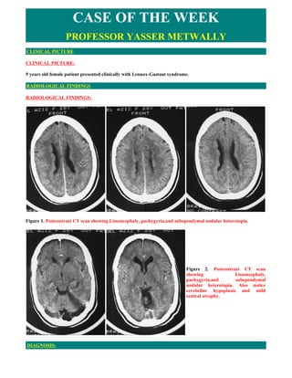
  • 1. CASE OF THE WEEK PROFESSOR YASSER METWALLY CLINICAL PICTURE CLINICAL PICTURE: 9 years old female patient presented clinically with Lennox-Gastaut syndrome. RADIOLOGICAL FINDINGS RADIOLOGICAL FINDINGS: Figure 1. Postcontrast CT scan showing Lissencephaly, pachygyria,and subependymal nodular heterotopia. Figure 2. Postcontrast CT scan showing Lissencephaly, pachygyria,and subependymal nodular heterotopia. Also notice cerebellar hypoplasia and mild central atrophy. DIAGNOSIS:
  • 2. DIAGNOSIS: CORTICAL DYSPLASIA DISCUSSION DISCUSSION: Abnormalities have long been recognized in the brains of children with developmental delay and seizures. The advent of newer imaging techniques, such as high-resolution (thin slice three-dimensional gradient recalled echo [GREI) magnetic resonance (MR) imaging and surface reconstructions of three-dimensional data sets, has led to a greater in vivo understanding of these malformations. There has been reclassification of the disorders of cortical malformations according to embryologic stages of brain cortex development at which these malformations are proposed to have occurred. These stages are cellular proliferation, cellular migration to the developing cortex, and cortical organizations. MALFORMATIONS OF ABNORMAL CELL PROLIFERATION  Nonneoplastic The earliest lesions resulting in cortical malformations are those with onset during the period of cellular proliferation in the germinal zones. During the seventh fetal week, germinal layers begin to form in the vesicle walls of telencephalic outpouchings (vesicles) at the foramina of Monro. Cortical malformations occurring during this stage of neuronal and glial proliferation may be generalized, multifocal, or focal. Generalized malformations of abnormal cell proliferation include microcephaly with diminished cortical thickness or with diminished sulcation. Commonly imaged focal or multifocal lesions include nonneoplastic disorders, such as hemimegalencephaly, focal cortical dysplasia (with balloon cells), and forme fruste tuberous sclerosis. Hemimegalencephaly may be associated with neurocutaneous or hemiovergrowth syndromes, such as hypomelanosis of Ito, epidermal nevus syndrome, or neurofibromatosis type 1. Proposed causes for hemispheric overgrowth in hemimegalencephaly include abnormal cellular proliferation and heteroploidy, defective cellular metabolism, or possibly an insult to the developing brain in the mid to late second trimester. In the event of a later insult to the developing brain, brain plasticity allows for the development of new synapses in the damaged brain, permitting the persistence of supernumerary axons and the potential for white matter overgrowth. Neuropathologic and neuroimaging features in hemimegalencephaly are abnormal neurons, lack of gray-white matter demarcation, disarrayed cortical lamination, gray matter heterotopias, broad gyri, and signal changes reflecting hypomyelination and gliosis. It is postulated that foci of agyria, with macroscopic heterotopias, extensive white matter gliosis, and less hemispheric white matter overgrowth, likely result from an earlier, more severe insult with destruction of the radial glial fibers. The variable patterns of cortical and white matter involvement demonstrated with neuroimaging likely reflect the variability in severity and timing of the precipitating insult. [1,5,19,23] Figure 1. Tl -weighted (TR 600/TE 15) and T2-weighted (I R 2800/ TE 90) axial images (A and B) demonstrate overgrowth of the right cerebral hemisphere in a patient with developmental delay, seizures, and
  • 3. hemimegalencephaly. Note the abnormal white matter signal and the unilateral ventriculomegaly. Focal cortical dysplasia is characterized by neocortical abnormalities. The pathologic spectrum of focal cortical dysplasia includes significant abnormalities of neuronal size, shape, orientation, and lamination; indistinctness of the gray-white junction; and variability in cortical thickness. Large bizarre neurons with abnormal Nissl patterns, giant neurons, binucleate neurons, a variable degree of cortical gliosis, and balloon cells with abundant pale eosinophilic cytoplasm are found. These balloon cells are similar to those seen in the cortical tubers and white matter lesions of tuberous sclerosis, and there is much overlap in the pathology of focal cortical dysplasia and tuberous sclerosis. Failure of myelin arborization, blurring of gray-white margins, variable sulcal depth and cortical thickness, and occasional hazy or dystrophic calcification may be appreciated on neuroimaging. Evidence of dual pathology, or focal cortical dysplasia in association with hippocampal sclerosis, should be sought. [8,14,17,21,26,28,32] Figure 2. Tl -weighted (TR 600/TE 15) sagittal (A) and axial images (B), and T2-weighted (TR 3000/ TE 120) axial image (C) in a child with severe microcephaly showing a thin cortical ribbon. A Tl -weighted axial image (D) in a second child with a similar degree of microcephaly reveals fewer gyri and less well developed sulcation. Imaging features in limited or forme fruste tuberous sclerosis include demonstration of a solitary smooth pyramidal-shaped gyri with a central depression and abnormal signal of the subcortical white matter. The cortical and subcortical lesions are uncommonly calcified, tend not to enhance, and are best demonstrated on MR. These children lack the calcified subependymal nodules of tuberous sclerosis as well as other organ system lesions. The cortical tubers are characterized by aberrant neurons, scattered balloon cells, and gliosis. The white matter lesions of tuberous sclerosis are characterized by decreased myelin and accumulations of balloon cells. Although there are similarities in the pathology of focal cortical dysplasia and the form fruste of tuberous sclerosis, the latter has, in general, more abundant balloon cells. [15,17, 18,22,24]  Neoplastic Lesions of proliferation after an abnormal induction event during brain development may be malformative, hamartomatous, neoplastic, or a combination. Malformative disorders consisting of neoplasias on a background of
  • 4. disordered cortex or in association with focal cortical dysplasia include dysembryoplastic neuroepithelial tumor and ganglioglioma. [5,7]  Dysembryoplastic neuroepithelial tumors Dysembryoplastic neuroepithelial tumors are supratentorial, predominantly temporal lobe tumors that are typically multinodular with a heterogeneous cell composition, including oligodendrocytes, neurons, astrocytes, and other cells. These lesions are typically fairly well-demarcated, wedge-shaped lesions extending from the cortex to the ventricle. Calcification, enhancement, and peritumoral edema are lacking on neuroimaging studies. These low- attenuation lesions may suggest an infarct on computed tomography (CT), although there is no volume loss over time, and scalloping of the inner table or calvarial bulging suggests slow growth. Lesions are low in signal on Tl- weighted images and high in signal on T2- weighted images and often have a multinodular or pseudocystic appearance. There is a spectrum of pathology in dysembryoplastic neuroepithelial tumors. On one end of the spectrum are multinodular lesions with intervening malformed cortex, in which there is some hesitation to use the designation tumor. On the other end are lesions, which are clearly neoplastic and have clinically demonstrated some growth potential. Because the term dysembryoplastic neuroepithelial tumor has only recently been introduced, such malformative and neoplastic lesions were previously labeled as hamartomas, gangliogliomas, or mixed gliomas. [9,17,20] Figure 3. A, Dysembryoplastic neuroepithelial tumour (frontobasal surgical specimen). Irregular thickening of the cerebral cortex with multiple cortical and subcortical nodules. This complex tumour consists of a mixture of glial cells and nerve cells and is often associated with dysplastic cortical foci. It can occur in any part of the brain and generally causes long-standing focal seizures. B, Dysembryoplastic neuroepithelial tumour (MT x40). Oligodendroglial-like cells arranged in groups or columns within a loose, microcystic matrix containing several solitary "floating" neurons and numerous congestive capillaries. Calcifications are possible.
  • 5. Figure 4. Dysembryoplastic neuroepithelial tumor. Wedge-shaped, nonenhancing, very low signal in- tensity lesion on Tl -weighted (TR 600/TE 15) axial image (A) demonstrating very high signal intensity lesion on the T2-weighted (TR 2800/TE 90) (B) axial image. Note the scalloping of the inner table, the multiple septations, and the lack of vasogenic edema or mass effect on the adjacent brain and ventricle. Pathologic specimen (Figure 3A) demonstrates nodules on an abnormal background of cortical dysplasia, and a specimen of one of the nodules seen on high power (Fig.3B) demonstrates oligodendroglial-like cells, some of which are actually immature neurons.  Gangliogliomas Gangliogliomas are typically demonstrated within the temporal lobe. In one large series of 51 gangliogliomas, 84% were found in the temporal lobe, 10% were found in the frontal lobe, 2% were found in the occipital lobe, and 4% were found in the posterior fossa. These lesions are typically hypodense (60% to 70%) on CT, with focal calcifications seen in 35% to 40%, contrast enhancement in 45% to 50%, and cysts in nearly 60%. The reported incidence of calcifications demonstrated on imaging in pediatric gangliogliomas is higher, seen in 61% of one series of 42 children. Features on MR imaging are less specific, with solid components isointense on T1-weighted images, bright on proton density images, and slightly less bright on T2-weighted images. Although imaging features are not specific, an enhancing, cystic temporal lobe lesion with focal calcification should suggest the diagnosis of ganglioglioma. The pathologic features that suggest the diagnosis of ganglioglioma include a neoplastic glial and neuronal component and calcification. Because calcifications are often poorly demonstrated on MR imaging and because they increase specificity of imaging findings, documentation of calcium should be sought on CT after MR imaging demonstration of a temporal lobe tumor. [11,27,33] Figure 5. A large,calcified, enhancing ganglioglioma with heterogeneous signal is demonstrated within the occipital lobe of a macrocephalic child on CT (A), and Tl -weighted (TR 600/TE 15) axial images before precontrast (B) and after (C) contrast administration. Location of ganglioglioma in this child is atypical.
  • 6. DISORDERS OF CELLULAR MIGRATION TO THE CORTEX The onset of neuronal migration to the cortex occurs during the eighth fetal week. Initially, cells in the germinal zone elongate with the nucleus remaining in the portion of cell that is furthest from the ventricular surface. After mitosis, the newly formed cells are distant from the ventricular surface. Later in neuronal migration, as distance to travel increases, migration occurs along the radial glial fibers (RGF), which span the distance from the ventricular surface to pia. Disorders of neuronal migration occur when migration is halted. Abnormalities of neuronal migration may occur with damage to the RGF by placental ischemia, infection (cytomegalovirus), or maternal trauma or with altered chemotaxis of neurons along the fibers from toxin exposure or inborn errors of metabolism. Diffuse disorders of disruption of neuronal migration include band heterotopia, classic (type 1) lissencephaly, and cobblestone (type 2) lissencephaly. Marginal glioneuronal heterotopia and nodular cortical dysplasias, nests of ectopic glial elements and gray matter within the leptomeninges and at the crown of the gyri, are believed to be the result of overmigration, possibly through areas of superficial necrosis or disruption of the external glial limitans. These findings, recognizable only on pathologic specimens, are believed to give rise to the lacy or cobblestoned cortex in type 2 lissencephaly. More focal disorders of disrupted neuronal migration include subependymal or subcortical neuronal ectopia and heterotopias. Disorders of neuronal migration are characterized on imaging studies by gray matter localized along the pathways of migration to the cortex. These gray matter rests, nodules, or masses share the attenuation (CT) and signal characteristics (MR imaging) of normal gray matter on all imaging sequences. Small deposits of heterotopia may be identified with high-resolution gradient volume acquisitions, which augment tissue contrast between gray and white matter. [3,5] Figure 6. Pachygyric (A), and agyric (B) lissencephalic brain Figure 7. A, Type II lissencephaly, Cobblestone lissencephaly (Walker-Warburg syndrome). The cortex is lissencephalic and thickened, with an irregular gray matter-white matter junction that probably represents the bundles of disorganized cortex surrounded by fibroglial tissue. The patient has been shunted for hydrocephalus. The brain is hypomyelinated. B, Presumed microcephalia vera. The brain is completely smooth (lissencephaly) with a very thin cortex. No layer of arrested neurons is present in the white matter. C, Presumed radial microbrain. Axial T2- weighted image shows an immature gyral pattern and hypomyelination. Cortical thickness is normal. This patient was profoundly microcephalic (head circumference 19 cm).
  • 7. Band heterotopia results from an early arrest of neuronal migration and gives the appearance of a continuous double cortex. The appearance has also been likened to a three-layer cake with the cortex and bilaterally symmetric, circumferential, subcortical layers of band heterotopia separated from each other by a thin white matter band. The cortex may be relatively normal or pachygyric. Shallow sulci are common. Band heterotopia has been reported to be an X-linked disorder with heterozygous females demonstrating band heterotopia and hemizygous males having classic lissencephaly. Seizures are common in band heterotopia, and mental retardation may be mild or moderate. Severity of symptoms correlates with the degree of disorganization of the overlying cortex, thickness of the continuous band of heterotopic gray matter, amount of T2 prolongation, and degree of ventriculomegaly The brain in lissencephaly type I may have a smooth surface (complete lissencephaly) or may have a nearly smooth surface with some gyral formation along the inferior frontal and temporal lobes. The thick cortex has a four-layered cortex composed of a molecular outer layer, an outer cellular layer, a cell sparse layer, and an inner cellular layer composed of arrested neurons. The arrest relates to a disruption of neuronal travel along the RGF, either from laminar necrosis or from disrupted chemotaxis. Imaging demonstrates broad, flat gyri with a thickened cortex and scanty white matter. Sylvian fissures are primitive, leading to an hourglass configuration of the brain. Type 2 or cobblestone lissencephaly is recognizable by its irregular surface, abnormal myelin, and the accompanying orbital and cerebellar anomalies. [2,10,29] Figure 8. A, Band heterotopia with pachygyria. B, band heterotopia with mild periventricular nodular heterotopia Figure 9. Subependymal nodular heterotopia
  • 8. Periventricular nodular heterotopia may be solitary, isolated lesions or may diffusely line the walls of both lateral ventricles. These lesions mimic the appearance of the subependymal tubers in tuberous sclerosis, although they do not calcify. When diffuse and bilateral, periventricular nodular heterotopia may be associated with mild cerebellar hypoplasia. Patients with periventricular or subependymal heterotopias may present with late-onset seizures, acquire normal early milestones, have normal motor development, and be of average or above-average intelligence. This disorder has been linked to markers in distal Xq28. Subcortical gray matter heterotopias also may be focal or diffuse. The greater the heterotopic mass, the more dysplastic the overlying cortex. Those patients with thick heterotopias and overlying gyral anomalies are more likely to have associated psychomotor delay. Deep infolding of thickened cortex, or deep clefts lined by heterotopia, often frontal, may also occur and are frequently associated on imaging with primitive vertical venous structures. [3,4,10,12,13,16] DISORDERS OF CORTICAL ORGANIZATION The primitive sylvian fissure is the first sulcus to form, at approximately 14 to 20 weeks, whereas the rolandic fissure, the parieto-occipital, and the superior temporal gyri form later. By 32 to 33 weeks, large numbers of cortical sulci are visible, and by 38 to 40 weeks, there is a nearly normal adult sulcal pattern. After full-term birth, the sulci continue to deepen over the next weeks. The final group of disorders are those that are associated with disruption of the process of gyral formation and subsequent cellular organization of the cortex. These disorders include generalized polymicrogyria (PMG) or focal and multifocal disorders such as focal PMG, bilateral symmetric PMG, and schizencephaly with or without PMG. The imaging appearance of dysplasias of cortical organization includes abnormalities of the cortical gyral pattern without radiographically evident subjacent heterotopias. The imaging appearance of generalized or focal polymicrogyria can be quite variable. Typically, there is a thick cortex with many small gyri separated by shallow sulci. The gyri may, however, be so small that they are difficult to discern on imaging. The appearance then is of a flat thickened cortex, simulating pachygyria or agyria. Small areas of cortical thickening may be better defined with adjunctive three-dimensional MR reformatted images. Bilateral opercular or perisylvian syndrome is a bilateral symmetric PMG disorder consisting of primitive sylvian fissures, primitive draining veins, and symmetric involvement of the operculum with polymicrogyria or pachygyria. Patients may present with seizures, motor and speech disorders, mental retardation, and a congenital pseudobulbar syndrome. Schizencephaly, or gray matter lined clefts of the brain, occurs after disruption of the RGF units from the ventricle to the pial surface. Smaller clefts may have coapted walls or closed lips. Larger clefts, or those with open lips, may allow free communication of the ventricles with the pericerebral spaces. [4,5] Figure 10. CT scan showing Subependymal nodular heterotopia with cerebellar hypoplasia Figure 11. MRI showing Subependymal nodular heterotopia   Also included in disorders of cortical organization are cortical dysplasia without balloon cells, with imaging
  • 9. features similar to the previously described focal cortical dysplasia with balloon cells, and microdysgenesis. Small foci of cortical dysplasia may require serial imaging over time, becoming apparent only when myelin maturation is complete. Other foci may be apparent only with high-resolution, high-contrast imaging, such as is possible with GRE sequences. Microdysgenesis is radiographically undetectable with pathologic features consisting of subtle neocortical abnormalities: neuronal ectopias within the white matter, abnormal neurons within the molecular layer, neuronal clustering; bare areas within cortical layers two to six, and Chaslin's subpial gliosis. [6,17,31] Figure 12. Lissencephaly Type 1. Tl -weighted (TR 600/ TE 15) (A) and T2-weighted (TR 3000/TE 120) (B) coronal images in a severely delayed patient with Miller-Dieker syndrome show a smooth cortical surface. Thickened cortex with rare gyri along the inferior temporal lobes is seen on Tl-weighted (C) and T2-weighted (D) coronal images in a patient with agyria-pachygyria complex and seizures. Figure 13. Bilateral opercular dysplasia is present in a 14-year-old with a history of spastic diplegia and recent onset of seizures. Primitive Sylvian fissures lined by thickened cortex are seen on Tl -weighted (TR 600/TE 15) (A) and T2-weighted (TR 2800/TE 90) (B) axial images.
  • 10. Figure 14. Closed-lip schizencephaly is seen on the right, and a deep cleft on the left on T1 -weighted (TR 600/TE 15) (A) and T2-weighted (TR 2800/TE 90) (B) axial images in a 2-year-old with psychomotor delay and microcephaly. Open-lip schizencephaly is documented on Tl -weighted (C) and T2-weighted (D) coronal views in a 2-year-old presenting with spastic quadriparesis. Figure 15. Open-lip schizencephaly with cortical dysplasia
  • 11. Figure 16. Bilateral open lip schizencephaly SUMMARY SUMMARY Lissencephaly is the most severe of the disorders discussed in this case recport and the most easily diagnosed by magnetic resonance imaging (MRI). In all types of lissencephaly the surface of the brain is abnormally smooth, with shallow sulci and an abnormal gyral pattern. In some types of lissencephaly the gyri are also abnormally broad and flat. lissencephaly can be divided by MRI criteria into five categories.  Type I Lissencephaly In type I lissencephaly, neuronal migration to the cortex has stopped before completion. Therefore, a band of neurons lies in the subcortical region, separated from an abnormally thin cortex by a layer of white matter. The cortex may be completely smooth (completely lissencephaly or agyria) or may have a few broad, flat gyri separated by shallow sulci (pachygyria), a pattern that may be present in parts of otherwise completely lissencephalic brains. Band heterotopias in which the cortex appears nearly normal have been arbitrarily included in a separate section in this chapter. However, the reader should recognize that these are most likely a mild form of type I lissencephaly. Depending on the severity of the lissencephaly, affected children may be microcephalic or normocephalic and may range from profoundly retarded to nearly normal. Similarly, the neurologic findings and the occurrence of associated anomalies vary with the severity of the migration defect. On imaging studies, patients with type I lissencephaly have a smooth cortex that may be agyric, pachygyric, or both, with an underlying layer of arrested neurons. The white matter is typically diminished in volume and the Sylvian fissures are most commonly shallow and vertically oriented. The layer of white matter separating the cortex from the layer of arrested neurons has a variable appearance depending on the severity of the lissencephaly and the state of myelination, which are, in turn, dependent on the patient's age. Patients with severe lissencephaly have a thick layer of white matter separating the cortex from the layer of arrested neurons, whereas those with milder lissencephaly (mild pachygyria) have thinner layers of arrested neurons and thinner layers of white matter separating the cortex from the arrested neurons. The gross appearance of the brain resembles that of the fetus before 23 or 24 weeks of gestation, when sulci normally begin to form. The middle cerebral arteries typically lie close to the inner table of the skull because there are no sulci in which they can lie. The severely affected cerebrum has a figure-8 appearance on axial images as a result of the shallow vertical sylvian fissures. In severe agyria, sagittal images may show a hypoplastic corpus callosum, with a small splenium and absent rostrum. The brainstem
  • 12. often appears hypoplastic, probably because many of the corticospinal and corticobulbar tracts do not form. Areas of pachygyria also have a thickened cortex, but broad gyri and shallow sulci are present. As stated earlier, the layer of arrested neurons and the layer of white matter separating it from the cortex are thinner in pachygyric areas than in agyric sections. Pachygyria can be focal or diffuse. When focal, it can occur in any part of the brain but is most common, in my experience, in the parieto-occipital regions. When diffuse, it is often associated with regions of agyria and tends to be more severe in the parieto-occipital region of the brain and least severe in the frontal and temporal lobes.  Type II Lissencephaly Type 11 lissencephaly occurs in the Walker-Warburg syndrome and in Fukuyama's congenital muscular dystrophy. It is characterized pathologically by lissencephaly, microphthalmia with retinal dysplasia, callosal hypogenesis, cerebellar cortical dysplasia, cerebellar vermian hypoplasia, hypomyelination, and hydrocephalus, which may result from obliteration of the subarachnoid spaces by thickened, fibrous meninges (3,11-14). Histologically, the cortex is characterized by a lack of lamination and disruption by penetrating vessels and fibroglial bundles that are continuous with the fibroglial tissue obliterating the subarachnoid spaces. The cortex is thicker than normal but not as thick as the combined cortex, layer of white matter, and layer of arrested neurons of type I lissencephaly. MR imaging studies mirror the pathologic findings. The cortex is lissencephalic and thick, with an irregular gray- white matter junction that probably represents the bundles of disorganized cortex surrounded by fibroglial tissue. Ventricles are large, and the patient has often been shunted before CT or MRI scans are obtained. The corpus callosum is hypogenetic or absent and the cerebellar vermis is always hypogenetic-hypoplastic with agenesis of the inferior vermis. The white matter is almost completely unmyelinated. About half of patients have an occipital encephalocele or evidence of a repaired encephalocele.  Type III Lissencephaly (Microcephalia Vera) The term "microcephalia vera" has been used to describe several genetic and sporadic developmental brain anomalies. Patients typically present with microcephaly and moderate developmental delay but no focal neurologic findings. Histopathologic examination of one brain of an affected 26-week gestational age fetus showed no migratory disorder. However, the germinal zone was completely depleted at an age when the normal germinal zone is of maximal volume. Pathologic examination shows a thin cerebral, cortex, diminished cerebral white matter, shallow sulci, and markedly diminished callosal axons. The only MR study published of a presumed case of microcephalia vera shows a very small brain with complete lissencephaly and an extremely thin cortex. No layer of arrested neurons was identified.  Type IV Lissencephaly (Radial Microbrain) The term "radial microbrain" has been used to describe patients born at term with markedly reduced brain size despite normal gyral patterns, normal cortical thickness, normal cortical lamination, and absence of destruction or gliosis (18). However, the number of neocortical neurons was only 30% of normal. All affected patients had profound microcephaly and non-CNS anomalies, such as nephropathy and acromicria. We have performed MRI on three infants that we believe fulfill the criteria for radial microbrain. All had profound microcephaly, delayed myelination, and a slightly immature gyral pattern. We presume that the myelination delay is due to lack of production of oligodendrocyte precursors by the depleted germinal zones.  Type V Lissencephaly (Diffuse Polymicrogyria) Diffuse polymicrogyria is a type of smooth brain that involves the entire cerebrum. Patients with this severe condition typically are microcephalic and present with developmental delay or the onset of seizures in the first 6-8 months of postnatal life. In our experience, most patients with diffuse polymicrogyria have congenital cytomegalovirus infections. MR images show a slightly thickened cortex (typically 5-7 mm) with irregularly "bumpy" outer and inner surfaces. The ventricles are typically enlarged and the white matter hypomyelinated. Patients with diffuse polymicrogyria are differentiated from those with Walker-Warburg syndrome by the absence of callosal and ocular anomalies and by the "bumpy" outer cortical surface. The outer cortical surface in the Walker- Warburg syndrome is typically smooth on MRI studies.  Addendum
  • 13. A new version of this PDF file (with a new case) is uploaded in my web site every week (every Saturday and remains available till Friday.)  To download the current version follow the link "http://pdf.yassermetwally.com/case.pdf".  You can also download the current version from my web site at "http://yassermetwally.com".  To download the software version of the publication (crow.exe) follow the link: http://neurology.yassermetwally.com/crow.zip  The case is also presented as a short case in PDF format, to download the short case follow the link: http://pdf.yassermetwally.com/short.pdf  At the end of each year, all the publications are compiled on a single CD-ROM, please contact the author to know more details.  Screen resolution is better set at 1024*768 pixel screen area for optimum display.  For an archive of the previously reported cases go to www.yassermetwally.net, then under pages in the right panel, scroll down and click on the text entry "downloadable case records in PDF format"  Also to view a list of the previously published case records follow the following link (http://wordpress.com/tag/case-record/) or click on it if it appears as a link in your PDF reader REFERENCES References I. Barkovich Aj, Chuang SH: Unilateral megalencephaly: Correlation of MR imaging and pathologic characteristics. AJNR Am j Neuroradiol 11:525-531, 1990 2. Barkovich Aj, Guerrini R, Battaglia, et al: Band heterotopia: Correlation of outcome with magnetic resonance imaging parameters. Ann Neurol 36:609-617, 1994 3. Barkovich Aj, Knos BO: Gray matter heterotopias: MR characteristics and correlation with developmental and neurologic manifestations. Radiology 182:493- 499,f992 4. Barkovich Aj, Kjos BO: Nonlissencephalic cortical dysplasias: Correlation of imaging findings with clinical deficits. AJNR Am J Neuroradiol 13:95-103, 1992 5. Barkovich Aj, Kuzniecky RI, Dobyns WB, et al: A classification scheme for malformations of cortical development. Neuropediatrics 27:59-63, 1996 6. Bastos AC, Korah IP, Cendes F, et al: Curvilinear reconstruction of 3D magnetic resonance imaging in patients with partial epilepsy: A pilot study. Magn Reson Imaging 13:1107-1112, 1995 7. Becker LE: Central neuronal tumors in childhood: Relationship to dysplasia. J Neurooncol 24:13-19, 1995 8. Cochrane DD, Poskitt Kj, Norman MG: Surgical implications of cerebral dysgenesis. Can J Neurol Sci 18:181- 195, 1991 9. Daumas-Duport C, Scheithauer BW, Chodkiewicz JP: Dysembryoplastic neuroepithelial tumor: A surgically curable tumor of young patients with intractable partial seizures. Neurosurgery 23:545-556, 1988 10. Dobyns WB, Truwit CL: Lissencephaly and other malformations of cortical development: 1995 update. Neuropediatrics 26:132-147, 1995 11. Dome HL, O'Gorman AM, Melanson D: Computed tomography of intracranial gangliogliomas. AJNR Am j Neuroradiol 7:281-285, 1986 12. Dubeau F, Tampieri D, Lee N, et al: Periventricular and subcortical nodular heterotopia: A study of 33 patients. Brain 118:1273-1287, 1995 13. Eksioglu YZ, Scheffer IE, Cardenas P, et al: Periventricular heterotopia: An X-linked dominant epilepsy locus causing aberrant cerebral cortical development. Neuron 16:77-87,1996 14. Farrell MA, DeRosa Mj, Curran JG, et al: Neuropathologic findings in cortical resections (including
  • 14. hemispherectomies) performed for the treatment of intractable childhood epilepsy. Acta Neuropathol 83:246- 259,1992 15. Freide RL: Dysplasias of cerebral cortex. In Developmental Neuropathology, ed 2. Berlin, Springer-Verlag, 1989 16. Jardine PE, Clarke MA, Super M: Familial bilateral periventricular nodular heterotopia mimics tuberous sclerosis. Arch Dis Child 74:244-246, 1996 17. Jay V, Becker LE, Otsubo H, et al: Pathology of temporal lobectomy for refractory seizures in children: Review of 20 cases including some unique malformative lesions. J Neurosurg 79:53-61, 1993 18. Jay V, Edwards V, Rutka JT. Crystalline inclusions in a subependymal giant cell tumor in a patient with tuberous sclerosis. Ultrastruct Pathol 17:503-513, 1993 19. Yimura M, Yoshino K, Maeoka Y, et al: Hypomelanosis of Ito: MR findings. Pediatr Radiol 24:68-69,1994 20. Kuroiwa T, Bergey GK, Rothman MI, et al: Radiologic appearance of the dysembroplastic neuroepithelial tumor. Radiology 197:233-238,1995 21. Kuzniecky R, Garcia JH, Gaught E, et al: Cortical dysplasia in temporal lobe epilepsy: Magnetic resonance imaging correlations. Ann Neurol 29:293-298,1991 22. Martin N, Debussche C, De Broucker, et al: Gadolinium-DTPA enhanced MR imaging in tuberous sclerosis. Neuroradiology 31:492-497, 1990 23. Manz Hj, Phillips TM, Rowden G, et al: Unilateral megalencephaly, cerebral cortical dysplasia, neuronal hypertrophy, and heterotopia. Acta Neuropathol 45:97- 103, 1979 24. Menor F, Marti-Bonmati L, Mulas F, et al: Neuroimaging in tuberous sclerosis: A clinico-radiological evaluation in pediatric patients. Pediatr Radiol 22:485- 489, 1992 25. Miller DC, Lang FF, Epstein Fj: Central nervous system gangliogliomas: Part 1. Pathology. f Neurosurg 79: 859-866,1993 26. Moreland DB, Glasauer FE, Egnatchik JG, et al: Focal cortical dysplasia. J Neurosurg 68:487-490, 1988 27. Otsubo H, Hoffman Hj, Humphreys RP, et al: Detection and management of gangliogliomas in children. Surg Neurol 38:371-378,1992 28. Otsubo H, Hwang P, Jay V, et al: Focal cortical dysplasia in children with localization related epilepsy: EEG, MRI and SPECT findings. Pediatr Neurol 9:101-107, 1993 29. Paimini A, Andermann F, Aicardi J, et al: Diffuse cortical dysplasia, or the 'double cortex' syndrome: The clinical and epileptic spectrum in 10 patients. Neurology 41:1656-1662, 1991 30. Raymond AA, Fish DR, Stevens JM, et al: Subependymal heterotopia: A distinct neuronal migration disorder associated with epilepsy. J Neurol Neurosurg Psychiatry 57:1195-1202, 1994 31. Rolland Y, Adamsbaum C, Sellier N, et al: Opercular malformations: Clinical and MRI features in 11 children. Pediatr Radiol 25(suppl 1):S2-8,1995 32. Taylor DC, Falconer MA, Bruton Cj, et al: Focal dysplasia of the cerebral cortex in epilepsy. J Neurol Neurosurg Psychiatry 34:369-387, 1971 33. Zentner J, Wolf HK, Ostertun B, et al: Gangliogliomas: Clinical, radiological, and histopathological findings in 51 patients. I Neurol Neurosurg Psychiatry 57:1497- 1502,1994 34. Metwally, MYM: Textbook of neuroimaging, A CD-ROM publication, (Metwally, MYM editor) WEB-CD agency for electronic publication, version 11.1a. January 2010