Muscles Important In Tendon Jerks
•Als DOCX, PDF herunterladen•
0 gefällt mir•679 views

This document lists several muscles, their proximal and distal attachments, innervation, and main actions. It includes the biceps brachii, triceps brachii, supinator, brachioradialis, gastrocnemius, quadriceps femoris, and its components - rectus femoris, vastus lateralis, vastus medialis, and vastus intermedius. For each muscle it provides details on origin, insertion, nerve supply, and functional role.

Empfohlen
Weitere ähnliche Inhalte
Was ist angesagt?
Was ist angesagt? (20)
Andere mochten auch
Andere mochten auch (15)
Ähnlich wie Muscles Important In Tendon Jerks
Ähnlich wie Muscles Important In Tendon Jerks (20)
Mehr von Yapa
Mehr von Yapa (20)
Kürzlich hochgeladen
Kürzlich hochgeladen (20)
Muscles Important In Tendon Jerks
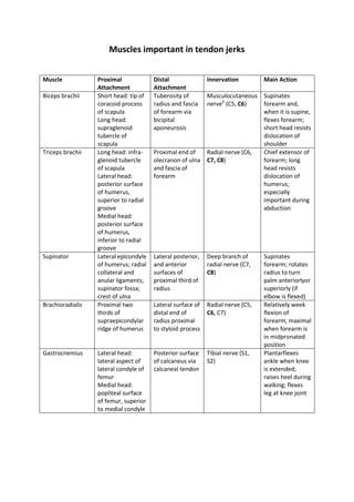
- 1. MuscleProximal AttachmentDistal AttachmentInnervationMain ActionBiceps brachii Short head: tip of coracoid process of scapula Long head: supraglenoid tubercle of scapulaTuberosity of radius and fascia of forearm via bicipital aponeurosisMusculocutaneous nerveb (C5, C6)Supinates forearm and, when it is supine, flexes forearm; short head resists dislocation of shoulderTriceps brachiiLong head: infra-glenoid tubercle of scapulaLateral head: posterior surface of humerus, superior to radial grooveMedial head: posterior surface of humerus, inferior to radial grooveProximal end of olecranon of ulna and fascia of forearmRadial nerve (C6, C7, C8)Chief extensor of forearm; long head resists dislocation of humerus; especially important during abductionSupinatorLateral epicondyle of humerus; radial collateral and anular ligaments; supinator fossa; crest of ulnaLateral posterior, and anterior surfaces of proximal third of radiusDeep branch of radial nerve (C7, C8)Supinates forearm; rotates radius to turn palm anteriorlyor superiorly (if elbow is flexed)BrachioradialisProximal two thirds of supraepicondylar ridge of humerusLateral surface of distal end of radius proximal to styloid processRadial nerve (C5, C6, C7)Relatively week flexion of forearm, maximal when forearm is in midpronated positionGastrocnemiusLateral head: lateral aspect of lateral condyle of femurMedial head: popliteal surface of femur, superior to medial condylePosterior surface of calcaneus via calcaneal tendonTibial nerve (S1, S2)Plantarflexes ankle when knee is extended; raises heel during walking; flexes leg at knee joint<br />Muscles important in tendon jerks<br />MuscleProximal AttachmentDistal AttachmentInnervationMain ActionQuadriceps femorisRectus femorisAnterior inferior iliac spine and ilium superior to acetabulumVia common tendinous (quadriceps tendon) and independent attachments to base of patella; indirectly via patellar ligament to tibial tuberosity; medial and lateral vasti also attach to tibia and patella via aponeuroses (medial and lateral patellar retinacula)Femoral nerve (L2, L3, L4)Extend leg at knee joint; rectus femoris also steadies hip joint and helps iliopsoas flex thighVastus lateralisGreater trochanter and lateral lip of linea aspera of femurVastus medialisIntertrochanteric line and medial lip of linea aspera of femurVastus intermediusAnterior and lateral surfaces of shaft of femur<br />