Beginners Guide to TikTok for Search - Rachel Pearson - We are Tilt __ Bright...
Cryptosporidium_8301-3 (06-29-2016).pdf
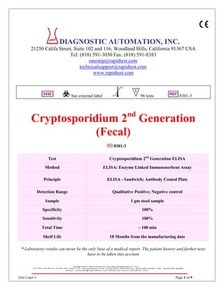
1. DAI Code# 3 Page 1 of 9
DIAGNOSTIC AUTOMATION, INC.
21250 Califa Street, Suite 102 and 116, Woodland Hills, California 91367 USA
Tel: (818) 591-3030 Fax: (818) 591-8383
onestep@rapidtest.com
technicalsupport@rapidtest.com
www.rapidtest.com
See external label 96 tests 8301-3
Cryptosporidium 2nd
Generation
(Fecal)
8301-3
* Laboratory results can never be the only base of a medical report. The patient history and further tests
have to be taken into account
Test Cryptosporidium 2nd
Generation ELISA
Method ELISA: Enzyme Linked Immunosorbent Assay
Principle ELISA - Sandwich; Antibody Coated Plate
Detection Range Qualitative Positive; Negative control
Sample 1 gm stool sample
Specificity 100%
Sensitivity 100%
Total Time ~ 100 min
Shelf Life 18 Months from the manufacturing date
2. DAI Code# 3 Page 2 of 9
Intended Use
This ELISA is an in vitro immunoassay for the qualitative determination of Cryptosporidium antigen in feces.
Summary
Cryptosporidium is a coccidian parasite that is recognized as an important enteric pathogen. The organism
causes an acute, though self-limiting infection in immunocompetent individuals. Incubation periods of 1 to
12 days have been reported with most oocyst shedding ending by day 21. Symptoms range from mild to
severe diarrhea with a variety of complications. 1,8,9,10,11,13
The infection in immunocompromised patients is much more severe and may often be life threatening.
Passage of fluid, up to 12 liters per day, has been reported. 1,2,3,12,14,16
Multiple pathways of Cryptosporidium transmission have been implicated. These include animal to human,
water contamination and person-to-person. The latter may include contact between members of the same
household, day care centers, and homosexual men. 1,2,12,14,16
Diagnosis of Cryptosporidium infections was done originally by direct detection techniques. Of these,
microscopic examination of stools using stains or fluorescence labeled antibodies has been the most
common. However, this method relies on an experienced technician and subsequent observation of intact
organisms. Because of the historically low proficiency of correct microscopic examinations, alternative
diagnostic methods have been investigated. 4,5,16,17
One important alternative has been the development of an antigen capture enzyme linked immunosorbent
assay (ELISA) for use with stools. These tests, which have shown comparable sensitivity to experienced
microscopic examinations, are fairly simple to perform and do not require the observation of intact
organisms.6,7
Principle of Procedure
During the first incubation, Cryptosporidium specific antigen present in the stool specimens are captured by
antibodies attached to the microwells. The wells are incubated and washed before anti-Cryptosporidium
antibodies conjugated to horseradish peroxidase are added. The enzyme conjugate will “sandwich” any
antigen bound to the wells. After washings to remove unbound enzyme, a chromogen is added which
develops a blue color in the presence of the enzyme complex. The stop solution ends the reaction and turns
the blue color to yellow. If no antigen is captured, or if there is an insufficient level of antigen, no colored
reaction will take place.
3. DAI Code# 3 Page 3 of 9
Reagents
Item Description Symbol
Test Strips
Microwells containing anti-Cryptosporidium polyclonal
antibodies - 96 test wells in a test strip holder.
Enzyme Conjugate
One (1) bottle containing 11 ml of anti-Cryptosporidium
antibodies conjugated to horseradish peroxidase with
thimerosal.
Positive Control
One (1) vial containing 2 ml of a diluted
Cryptosporidium positive formalinized stool
supernatant.
Negative Control
One (1) vial containing 2 ml of a buffered protein
solution with thimerosal.
Chromogen
One (1) bottle containing 11 ml of the chromogen
tetramethylbenzidine (TMB) and peroxide.
Wash Concentrate (20X)
Two (2) bottles containing 25 ml of concentrated buffer
with detergent and thimerosal.
Dilution Buffer
Four (4) bottles containing 30 ml buffered protein
solution containing Thimerosal
Stop Solution One (1) bottle containing 11 ml of 5% phosphoric acid.
Precautions
Do not deviate from the specified procedures when performing this assay. All specimen dilutions,
incubation times/temperatures and washings have been optimized for the best performance
characteristics. Deviations from the specified procedures may affect the sensitivity and specificity of
the assay.
• For In Vitro Diagnostic Use Only.
• Do not interchange reagents between kits with different lot numbers.
• Do not use reagents that are beyond their expiration dates. Expiration dates are on each reagent label.
Use of reagents beyond their expiration dates may affect results.
• Unused microwells should be stored in the desiccated pouch to protect them from moisture.
• Do not use solutions if they precipitate or become cloudy.
Exception: Wash concentrate may precipitate during refrigerated storage, but will dissolve upon
warming.
• Do not add azides to the samples or any of the reagents.
• Controls and some reagents contain thimerosal as a preservative, which may be irritating to skin, eyes
and mucous membranes. In case of contact, flush eyes or rinse skin with copious amounts of water.
• Treat all reagents and samples as potentially infectious materials. Use care to prevent aerosols and
decontaminate any spills of samples.
• Stop solution is a 5% solution of phosphoric acid in water. If spilled on the skin, wash with copious
amounts of water. If acid gets into the eyes, wash with copious amounts of water and seek medical
attention.
4. DAI Code# 3 Page 4 of 9
• Persons who are color blind or visually impaired may not be able to read the test visually and should
use spectrophotometric readings to interpret results.
Storage Conditions
Reagents, strips and bottled components:
Store between 2 – 8 ºC.
Squeeze bottle containing diluted wash buffer may be stored at room temperature (15-25ºC).
Preparation
• Before use, bring all reagents and samples to room temperature (15-25 °C) and mix.
• (20X) Wash Concentrate may precipitate during refrigerated storage, but will go back into solution when
brought to room temperature (15-25°C) and mixed. Ensure that (20X) wash concentrate is completely in
solution before diluting to working concentration. To dilute (20X) wash concentrate to working dilution,
remove cap and add contents of one bottle of Wash Concentrate to a squeeze bottle containing 475 ml of DI
water. Swirl to mix. Squeeze bottle should have a narrow tip to optimize washings.
Collection of Stool (Feces)
1. No modification of collection techniques used for standard microscopic O&P examinations is needed.
2. Stool samples may be used as unpreserved or frozen, in Cary-Blair Transport Medium or in
preservation media of 10% formalin or SAF.
3. Unpreserved samples should be kept at 2-8°C and tested within 24 hours of collection. Samples that
cannot be tested within this time should be frozen at -20°C or lower until used. Avoid multiple
freeze/thaw cycles.
4. Samples preserved in Formalin and SAF may be kept at room temperature (15-25°C) or at 2-8°C and
tested within 18 months of collection. DO NOT freeze preserved samples.
5. Samples in Cary-Blair should be kept at 2-8°C or -20°C and tested within 1 week of collection. Avoid
multiple freeze/thaw cycles.
Procedure
Materials Provided
Cryptosporidium Stool Antigen Microwell ELISA Kit
Materials Required But Not Provided
Transfer Pipettes
Squeeze bottle for washing strips (narrow tip is recommended)
Graduated Cylinder
Reagent grade (DI) water
Sample dilution tubes
Applicator sticks (recommended) or swabs for sample preparation
Micropipette
Suggested Equipment
ELISA plate reader with 450 and 620-650 nm filters
5. DAI Code# 3 Page 5 of 9
Test Procedure
Notes:
• All incubations are to be done at room temperature (15 to 25 °C)
• Ensure all samples and reagents are at room temperature (15-25°C) before use. Frozen samples must be
thawed completely before use.
• All dilutions of stools must be made with the Dilution Buffer provided. Do not use dilution buffer from a
kit with a different lot number.
• If needed, prepared samples can be centrifuged at 2000-3000 g for 5-10 minutes. Ensure supernatant is
clear before use.
• When running the assay, try to avoid the formation of bubbles in the wells. Bubbles may affect overall
performance and reading of end results. Slapping the wells out on a clean absorbent towel after each wash
step should help to minimize bubbles in the wells.
• Controls must be included each time the kit is run. Controls are provided ready to use. DO NOT dilute
further.
• Unpreserved and Preserved specimens have different testing procedures. See below for specific
instructions on how to run the assay using each procedure.
Preserved Specimen Procedure:
1. For samples in SAF, 10% Formalin or Cary-Blair, mix contents thoroughly inside container. No
further processing is required.
2. Break off the required number of wells needed (number of samples plus 2 for controls) and place in
holder.
3. Using a micropipette, add 100 μl of negative control to well # 1 and 100 μl of positive control to
well# 2.
4. Using a micropipette, add 50 μl of Dilution Buffer to each sample well. DO NOT add Dilution
Buffer to control wells.
5. Add 50 μl of sample to each sample well with Dilution Buffer.
6. Incubate for 60 minutes at room temperature (15-25°C), then wash.* After last wash, slap the wells
out on a clean absorbent towel to remove excess wash buffer.
7. Add 2 drops of Enzyme Conjugate to each well.
8. Incubate for 30 minutes at room temperature (15-25°C), then wash.* After last wash, slap the wells
out on a clean absorbent towel to remove excess wash buffer.
9. Add 2 drops of Chromogen to each well.
10. Incubate for 10 minutes at room temperature (15-25°C).
11. Add 2 drops of Stop Solution to each well. Mix wells by gently tapping the side of the strip holder
with index finger for approximately 15 seconds. Read reaction within 5 minutes after adding stop
solution.
12. Read results visually or using an ELISA plate reader (see instructions below).
Unpreserved Specimen Procedure:
1. Prepare sample dilutions in tubes using 0.7 ml of Dilution Buffer and 0.1 g, about the size of a small
pea, of fecal sample using an applicator stick. Mix thoroughly before using.
6. DAI Code# 3 Page 6 of 9
-IF USING SWABS, add 1 ml of dilution buffer to dilution tube. Coat the swab with a thin layer of
specimen and mix into dilution buffer, expressing as much fluid as possible. Mix thoroughly before
using
2. For watery unpreserved specimens, mix contents then add 0.1 ml of sample to 0.7 ml of Dilution
Buffer in dilution tubes. Mix thoroughly before using.
3. Break off the required number of wells needed (number of samples plus 2 for controls) and place in
holder.
4. Using a micropipette, add 100 μl of negative control to well # 1.
5. Using a micropipette, add 100 μl of positive control to well # 2.
6. Add 100 μl of diluted sample to each well.
7. Incubate for 60 minutes at room temperature (15-25°C), then wash.* After last wash, slap the wells
out on a clean absorbent towel to remove excess wash buffer.
8. Add 2 drops of Enzyme Conjugate to each well.
9. Incubate for 30 minutes at room temperature (15-25°C), then wash.* After last wash, slap the wells
out on a clean absorbent towel to remove excess wash buffer.
10. Add 2 drops of Chromogen to each well.
11. Incubate for 10 minutes at room temperature (15-25°C).
12. Add 2 drops of Stop Solution to each well. Mix wells by gently tapping the side of the strip holder
with index finger for approximately 15 seconds. Read reaction within 5 minutes after adding stop
solution.
13. Read results visually or using an ELISA plate reader (see instructions below).
* Washings consist of vigorously filling each well to overflowing and decanting contents five (5)
separate times. When possible, avoid formation of bubbles in the wells as this may affect the end
results.
Interpretation of Results – Visual
Reactive: Any sample well that is obviously more yellow than the negative control well.
Non-reactive: Any sample well that is not obviously more yellow than the negative control well.
NOTE: The negative control, as well as some samples, may show some slight color. A sample well must
be obviously darker than the negative control well to be called a positive result.
Interpretation of Results - ELISA Reader
Zero reader on air. Read all wells at 450/620-650 nm.
Reactive: Absorbance reading of 0.08 OD units and above indicates the sample contains Cryptosporidium
antigen.
Non-reactive: Absorbance reading less than 0.08 OD units indicates the sample does not contain detectable
levels of Cryptosporidium antigen.
Test Limitations
Test results should be used as an aid in diagnosis and should not be interpreted as diagnostic by themselves.
DO NOT concentrate stool samples. Assay will not give accurate results on a concentrated sample.
7. DAI Code# 3 Page 7 of 9
A negative result can occur from an antigen level lower than the detection limits of this assay. Multiple
samples over time may be indicated for those patients that are suspected of being positive for
Cryptosporidium.
Expected Results
Normal healthy individuals should be free of Cryptosporidium and should test negative. A positive reaction
indicates that the patient is shedding detectable amounts of Cryptosporidium antigen. Certain populations,
such as homosexual men and children in day care settings, have shown higher rates of infection with
Cryptosporidium than the normal population.
Performance Characteristics
Study 1
A study was performed with the Diagnostic Automation, Inc. Cryptosporidium assay using fresh/frozen
specimens, specimens preserved in 10% Formalin and SAF and specimens in Cary-Blair Transport media.
There were a total of 94 specimens used in the study that were confirmed positive or negative for
Cryptosporidium by microscopy. Of the 94 specimens there were 16 that were positive for
Cryptosporidium, and 78 that were negative for Cryptosporidium. The results from the study are shown in
the following table.
Microscopy
+ -
DAI Cryptosporidium
+ 16 0
- 0 78
Sensitivity: 100% (16/16)
Specificity: 100% (78/78)
Study 2
Another study was performed comparing the Diagnostic Automation, Inc. Cryptosporidium assay with
another commercially available ELISA. The study was performed using fresh/frozen specimens and
specimens preserved in 10% Formalin and SAF. There were a total of 94 specimens used in the study that
were confirmed positive or negative for Cryptosporidium. Of the 94 specimens there were 16 that were
positive for Cryptosporidium, and 78 that were negative for Cryptosporidium. The results from the study
are shown in the following table.
DAI
+ -
Other Commercial
ELISA
+ 16 0
- 0 78
Positive Agreement: 100% (16/16)
Negative Agreement: 100% (78/78)
8. DAI Code# 3 Page 8 of 9
Reproducibility
• The intra-assay (well to well) CV was calculated using 4 positive and 4 negative samples assayed 24
times in a single run. The mean CV was 5.96% with the highest being 9.83%.
• The inter-assay (run to run) CV was calculated using 4 positive and 4 negative samples assayed on
three separate days. The mean CV was 4.48% with the highest being 7.3%.
Cross Reactivity
No cross-reactions were seen with the following organisms:
Entamoeba hartmanni, Endolimax nana, Entamoeba histolytica/dispar, Entamoeba coli, Blastocystis
hominis, Dientamoeba fragilis, Chilomastix mesnili, Strongyloides stercoralis, Ascaris lumbricoides,
Enterobius vermicularis, Diphyllobothrium species, Hymenolepis nana, Clonorchis sinensis, Enteromonas
hominis, Trichuris trichiura, Iodamoeba buetschlii, Hookworm, Schistosoma mansoni, Giardia lamblia,
Rotavirus, Taenia eggs, Fasciola eggs, Isospora belli, Entamoeba polecki, Adenovirus, & 33 bacterial
species (list available on request).
Quality Control
The positive and negative control must be included each time the assay is run. The use of a positive and negative
control allows easy validation of kit stability.
Negative control should appear colorless when read visually and should read less than 0.08 OD when read at
a dual wavelength of 450/620-650 nm.
Positive control should be a clearly visible yellow color and read at greater than 0.5 OD when read at a dual
wavelength of 450/620-650 nm.
Troubleshooting
Problem: Negative control has excessive color after development.
Reason: Inadequate washings
Correction: Wash more vigorously. Remove excessive liquid from the wells by tapping against an
Absorbent towel. Do not allow test wells to dry out.
References
1. Chapman, P.A. “Cryptosporidiosis: Recent Trends in Epidemiology, Diagnosis, and Treatment.” Serodiag &
Immunother Infect Dis #2, 1988, pp. 311-317.
2. Meyer, E.A. “Waterborne Giardia and Cryptosporidium.” Parasit Today. Vol. 4, #7, 1988, pp. 200-201.
3. Garcia, L., Bruckner, D., Brewer, T., “Cryptosporidiosis in Patients with AIDS.” ACPR, May 1988, pp. 38-41.
4. Stibbs, H., Ongerth, J. “Immunofluorescence Detection of Cryptosporidium Oocysts in Fecal Smears.” J Clin
Micro, Vol 24 #4, Oct. 1986, pp.517-521.
5. McLaughlin, J. et al. “Indentification of Cryptosporidium Oocysts by Monoclonal Antibody.” Lancet, January
3, 1987, pp.51.
6. Ungar, B. “Enzyme-Linked Immunoassay for Detection of Cryptosporidium Antigens in Fecal Specimens.” J
Clin Micro, Vol. 28 #11, Nov 1990, pp. 2491-2495.
7. Anusz, K., et al. “Detection of Cryptosporidium parvum Oocysts in Bovine Feces by Monoclonal Antibody
Capture Enzyme-Linked Immunosorbent Assay.” J. Clin Micro, Vol. 28 #12, dec. 1990, pp. 2770-2774.
8. Jokipii, L., et al. “Cryptosporidium: A Frequent Finding In Patients With Gastrointestinal Symptoms.” Lancet,
August 13, 1983, pp. 358-360.
9. Shephard, R., et al. “Shedding of Oocysts of Cryptosporidium in Immunocompetent Patients.” J Clin Pathol,
Vol. 41, 1988, pp. 1104-1106.
9. DAI Code# 3 Page 9 of 9
10. Holten-Anderson, W., et al. “Prevelence of Cryptosporidium Among Patients with Acute Enteric Infection.” J.
Infect, Vol. 9, 1984, pp. 277-282.
11. Jokipii, L. and Jokipii, M. “Timing of Symptoms and Oocyst Excretion in Human Cryptosporidiosis.” N Engl J
Med, Vol. 315 #26, 1986, pp.1643-1647.
12. Egger, M., et al. “Symptoms and Transmission of Intestinal Cryptosporidiosis.” Arch Dis Child, Vol 65, pp
445-447.
13. Hart, M., et al. “Acute Self-Limited Colitis Associated with Cryptosporidium in an Immunocompetent Patient.”
J Ped Gastro Nutr, Vol. 8, 1989, pp. 401-403.
14. Nwanyanwu, O., et al. “Cryptosporidiosis in a Day-Care Center.” Texas Med, Vol. 85, June 1989, pp. 40-43.
15. Sloan, L.M., and Rosenblatt, J.E. “Evaluation of Enzyme-Linked Immunosorbent Assay for Detection of
Cryptosporodium spp. in Stool Specimens.” J Clin Micro, Vol. 31 #6, June 1993, pp. 1468-1471.
16. Current, W. and Garica, L. “Cryptosporidiosis.” Clin Micro Rev, Vol. 4 #3, July 1991, pp. 325-358.
17. Weber, R. et al. “Threshold of Detection of Cryptosporidium Oocysts in Human Stool Specimens; Evidence for
Low Sensitivity of Current Diagnostic Methods.” J Clin Micro, Vol. 29 #7, July 1991, pp. 1323-1327.
Date Adopted 2016-06-29
8301-3 DA-Cryptosporidium
DIAGNOSTIC AUTOMATION, INC.
21250 Califa Street, Suite 102 and 116, Woodland Hills, California 91367 USA
Tel: (818) 591-3030 Fax: (818) 591-8383
ISO 13485-2003
Revision Date: 2010-11-20