
Empfohlen
Weitere ähnliche Inhalte
Was ist angesagt?
Was ist angesagt? (20)
Ähnlich wie Airway manegement
Ähnlich wie Airway manegement (20)
Mehr von wanted1361
Mehr von wanted1361 (20)
Kürzlich hochgeladen
Kürzlich hochgeladen (20)
Airway manegement
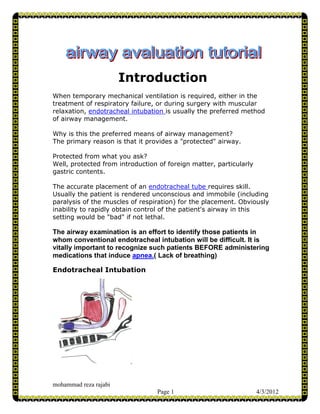
- 1. Introduction When temporary mechanical ventilation is required, either in the treatment of respiratory failure, or during surgery with muscular relaxation, endotracheal intubation is usually the preferred method of airway management. Why is this the preferred means of airway management? The primary reason is that it provides a "protected" airway. Protected from what you ask? Well, protected from introduction of foreign matter, particularly gastric contents. The accurate placement of an endotracheal tube requires skill. Usually the patient is rendered unconscious and immobile (including paralysis of the muscles of respiration) for the placement. Obviously inability to rapidly obtain control of the patient's airway in this setting would be "bad" if not lethal. The airway examination is an effort to identify those patients in whom conventional endotracheal intubation will be difficult. It is vitally important to recognize such patients BEFORE administering medications that induce apnea.( Lack of breathing) Endotracheal Intubation mohammad reza rajabi Page 1 4/3/2012
- 2. Endotracheal Tube Acknowledgements This educational site was developed by Tammy Euliano, MD, Associate Professor of Anesthesiology with the assistance of future doctor Amy Lee programmers Karthik Paladugu and Rick Lockwood graphic artist future doctor Christopher Hurt. Major contributions were provided by Ilona Schmalfuss, MD, Assistant Professor of Radiology Jeremy Melker, MD, Otolaryngology Resident. Funding was provided by the University of Florida College of Medicine Education Committee. mohammad reza rajabi Page 2 4/3/2012
- 3. Aspiration of Gastric Contents The risk of passive reflux of gastric contents into the pharynx is increased when the stomach is full. If the gag reflex has been blunted (by alcohol ingestion, decreased mental status or medications), the acidic volume can make its way into the trachea causing potentially extensive damage. Aspiration Prevention In this case, which of the following could reduce the risk of aspiration and its consequences? Wait 6 hours before proceeding keeping the patient NPO Yes No (yes) Incorrect! - Nil per os While this will help for elective surgery patients, trauma patients and those with acute GI problems will not empty their stomachs well. In addition this operation should not be postponed for any length of time due to the risk of appendix rupture and/or sepsis. (no)Correct! While a 6-hour NPO period is ideal, this surgery should not be postponed. Administration of a "non-particulate" antacid Yes No (yes)Correct ! - Non-particulate antacid Many would advocate having the patient drink 15-30cc sodium citrate or bicitra within 30 minutes of induction of anesthesia. Though this increases the stomach volume, it is actually protective mohammad reza rajabi Page 3 4/3/2012
- 4. as it raises the pH of the stomach contents, reducing injury to the lung in the event of an aspiration. (no)Incorrect ! - Non-particulate antacid Many would advocate having the patient drink 15-30cc sodium citrate or bicitra within 30 minutes of induction of anesthesia. Though this increases the stomach volume, it is actually protective as it raises the pH of the stomach contents, reducing injury to the lung in the event of an aspiration Administration of H2 blockers Yes No (yes)Correct ! - H2 Blockers The onset time of these medications is 30+ minutes, and even then they do not affect the pH of the volume already in the stomach. However, new fluid will be secreted into the stomach at a higher pH, perhaps increasing the overall pH by the time of emergence from anesthesia (the other time at which patients are at risk for aspiration). (no)Incorrect ! - H2 Blockers The onset time of these medications is 30+ minutes, and even then they do not affect the pH of the volume already in the stomach. However, new fluid will be secreted into the stomach at a higher pH, perhaps increasing the overall pH by the time of emergence from anesthesia (the other time at which patients are at risk for aspiration). Administration of metoclopramide Yes No mohammad reza rajabi Page 4 4/3/2012
- 5. (yes)Correct! - Metoclopramide Metoclopramide speeds gastric emptying and increases the lower esophageal sphincter (LES) pressure. While the latter is helpful at reducing the risk of aspiration within minutes, stomach volume reduction takes more time. This emptying should occur, however, and can reduce the risk of aspiration during emergence and extubation at the end of the operation. There are some risks to metoclopramide so, as with everything, a risk:benefit evaluation must be performed. (no)Incorrect! - Metoclopramide Metoclopramide speeds gastric emptying and increases the lower esophageal sphincter (LES) pressure. While the latter is helpful at reducing the risk of aspiration within minutes, stomach volume reduction takes more time. This emptying should occur, however, and can reduce the risk of aspiration during emergence and extubation at the end of the operation. There are some risks to metoclopramide so, as with everything, a risk:benefit evaluation must be performed. Rapid Sequence Induction Yes No (yes)Correct! - Rapid Sequence Induction Following pre-oxygenation, the patient is put to sleep with a rapid acting IV induction agent such as sodium thiopental, immediately followed by succinylcholine (or other rapid-acting agent), application of cricoid pressure , and intubation of the trachea. Positive pressure mask ventilation is not performed to avoid increasing gastric volume. The purpose of this technique is to minimize the duration of impaired gag reflex prior to intubation Cricoid Pressure during intubation Yes No mohammad reza rajabi Page 5 4/3/2012
- 6. (no) Incorrect! - Cricoid Pressure during Intubation An assistant identifies the cricoid ring and applies pressure, compressing the esophagus against the underlying vertebral body. This prevents passive reflux of gastric contents into the lung. How much pressure to apply is a continuing question, current recommendations suggest approximately 10 Newtons (1 kg) of force (mild discomfort for the patient), as the induction medications are being administered. Once the patient loses consciousness, the cricoid pressure should be increased to approximately 30 Newtons (3 kg). It is possible for this pressure to make intubation more difficult and some reduction in force may be necessary. (yes)Correct! - Cricoid Pressure during Intubation An assistant identifies the cricoid ring and applies pressure, compressing the esophagus against the underlying vertebral body. This prevents passive reflux of gastric contents into the lung. How much pressure to apply is a continuing question, current recommendations suggest approximately 10 Newtons (1 kg) of force (mild discomfort for the patient), as the induction medications are being administered. Once the patient loses consciousness, the cricoid pressure should be increased to approximately 30 Newtons (3 kg). It is possible for this pressure to make intubation more difficult and some reduction in force Endotracheal Intubation Intubation is typically performed under direct visualization. That is, by looking through the mouth directly at the vocal cords (direct laryngoscopy), and watching the endotracheal tube pass through the cords and into the trachea. However, there is no direct line-of-sight from the mouth to the vocal cords. Check in a mirror or examine a friend (preferably one who has not eaten onions recently), even with the mouth maximally opened and tongue extended you cannot see the vocal cords, in fact only rarely can you see the epiglottis. mohammad reza rajabi Page 6 4/3/2012
- 7. Mallampati Classification Actually, the amount of the posterior pharynx you can visualize is important and correlates with the difficulty of intubation. Visualization of the pharynx is obscured by a large tongue (relative to the size of the mouth), which also interferes with visualization of the larynx on laryngoscopy. The Mallampati Classification is based on the structures visualized with maximal mouth opening and tongue protrusion in the sitting position (originally described without phonation, but others have suggested minimum Mallampati Classification with or without phonation best correlates with intubation difficulty). mohammad reza rajabi Page 7 4/3/2012
- 8. Class I: soft palate, fauces, uvula, pillars Class II: soft palate, fauces, portion of uvula Class III: soft palate, base of uvula Class IV: hard palate only Other Predictors of Difficult Intubation Obesity – body weight > 110kg Mouth opening – inter-incisor distance < 4cm in an adult Ability to prognath – a large overbite, or the inability to shift the lower incisors in front of the upper incisors Thyromental distance – The distance from the thyroid cartilage to the mentum (tip of the chin) should be > 6.5-7 cm. Mentum-Hyoid distance – Similar to thyromental distance, and should be at least 3-4 finger-breadths. Many other factors have been investigated with variable results. Other factors that may indicate a difficult intubation Sternomental distance – Similar to above, measured from the sternum to the tip of the mandible with the head extended. This measure is influenced by neck extension. Should be >12.5cm. Mandibulohyoid distance – the vertical distance between the mandible and the hyoid bone, determined radiographically. This may be increased with a short mandibular ramus or a caudally located hyoid bone. Such an increase in this distance may be associated with difficult intubation {Chou 1993} Thyrosternal distance – <8cm may suggest difficulty, probably related to the caudally located hyoid as above. mohammad reza rajabi Page 8 4/3/2012
- 9. Various radiographic measurements of the cervical spine, its alignment with airway structures and the atlanto-occipital joint. Positioning To obtain a direct line of sight, the patient is positioned in the "sniffing position." The neck is flexed at the lower cervical spine and extended at the atlanto-occipital joint. This flexion and extension is amplified during laryngoscopy. The patient’s neck mobility should be assessed preoperatively by having them flex and extend their head maximally. The range of motion should be more than 90°. Motion less than 80° may triple the risk of a poor view at laryngoscopy. mohammad reza rajabi Page 9 4/3/2012
- 10. Direct Laryngoscopy Then a laryngoscope is used to pull the lower jaw and tongue up and out of the way. The metal blade is passed into the mouth to the level of the epiglottis, then with an anterior and caudad motion (ie toward the edge of the ceiling across the room) , the lower jaw is elevated, allowing visualization of the glottic structures.( The glottis is the structures of phonation including the vocal cords and surrounding structures.) In most patients this results in a clear view of the larynx and the endotracheal tube is passed through the vocal cords under direct visualization. mohammad reza rajabi Page 10 4/3/2012
- 11. Laryngoscopy Grades In most patients Direct Laryngoscopy results in a clear view of the larynx. The laryngeal view has been classified by Cormack and Lehane as follows: Grade 1: Full view of the glottis Grade 2: Only the posterior commissure is visible Grade 3: Only the epiglottis is seen Grade 4: No epiglottis or glottis structure visible Airway Review What might make Direct Laryngoscopy and Intubation more difficult? Inability to open the mouth Yes No (yes)Correct! There must be room to place the laryngoscope in the mouth…usually at least 3 finger breadths in the adult. (no)Incorrect! There must be room to place the laryngoscope in the mouth…usually at least 3 finger breadths in the adult. mohammad reza rajabi Page 11 4/3/2012
- 12. Inability to extend the neck Yes No (yes)Correct! The "sniffing position" requires significant neck extension. (no)Incorrect! The "sniffing position" requires significant neck extension. Inability to breathe through the nose Yes No (no)Correct! Unless a nasal intubation is planned. (yes)Incorrect! Unless a nasal intubation is planned. Large tongue Yes No (yes)Correct! Also if it is immobile, as from radiation therapy. (no)Incorrect! Also if it is immobile, as from radiation therapy. Redundant pharyngeal tissue Yes No (yes)Correct!! This occurs with obesity, and is often suggested by a history of snoring and/or obstructive sleep apnea. mohammad reza rajabi Page 12 4/3/2012
- 13. (no)Incorrect! This occurs with obesity, and is often suggested by a history of snoring and/or obstructive sleep apnea. Case 2 : Abnormal Exam A healthy 25-year-old man is scheduled to have a shoulder repair requiring general anesthesia. Let's review his airway examination. What would you like the patient to do: Open his mouth as wide as possible Extend his neck as far as possible without pain View from the side mohammad reza rajabi Page 13 4/3/2012
- 14. Open Mouth This patient's mouth opening is 2 finger-breadths, the soft palate is barely visible on maximal mouth opening. Neck mohammad reza rajabi Page 14 4/3/2012
- 15. View from the side 2 finger-breadths fit between the tip of the chin and the neck. Airway Examination Mouth opening Normal Reduced What is mouth opening? (normal)Incorrect it is less than 3 finger breadths. mohammad reza rajabi Page 15 4/3/2012
- 16. (reduced)Correct The mouth opening is less than 3 finger-breadths. Open Mouth The inter-incisor distance on maximal mouth opening. Should be >4 cm in an adult, or 3-4 of the patient's finger-breadths. This patient's mouth opening is 2 finger breadths, the soft palate is barely visible on maximal mouth opening. Mallampati Score I II III IV What is Mallampati Score? mohammad reza rajabi Page 16 4/3/2012
- 17. (I)Incorrect The uvula cannot be seen. (II)Incorrect Not even the top of the uvula is visible. (III)Yes All structures visible up to the soft palate is a Mallampati Class III. (IV)Incorrect The soft palate is visible. Mentum-Hyoid distance Normal Reduced What distance? (normal)Incorrect 3 finger-breadths is normal, this patient has only 2. (reduced)Yes this is less than the normal 3 finger-breadths. View from the side 2 finger-breadths fit between the tip of the chin and the neck. mohammad reza rajabi Page 17 4/3/2012
- 18. Neck Extension Normal Reduced What is neck extension? (normal)Correct The neck extends. (reduced)Incorrect The neck motion is > 90 degrees. Neck The range of motion should be more than 90°. Motion less than 80° may triple the risk of a poor view at laryngoscopy. Airway Evaluation Summary Because of the reduced mentum-hyoid distance, it may be difficult to visualize the larynx with traditional direct laryngoscopy. There are other options, including other blades and techniques that do not require a direct line-of-sight, which are beyond the scope of this site. Perhaps the most conservative method of securing the airway of a patient who is anticipated to have a "difficult airway" is with awake fiberoptic intubation. This technique requires substantial skill, but allows intubation in an awake, spontaneously breathing patient. The trachea is identified with a flexible fiberscope, and then the endotracheal tube is advanced over the fiberscope like a stylet. Such mohammad reza rajabi Page 18 4/3/2012
- 19. a procedure requires blockade of the sensory innervation to the airway, and blunting of the gag reflex. Innervation of the Upper Airway Awake fiberoptic intubation requires topical anesthesia for patient comfort, as well as to blunt the gag reflex that would prevent successful intubation of the trachea. Several nerves are involved in the sensation of the upper airway: Anterior 2/3 of the tongue - Trigeminal nerve (V). Posterior 1/3 of tongue to epiglottis - Glossopharyngeal nerve (IX; afferent limb of gag reflex). Epiglottis to vocal cords - Internal branch of Superior Laryngeal Nerve (Vagus, X) mohammad reza rajabi Page 19 4/3/2012
- 20. Trachea below vocal cords - Recurrent Laryngeal Nerve (Vagus, X) MOTOR INNERVATION Motor innervation to the larynx is provided by the Vagus Nerve, but recall there are two branches involved. The Recurrent Laryngeal Nerve innervates all the muscles of the larynx EXCEPT the cricothyroid muscle, which is innervated by the External Branch of the Superior Laryngeal Nerve. Because the function of the cricothyroid muscle is to stretch and tense the vocal cords, unopposed action of the cricothyroid, as may occur with bilateral destruction of the recurrent laryngeal nerves, would lead to stridor, respiratory distress and possibly airway obstruction. GAG REFLEX So the sensory, afferent limb of the gag reflex is the glossopharyngeal nerve (IX), while the motor, efferent limb is the Vagus (X). It's not much of a mnemonic, but I remember this as a variant of TGIF: "Thank God it's Recurrent" I know, it's lame, perhaps just lame enough to be memorable! Airway Blocks Topical application of local anesthetics is usually sufficient for the tongue and oro/nasopharynx, though glossopharyngeal blocks are performed occasionally. Blunting of the gag reflex requires Transtracheal (really translaryngeal) with or without bilateral Superior Laryngeal Nerve blocks as shown below. mohammad reza rajabi Page 20 4/3/2012
- 21. The superior laryngeal nerves are blocked by deposition of 1% lidocaine near where the nerves penetrate the thyrohyoid membrane. The transtracheal block is accomplished with 4% lidocaine injected directly into the tracheal lumen. Often this block alone, coupled with nebulized or atomized lidocaine is sufficient for awake intubation. mohammad reza rajabi Page 21 4/3/2012
- 22. Airway Structures The right panel displays images seen during fiberoptic bronchoscopy. The corresponding level on CT is displayed on the middle panel. Place the cursor over structures to learn their identity. mohammad reza rajabi Page 22 4/3/2012
- 23. mohammad reza rajabi Page 23 4/3/2012
- 24. Review of Airway Innervation Let's review the innervation of the upper airway: mohammad reza rajabi Page 24 4/3/2012
- 25. Purple Facial (VII) Trigeminal (V) Glossopharyngeal (IX) Vagus (X) (VII)No The Facial Nerve supplies only taste to the tongue. (V)Yes The maxillary branch (V2) supplies the nasal cavity and palate, while the mandibular branch (V3) supplies the anterior 2/3 of the tongue. (IX)Incorrect The glossopharyngeal nerve supplies sensation to the posterior 1/3 of the tongue and its overlying structures including the soft palate. (X)No The vagus innervates the airway further distal. Green Facial (VII) Trigeminal (V) Glossopharyngeal (IX) Vagus (X) (VII)No The Facial Nerve supplies only taste to the tongue. (IX)Yes The glossopharyngeal nerve supplies sensation to the posterior 1/3 of the tongue and it's overlying structures including the soft palate. (V)No The maxillary branch (V2) supplies the nasal cavity and palate, while the mandibular branch (V3) supplies the anterior 2/3 of the tongue. (X)No The vagus innervates the airway further distal. mohammad reza rajabi Page 25 4/3/2012
- 26. Blue Facial (VII) Trigeminal (V) Glossopharyngeal (IX) Vagus (X) (VII)No The Facial Nerve supplies only taste to the tongue. (IX)No The glossopharyngeal nerve supplies sensation to the posterior 1/3 of the tongue and it's overlying structures including the soft palate. (V)No The maxillary branch (V2) supplies the nasal cavity and palate, while the mandibular branch (V3) supplies the anterior 2/3 of the tongue. (x)Yes, but which branch Internal branch of superior laryngeal External branch of superior laryngeal Recurrent laryngeal (Yes The Internal Branch of the Superior Laryngeal Nerve provides sensory innervation to the mucous membrane from the epiglottis to and including the vocal cords. Incorrect The External Branch of the Superior Laryngeal nerve provides motor innervation to the cricothyroid muscle only. Incorrect The Recurrent Laryngeal Nerve supplies sensory innervation to the trachea below the vocal cords, as well as motor innervation to all the intrinsic muscles of the larynx except the cricothyroid muscle.. ) mohammad reza rajabi Page 26 4/3/2012
- 27. Red Facial (VII) Trigeminal (V) Glossopharyngeal (IX) Vagus (X) (VII)No The Facial Nerve supplies only taste to the tongue. (IX)No The glossopharyngeal nerve supplies sensation to the posterior 1/3 of the tongue and it's overlying structures including the soft palate. (V)No The maxillary branch (V2) supplies the nasal cavity and palate, while the mandibular branch (V3) supplies the anterior 2/3 of the tongue. (x)Yes, but which branch Internal branch of superior laryngeal External branch of superior laryngeal Recurrent laryngeal (Incorrect The Internal Branch of the Superior Laryngeal Nerve provides sensory innervation to the mucous membrane from the epiglottis to and including the vocal cords. Incorrect The External Branch of the Superior Laryngeal nerve provides motor innervation to the cricothyroid muscle only. Yes The Recurrent Laryngeal Nerve supplies sensory innervation to the trachea below the vocal cords, as well as motor innervation to all the intrinsic muscles of the larynx except the cricothyroid muscle..) mohammad reza rajabi Page 27 4/3/2012
- 28. Case 3: Spine Evaluation A previously healthy 40-year-old male presents with an open femur fracture from a Motor Vehicle Accident (MVA) that needs to be repaired under general anesthesia. He is currently on a backboard with a cervical collar in place and is hemodynamically stable. Examination of this patient's airway is complicated by the presence of the cervical collar, which both inhibits mouth opening and by definition prevents neck extension. As you have seen above, neck extension is required for direct laryngoscopy. So what shall we do? Remove the neck collar and intubate as usual. Intubate with a technique that does not require neck movement. Avoid general anesthesia and perform a regional block for the procedure. Perform studies to "clear" the cervical spine. First a basic review of the anatomy is helpful. Recall that the cervical spine consists of 7 vertebrae, the first two of which are highly specialized. (Should this patient have an unstable cervical spine, the movement resulting from laryngoscopy could permanently damage the spinal cord, likely resulting in quadriplegia.) (There are numerous techniques (retrograde intubation,…) purported to involve less cervical spine motion, each of which requires substantial skill and experience. These should only be attempted by experienced practitioners. Some advocate "in-line stabilization" where a second person attempts to hold the cervical spine still while the primary person attempts direct laryngoscopy. This technique makes intubation more difficult, and is inadequate for stabilization.) mohammad reza rajabi Page 28 4/3/2012
- 29. (While an attractive option, many would argue that anytime a regional anesthetic is planned, immediate endotracheal intubation must be possible. Complications may occur during the regional block, or it may be inadequate for the operation, or wear off before the surgeons are done. Therefore, inability to emergently intubate a patient is a relative contraindication to regional anesthesia and should be considered in this patient with a possible unstable neck.) (Great idea!) Cervical Spine Anatomy-Atlas C1: The Atlas is a ring that interacts with the skull base above and C2 shown on next page. It is unique in that it lacks a vertebral body and spinous process. The articulation of C1 with the occiput is very tight, providing little of the flexion of the cervical spine and only about 20 degrees of extension. mohammad reza rajabi Page 29 4/3/2012
- 30. Cervical Spine Anatomy-Axis C2: The Axis has an unusual thumb-like extension of its vertebral body that passes through the arch of C1. This process is called the dens or odontoid. The odontoid process is normally held very tightly against the anterior arch of C1 by the transverse ligament. Meanwhile the spinal cord travels behind the odontoid within the arch of C1. mohammad reza rajabi Page 30 4/3/2012
- 31. Atlanto Axial Joint This atlanto-axial joint provides the majority of the rotational motion of the cervical spine. Meanwhile flexion and extension are primarily accomplished at C2 and below, and particularly between C4 and C6. Neck Movement with DL What happens to the neck during direct laryngoscopy and intubation? As you have seen, the sniffing position involves neck flexion in the lower cervical spine with extension superiorly. In the process of direct laryngoscopy this motion is accentuated. As the laryngoscope is lifted upward, the occiput is extended primarily at the atlanto- occipital joint (occiput-C1), while flexion occurs at C2-3 and below. Therefore, any intervention that impedes this flexion and extension will make visualization of the glottis more difficult. In someone with a cervical fusion up to the occiput it is pretty much impossible to perform direct laryngoscopy. Similarly, a patient with external stabilization such as a c-collar in this case will (SHOULD) have neck movement reduced sufficient to make visualization difficult if not impossible. mohammad reza rajabi Page 31 4/3/2012
- 32. Clearing the C-Spine How does one rule out damage to the cervical spine? At present history is our greatest ally. If the healthy patient has no history of neck problems and no symptoms on maximal flexion and extension, they are unlikely to have cervical spine disease. On the other hand there are many patients whose cervical spine SHOULD be radiographically evaluated pre-operatively including certain trauma patients, as well as those with disease states that affect the cervical spine including rheumatoid arthritis and Down's Syndrome. These diseases may affect the transverse ligament and thus the stability at the atlanto-axial joint. Nexus Criteria Which trauma patients require cervical spine films prior to surgery or intubation? There is a set of criteria identified by the National Emergency X- Radiography Utilization Study (NEXUS) that attempt to identify patients with a low probability of injury, thereby reducing the number of negative cervical spine radiographs taken. The criteria include No midline cervical tenderness No focal neurologic deficit Normal alertness No intoxication No painful, distracting injury that might make them ignore their neck pain For those patients whose cervical spine is not cleared, the anesthesiologist must consider the risks of cervical spine damage that can be worsened through direct laryngoscopy, versus the risk of alternative techniques that may minimize neck motion, including awake fiberoptic intubation. A description of these alternate techniques is beyond the scope of this site at present. mohammad reza rajabi Page 32 4/3/2012
- 33. Spine Film For the current case the following film is obtained. Patient's Film Normal for Comparison mohammad reza rajabi Page 33 4/3/2012
- 34. Explanation Note the large step-off between C6 and C7. This subluxation causes entrapment of the spinal cord and damage. Therefore this patient requires an intubation technique with minimal neck motion and awake positioning,as well as some external stabilization or operative intervention to prevent damage to the spinal cord at the neck. mohammad reza rajabi Page 34 4/3/2012
- 35. C-Spine Review So which patients are at higher risk for neck injury during intubation? Trauma patients Yes No (yes)Correct! They may have trauma to the cervical spine as well. (no)Incorrect! They may have trauma to the cervical spine as well. Rheumatoid arthritis patients Yes No (yes)Correct! Approximately 30% of patients with severe disease will have some instability at C1-C2. All should have periodic flexion or extension xrays, particularly prior to surgery. (no)Incorrect! Approximately 30% of patients with severe disease will have some instability at C1-C2. All should have periodic flexion or extension xrays, particularly prior to surgery. Down's Syndrome patients Yes No (yes)Correct! About 15% of these patients have laxity in the transverse ligament that holds the odontoid against the anterior arch of C1. Xrays are also recommended in these patients prior to anticipated neck manipulation including laryngoscopy. mohammad reza rajabi Page 35 4/3/2012
- 36. (no)Incorrect! About 15% of these patients have laxity in the transverse ligament that holds the odontoid against the anterior arch of C1. Xrays are also recommended in these patients prior to anticipated neck manipulation including laryngoscopy. Osteoarthritic patients Yes No (yes)Incorrect They are not at higher risk. (no)Correct They are not at higher risk Patient with a prior cervical spine fusion Yes No (yes)Incorrect Assuming the repair is stable and there is no further disease there is little risk of damage. Such patients may be difficult to intubate, though, if their mobility is significantly limited. (no)Correct Assuming the repair is stable and there is no further disease there is little risk of damage. Such patients may be difficult to intubate, though, if their mobility is significantly limited. mohammad reza rajabi Page 36 4/3/2012
- 37. Airway References Cricoid Pressure Vanner RG, Asai T. Safe use of cricoid pressure. Anaesthesia 1999: 54: 1-3. A review of literature with recommendations. Sellick BA. Cricoid pressure to control regurgitation of stomach contents during induction of anaesthesia. Lancet 1961; 2: 404-6. The original description. Views and Grades Mallampati SR, Gatt SP, et al. A clinical sign to predict difficult tracheal intubation: a prospective study. Can Anaesth Soc J 1985;32(4):429-434. The original paper describing the classification system, but only 3 grades (III and IV combined). Samsoon GLT and Young JRB. Difficult tracheal intubation: a retrospective study. Anaesthesia 1987;42:487-490. Describes the addition of Mallampati class 4 Cormack RS and Lehane J. Difficult tracheal intubation in obstetrics. Anaesthesia 1984;39:1105-1111. Describes the laryngoscopy grades and correlates with difficult intubation. Also proposes a technique of attempting to intubate while intentionally achieving a suboptimal (Class III) view. Studies of Predictive Indices There are many studies, some which counter others. One difficulty is defining a difficult airway. Most use a Cormack-Lehane laryngoscopy grade of III-IV. Some investigate specific radiographic measurements that are impractical in daily clinical practice. Below are a few useful references: El-Ganzouri AR, McCarthy RJ, et al. Preoperative airway assessment: Predictive value of a multivariate risk index. Anesth Analg 1996;82:1197-1204. A logistic regression comparing examination tests and developing a risk index. Chou HC, Wu TL, et al. Mandibulohyoid distance in difficult laryngoscopy. Br J Anaesth 1993; 71:335-339. A single article sighting this distance as an important factor in an analysis of only 11 patients. Frerk CM. Predicting difficult intubation. Anaesthesia 1991;46:1005-1008. A study suggesting that a Mallampati Class III or IV with thyromental distance of <7cm is sensitive and specific for difficult intubation (laryngoscopy grade 3 or 4). C-Spine Evaluation mohammad reza rajabi Page 37 4/3/2012
- 38. Hoffman JR, Mower WR, et al. Validity of a set of clinical criteria to rule out injury to the cervical spine in patients with blunt trauma. N Engl J Med 2000;343:94- 99. Application of the NEXUS criteria. mohammad reza rajabi Page 38 4/3/2012