SlideShare ist ein Scribd-Unternehmen logo
1 von 24
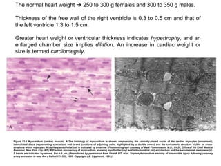
Figure 12-1 Myocardium (cardiac muscle).  A  The histology of myocardium is shown, emphasizing the centrally-placed nuclei of the cardiac myocytes (arrowhead), intercalated discs (representing specialized end-to-end junctions of adjoining cells; highlighted by a double arrow) and the sarcomeric structure visible as cross-striations within myocytes. A capillary endothelial cell is indicated by an arrow.  (Photomicrograph courtesy of Mark Flomenbaum, M.D., Ph.D., Office of the Chief Medical Examiner, New York City, NY.) B  Electron microscopy of myocardium, showing myofibrillar (my) and mitochondrial (mi) architecture and the sarcolemmal membrane (s). Z bands are indicated by arrows. Bar = 1 ÎŒm.  (Reproduced by permission from Vivaldi MT, et al. Triphenyltetrazolium staining of irreversible injury following coronary artery occlusion in rats. Am J Pathol 121:522, 1985. Copyright J.B. Lippincott, 1985.) The normal heart weight    250 to 300 g females and 300 to 350 g males.  Thickness of the free wall of the right ventricle is 0.3 to 0.5 cm and that of the left ventricle 1.3 to 1.5 cm.  Greater heart weight or ventricular thickness indicates  hypertrophy,  and an enlarged chamber size implies  dilation . An increase in cardiac weight or size is termed  cardiomegaly .
Figure 12-2 Aortic valve histology, shown as a low-magnification photomicrograph of cuspal cross-section in the systolic (nondistended) state, emphasizing three major layers (ventricularis [v], spongiosa [s], and fibrosa [f]). Superficial endothelial cells  (arrow)  and diffusely distributed deep interstitial cells are noted. The strength of the valve is predominantlyderived from the fibrosa, with its dense collagen  (yellow) . This section highlights the dense, laminated elastic tissue in the ventricularis  (double arrow) . The outflow surface is at top.  (Reproduced by permission from Schoen FJ: Aortic valve structure-function correlations: Role of elastic fibers no longer a stretch of the imagination. J Heart Valve Dis 6:1, 1997.)
TABLE 12-1  -- Changes in the Aging Heart Increased cross-sectional luminal area Tortuosity Epicardial Coronary Arteries Lambl excrescences Buckling of mitral leaflets toward the left atrium Fibrous thickening of leaflets Mitral valve annular calcific deposits Aortic valve calcific deposits Valves Sigmoid-shaped ventricular septum Decreased left ventricular cavity size Increased left atrial cavity size Chambers
Atherosclerotic plaque Elastic fragmentation and collagen accumulation Sinotubular junction calcific deposits Elongated (tortuous) thoracic aorta Dilated ascending aorta with rightward shift Aorta Amyloid deposits Basophilic degeneration Lipofuscin deposition Brown atrophy Increased subepicardial fat Increased mass Myocardium Atherosclerotic plaque Calcific deposits
‱  Failure of the pump .  In the most common circumstance, the cardiac muscle contracts weakly or inadequately, and the chambers cannot empty properly. In some conditions, however, the muscle cannot relax sufficiently to permit ventricular filling. ‱  An obstruction to flow ,  owing to a lesion preventing valve opening or otherwise causing increased ventricular chamber pressure (e.g., aortic valvular stenosis, systemic hypertension, or aortic coarctation). The increased pressure overworks the chamber that pumps against the obstruction. ‱  Regurgitant flow   causes some of the output from each contraction to flow backward, adding a volume workload to each of the chambers, which must pump the extra blood (e.g., left ventricle in aortic regurgitation; left atrium and left ventricle in mitral regurgitation). ‱  Disorders of cardiac conduction .  Heart block or arrhythmias owing to uncoordinated generation of impulses (e.g., atrial or ventricular fibrillation) lead to nonuniform and inefficient contractions of the muscular walls. ‱  Disruption of the continuity of the circulatory system   that permits blood to escape (e.g., gunshot wound through the thoracic aorta).
‱  The Frank-Starling mechanism , in which the increased preload of dilation (thereby increasing cross-bridges within the sarcomeres) helps to sustain cardiac performance by enhancing contractility ‱  Myocardial structural changes, including augmented muscle mass (hypertrophy)  with or without cardiac chamber dilation , in which the mass of contractile tissue is augmented ‱  Activation of neurohumoral systems , especially (1) release of the neurotransmitter norepinephrine by adrenergic cardiac nerves (which increases heart rate and augments myocardial contractility and vascular resistance), (2) activation of the renin-angiotensin-aldosterone system, and (3) release of atrial natriuretic peptide.
Figure 12-3 Left ventricular hypertrophy.  A,  Pressure hypertrophy due to left ventricular outflow obstruction. The left ventricle is on the lower right in this apical four-chamber view of the heart.  B,  Altered cardiac configuration in left ventricular hypertrophy without and with dilation, viewed in transverse heart sections. Compared with a normal heart ( center ), the pressurehypertrophied hearts ( left  and in  A ) have increased mass and a thick left ventricular wall, but the hypertrophied and dilated heart  (right)  has increased mass but a normal wall thickness.  (Reproduced by permission from Edwards WD: Cardiac anatomy and examination of cardiac specimens. In Emmanouilides GC, Riemenschneider TA, Allen HD, Gutgesell HP (eds): Moss and Adams Heart Disease in Infants, Children, and Adolescents: Including the Fetus and Young Adults, 5th ed. Philadelphia, Williams and Wilkins, 1995, p. 86.)
Figure 12-4 Schematic representation of the sequence of events in cardiac hypertrophy and its progression to heart failure, emphasizing cellular and extracellular changes.
The most frequent causes of the major functional valvular lesions are as follows: ‱  Aortic stenosis : calcification of anatomically normal and congenitally bicuspid aortic valves ‱  Aortic insufficiency : dilation of the ascending aorta, related to hypertension and aging. ‱  Mitral stenosis:  rheumatic heart disease ‱  Mitral insufficiency:  myxomatous degeneration (mitral valve prolapse)
Figure 12-22 Calcific valvular degeneration.  A,  Calcific aortic stenosis of a previously normal valve having three cusps (viewed from aortic aspect). Nodular masses of calcium are heapedup within the sinuses of Valsalva  (arrow) . Note that the commissures are not fused, as in postrheumatic aortic valve stenosis (see Fig. 12-24 E  ).  B,  Calcific aortic stenosis occurring on a congenitally bicuspid valve. One cusp has a partial fusion at its center, called a  raphe  (arrow).  C and D,  Mitral annular calcification, with calcific nodules at the base (attachment margin) of the anterior mitral leaflet  (arrows) .  C,  Left atrial view.  D,  Cut section of myocardium.
Figure 12-23 Myxomatous degeneration of the mitral valve.  A,  Long axis of left ventricle demonstrating hooding with prolapse of the posterior mitral leaflet into the left atrium  (arrow) . The left ventricle is on right in this apical four-chamber view.  (Courtesy of William D. Edwards, M.D., Mayo Clinic, Rochester, MN.) B,  Opened valve, showing pronounced hooding of the posterior mitral leaflet with thrombotic plaques at sites of leaflet-left atrium contact  (arrows). C , Opened valve with pronounced hooding from patient who died suddenly  (double arrows) . Note also mitral annular calcification  (arrowhead) .
Figure 12-12 Schematic representation of sequential progression of coronary artery lesion morphology, beginning with stable chronic plaque responsible for typical angina and leading to the various acute coronary syndromes.  (Modified and redrawn from Schoen FJ: Interventional and Surgical Cardiovascular Pathology: Clinical Correlations and Basic Principles. Philadelphia, W.B. Saunders Co., 1989, p. 63.)
Figure 12-11 Atherosclerotic plaque rupture.  A,  Plaque rupture without superimposed thrombus, in patient who died suddenly.  B,  Acute coronary thrombosis superimposed on an atherosclerotic plaque with focal disruption of the fibrous cap, triggering fatal myocardial infarction.  C,  Massive plaque rupture with superimposed thrombus, also triggering a fatal myocardial infarction (special stain highlighting fibrin in red). In both  A  and  B,  an arrow points to the site of plaque rupture.  (B, reproduced from Schoen FJ: Interventional and Surgical Cardiovascular Pathology: Clinical Correlations and Basic Principles. Philadelphia, W.B. Saunders, 1989, p. 61.)
Figure 12-13 Postmortem angiogram showing the posterior aspect of the heart of a patient who died during the evolution of acute myocardial infarction, demonstrating total occlusion of the distal right coronary artery by an acute thrombus  (arrow)  and a large zone of myocardial hypoperfusion involving the posterior left and right ventricles, as indicated by arrowheads, and having almost absent filling of capillaries, that is, less white. The heart has been fixed by coronary arterial perfusion with glutaraldehyde and cleared with methyl salicylate, followed by intracoronary injection of silicone polymer.  Photograph courtesy of Lewis L. Lainey. (Reproduced by permission from Schoen FJ: Interventional and Surgical Cardiovascular Pathology: Clinical Correlations and Basic Principles. Philadelphia, WB Saunders, 1989, p. 60.) TABLE 12-4 -- Approximate Time of Onset of Key Events in Ischemic Cardiac Myocytes ATP, adenosine triphosphate. >1 hr Microvascular injury 20–40 min Irreversible cell injury 40 min ‱‱ to 10% of normal 10 min ‱‱ to 50% of normal ATP reduced <2 min Loss of contractility Seconds Onset of ATP depletion Time Feature
Figure 12-14 Schematic representation of the progression of myocardial necrosis after coronary artery occlusion. Necrosis begins in a small zone of the myocardium beneath the endocardial surface in the center of the ischemic zone. This entire region of myocardium  (shaded)  depends on the occluded vessel for perfusion and is the area at risk. Note that a very narrow zone of myocardium immediately beneath the endocardium is spared from necrosis because it can be oxygenated by diffusion from the ventricle. The end result of the obstruction to blood flow is necrosis of the muscle that was dependent on perfusion from the coronary artery obstructed. Nearly the entire area at risk loses viability. The process is called  myocardial infarction,  and the region of necrotic muscle is a  myocardial infarct .
 
Figure 12-15 Acute myocardial infarct, predominantly of the posterolateral left ventricle, demonstrated histochemically by a lack of staining by the triphenyltetrazolium chloride (TTC) stain in areas of necrosis  (arrow) . The staining defect is due to the enzyme leakage that follows cell death. Note the myocardial hemorrhage at one edge of the infarct that was associated with cardiac rupture, and the anterior scar  (arrowhead) , indicative of old infarct. (Specimen the oriented with the posterior wall at the top.)
Figure 12-16 Microscopic features of myocardial infarction and its repair.  A,  One-day-old infarct showing coagulative necrosis along with wavy fibers (elongated and narrow), compared with adjacent normal fibers (at right). Widened spaces between the dead fibers contain edema fluid and scattered neutrophils.  B,  Dense polymorphonuclear leukocytic infiltrate in area of acute myocardial infarction of 3 to 4 days' duration.  C,  Nearly complete removal of necrotic myocytes by phagocytosis (approximately 7 to 10 days).  D,  Granulation tissue characterized by loose collagen and abundant capillaries.  E,  Well-healed myocardial infarct with replacement of the necrotic fibers by dense collagenous scar. A few residual cardiac muscle cells are present.
Grupo 1    Arritmias Grupo 2    Factores congĂ©nitos
Figure 12-5 Cardiac defects related to neural crest abnormalities.  A,  Biologic pathways for cardiac neural crest-related defects.  B,  Disease phenotypes. DORV, double-outlet right ventricle; TGA, transposition of the great arteries.  (Reproduced by permission from Chien KR: Genomic circuits and the integrative biology of cardiac diseases. Nature 407:227, 2000.)
Figure 12-6 Schematic diagram of congenital left-to-right shunts.  A,  Atrial septal defect (ASD).  B,  Ventricular septal defect (VSD). With VSD the shunt is left-to-right, and the pressures are the same in both ventricles. Pressure hypertrophy of the right ventricle and volume hypertrophy of the left ventricle are generally present.  C,  Patent ductus arteriosus (PDA).  D,  Atrioventricular septal defect (AVSD).  E,  Large VSD with irreversible pulmonary hypertension. The shunt is right-to-left (shunt reversal). Volume hypertrophy and pressure hypertrophy of the right ventricle are present. Arrow indicates the direction of blood flow. The right ventricular pressure is now sufficient to yield a right-to-left shunt (Ao, aorta; LA, left atrium; LV, left ventricle; PT, pulmonary trunk; RA, right atrium; RV, right ventricle.)
Figure 12-7 Gross photograph of a ventricular septal defect (membranous type); defect denoted by arrow.  (Courtesy of William D. Edwards, M.D., Mayo Clinic, Rochester, MN.)
Figure 12-8 Schematic diagram of the most important right-to-left shunts  (cyanotic congenital heart disease) .  A,  Tetralogy of Fallot. Diagrammatic representation of anatomic variants, indicating that the direction of shunting across the VSD depends on the severity of the subpulmonary stenosis. Arrows indicate the direction of the blood flow.  B,  Transposition of the great vessels with and without VSD. (Ao, aorta; LA, left atrium; LV, left ventricle; PT, pulmonary trunk; RA, right atrium; RV, right ventricle.)  (Courtesy of William D. Edwards, M.D., Mayo Clinic, Rochester, MN.)
Figure 12-9 Transposition of the great arteries.  (Courtesy of William D. Edwards, M.D., Mayo Clinic, Rochester, MN.) Figure 12-10 Diagram showing coarctation of the aorta with and without PDA. (Ao, aorta; LA, left atrium; LV, left ventricle; PT, pulmonary trunk; RA, right atrium; RV, right ventricle; PDA, persistent ductus arteriosus.)  (Courtesy of William D. Edwards, M.D., Mayo Clinic, Rochester, MN.)

Weitere Àhnliche Inhalte

Was ist angesagt?

Regulaion of Respiration (presentation)
Regulaion of Respiration (presentation)Regulaion of Respiration (presentation)
Regulaion of Respiration (presentation)Roshan Timilsina
 
Hemodynamic Disorders
Hemodynamic DisordersHemodynamic Disorders
Hemodynamic Disordersmvraveendrambbs
 
Respiratory Physiology
Respiratory PhysiologyRespiratory Physiology
Respiratory PhysiologyDang Thanh Tuan
 
Renal blood flow (The Guyton and Hall physiology)
Renal blood flow (The Guyton and Hall physiology)Renal blood flow (The Guyton and Hall physiology)
Renal blood flow (The Guyton and Hall physiology)Maryam Fida
 
Edema and-effusions
Edema and-effusionsEdema and-effusions
Edema and-effusionsAshim Gyawali
 
Nursing Bulletin Respiratory System
Nursing Bulletin Respiratory SystemNursing Bulletin Respiratory System
Nursing Bulletin Respiratory Systemseigfredo origenes
 
blood pressure.pptx
blood pressure.pptxblood pressure.pptx
blood pressure.pptxdeepamanandhar1
 
Anatomy & Physiology Lecture Notes - The senses
Anatomy & Physiology Lecture Notes - The sensesAnatomy & Physiology Lecture Notes - The senses
Anatomy & Physiology Lecture Notes - The sensesAmmedicine Medicine
 
Pulmonary circulation
Pulmonary circulationPulmonary circulation
Pulmonary circulationUphar Gupta
 
Cardiac muscle (The Guyton and Hall Physiology)
Cardiac muscle (The Guyton and Hall Physiology)Cardiac muscle (The Guyton and Hall Physiology)
Cardiac muscle (The Guyton and Hall Physiology)Maryam Fida
 
Hemodynamic Disorders, Thromboembolic Disease & SHOCK
Hemodynamic Disorders, Thromboembolic Disease & SHOCKHemodynamic Disorders, Thromboembolic Disease & SHOCK
Hemodynamic Disorders, Thromboembolic Disease & SHOCKDr. Roopam Jain
 
Respiratory system
Respiratory systemRespiratory system
Respiratory systemAHMED ALBAYATI
 
cardiac physiology
cardiac physiologycardiac physiology
cardiac physiologyHETA PATEL
 
Splanchnic circulation
Splanchnic circulationSplanchnic circulation
Splanchnic circulationSalsabil A.
 
Anatomy and physiology RESPIRATORY system
Anatomy and physiology  RESPIRATORY system Anatomy and physiology  RESPIRATORY system
Anatomy and physiology RESPIRATORY system Ayman Nasr
 
Cardiovascular system
Cardiovascular systemCardiovascular system
Cardiovascular systemNisha Mhaske
 
Central nervous system
Central nervous systemCentral nervous system
Central nervous systemShailesh Jaiswal
 

Was ist angesagt? (20)

Regulaion of Respiration (presentation)
Regulaion of Respiration (presentation)Regulaion of Respiration (presentation)
Regulaion of Respiration (presentation)
 
Renal system
Renal systemRenal system
Renal system
 
Hemodynamic Disorders
Hemodynamic DisordersHemodynamic Disorders
Hemodynamic Disorders
 
Respiratory Physiology
Respiratory PhysiologyRespiratory Physiology
Respiratory Physiology
 
Renal blood flow (The Guyton and Hall physiology)
Renal blood flow (The Guyton and Hall physiology)Renal blood flow (The Guyton and Hall physiology)
Renal blood flow (The Guyton and Hall physiology)
 
Edema and-effusions
Edema and-effusionsEdema and-effusions
Edema and-effusions
 
Circulation..
Circulation..Circulation..
Circulation..
 
Nursing Bulletin Respiratory System
Nursing Bulletin Respiratory SystemNursing Bulletin Respiratory System
Nursing Bulletin Respiratory System
 
blood pressure.pptx
blood pressure.pptxblood pressure.pptx
blood pressure.pptx
 
Anatomy & Physiology Lecture Notes - The senses
Anatomy & Physiology Lecture Notes - The sensesAnatomy & Physiology Lecture Notes - The senses
Anatomy & Physiology Lecture Notes - The senses
 
Pulmonary circulation
Pulmonary circulationPulmonary circulation
Pulmonary circulation
 
Cardiac muscle (The Guyton and Hall Physiology)
Cardiac muscle (The Guyton and Hall Physiology)Cardiac muscle (The Guyton and Hall Physiology)
Cardiac muscle (The Guyton and Hall Physiology)
 
Hemodynamic Disorders, Thromboembolic Disease & SHOCK
Hemodynamic Disorders, Thromboembolic Disease & SHOCKHemodynamic Disorders, Thromboembolic Disease & SHOCK
Hemodynamic Disorders, Thromboembolic Disease & SHOCK
 
Respiratory system
Respiratory systemRespiratory system
Respiratory system
 
cardiac physiology
cardiac physiologycardiac physiology
cardiac physiology
 
Splanchnic circulation
Splanchnic circulationSplanchnic circulation
Splanchnic circulation
 
Anatomy and physiology RESPIRATORY system
Anatomy and physiology  RESPIRATORY system Anatomy and physiology  RESPIRATORY system
Anatomy and physiology RESPIRATORY system
 
Gastrointestinal physiology
Gastrointestinal physiologyGastrointestinal physiology
Gastrointestinal physiology
 
Cardiovascular system
Cardiovascular systemCardiovascular system
Cardiovascular system
 
Central nervous system
Central nervous systemCentral nervous system
Central nervous system
 

Ähnlich wie 05 Cat. Cardiovascular I

Mitral valve disease
Mitral valve disease Mitral valve disease
Mitral valve disease RiyadhWaheed
 
23204940
2320494023204940
23204940radgirl
 
Cardiovascular System Pathology 2014 edited by @jennings argwing
Cardiovascular System Pathology 2014 edited by @jennings argwingCardiovascular System Pathology 2014 edited by @jennings argwing
Cardiovascular System Pathology 2014 edited by @jennings argwingJennings Agingu jenningsadd@gmail.com
 
Congenital heart diseases2020
Congenital heart diseases2020Congenital heart diseases2020
Congenital heart diseases2020RiyadhWaheed
 
23204931
2320493123204931
23204931radgirl
 
Anatomy of cardiovascular system
Anatomy of cardiovascular systemAnatomy of cardiovascular system
Anatomy of cardiovascular systembutterflyfairy
 
06 Cat. Cardiovascular II
06 Cat. Cardiovascular II06 Cat. Cardiovascular II
06 Cat. Cardiovascular IIunab.patologia
 
CARDIOMYOPATHY.pptx
CARDIOMYOPATHY.pptxCARDIOMYOPATHY.pptx
CARDIOMYOPATHY.pptxZellanienhd
 
Acquired heart disease II
Acquired heart disease IIAcquired heart disease II
Acquired heart disease IIabeerahameed1
 
Hemodynamic Disorders
Hemodynamic DisordersHemodynamic Disorders
Hemodynamic Disordersmvraveendrambbs
 
Mital regurge
Mital regurgeMital regurge
Mital regurgeRiyadhWaheed
 
Mcq in cardiology 2015 magdi sasi
Mcq  in cardiology  2015  magdi  sasiMcq  in cardiology  2015  magdi  sasi
Mcq in cardiology 2015 magdi sasicardilogy
 
Tumores cardiacos
Tumores cardiacosTumores cardiacos
Tumores cardiacosAbraham Flores
 
23204910
2320491023204910
23204910radgirl
 
Emergency cadiopulmonary imaging
Emergency cadiopulmonary imagingEmergency cadiopulmonary imaging
Emergency cadiopulmonary imagingAmir Mahmoud
 
Echo and CAD-2.pptx
Echo and CAD-2.pptxEcho and CAD-2.pptx
Echo and CAD-2.pptxAnayaAnaya14
 

Ähnlich wie 05 Cat. Cardiovascular I (20)

Mitral valve disease
Mitral valve disease Mitral valve disease
Mitral valve disease
 
23204940
2320494023204940
23204940
 
Pericarditis constrictiva
Pericarditis constrictivaPericarditis constrictiva
Pericarditis constrictiva
 
Cardiovascular System Pathology 2014 edited by @jennings argwing
Cardiovascular System Pathology 2014 edited by @jennings argwingCardiovascular System Pathology 2014 edited by @jennings argwing
Cardiovascular System Pathology 2014 edited by @jennings argwing
 
Congenital heart diseases2020
Congenital heart diseases2020Congenital heart diseases2020
Congenital heart diseases2020
 
23204931
2320493123204931
23204931
 
Anatomy of cardiovascular system
Anatomy of cardiovascular systemAnatomy of cardiovascular system
Anatomy of cardiovascular system
 
06 Cat. Cardiovascular II
06 Cat. Cardiovascular II06 Cat. Cardiovascular II
06 Cat. Cardiovascular II
 
CARDIOMYOPATHY.pptx
CARDIOMYOPATHY.pptxCARDIOMYOPATHY.pptx
CARDIOMYOPATHY.pptx
 
Acquired heart disease II
Acquired heart disease IIAcquired heart disease II
Acquired heart disease II
 
Hocm
HocmHocm
Hocm
 
Hemodynamic Disorders
Hemodynamic DisordersHemodynamic Disorders
Hemodynamic Disorders
 
Mital regurge
Mital regurgeMital regurge
Mital regurge
 
Mcq in cardiology 2015 magdi sasi
Mcq  in cardiology  2015  magdi  sasiMcq  in cardiology  2015  magdi  sasi
Mcq in cardiology 2015 magdi sasi
 
Tumores cardiacos
Tumores cardiacosTumores cardiacos
Tumores cardiacos
 
Heart attack
Heart attackHeart attack
Heart attack
 
23204910
2320491023204910
23204910
 
Emergency cadiopulmonary imaging
Emergency cadiopulmonary imagingEmergency cadiopulmonary imaging
Emergency cadiopulmonary imaging
 
Echo and CAD-2.pptx
Echo and CAD-2.pptxEcho and CAD-2.pptx
Echo and CAD-2.pptx
 
12 DAVID SUTTON PICTURES
12 DAVID SUTTON PICTURES12 DAVID SUTTON PICTURES
12 DAVID SUTTON PICTURES
 

Mehr von unab.patologia

02 Disert. Mecanismo - Factores y receptores de Crecimiento
02 Disert. Mecanismo - Factores y receptores de Crecimiento02 Disert. Mecanismo - Factores y receptores de Crecimiento
02 Disert. Mecanismo - Factores y receptores de Crecimientounab.patologia
 
07 Disert. Neumonia - Tuberculosis
07 Disert. Neumonia - Tuberculosis07 Disert. Neumonia - Tuberculosis
07 Disert. Neumonia - Tuberculosisunab.patologia
 
07 Disert. Enfermedades Intersticiales
07 Disert. Enfermedades Intersticiales07 Disert. Enfermedades Intersticiales
07 Disert. Enfermedades Intersticialesunab.patologia
 
07 Cat. Sistema Respiratorio
07 Cat. Sistema Respiratorio07 Cat. Sistema Respiratorio
07 Cat. Sistema Respiratoriounab.patologia
 
06 Disert. Vasculitis
06 Disert. Vasculitis06 Disert. Vasculitis
06 Disert. Vasculitisunab.patologia
 
06 Disert. Tumores Vasculares
06 Disert. Tumores Vasculares06 Disert. Tumores Vasculares
06 Disert. Tumores Vascularesunab.patologia
 
05 Disert. Factores Congenitos
05 Disert. Factores Congenitos05 Disert. Factores Congenitos
05 Disert. Factores Congenitosunab.patologia
 
04 Cat. Sistema Endocrino II
04 Cat. Sistema Endocrino II04 Cat. Sistema Endocrino II
04 Cat. Sistema Endocrino IIunab.patologia
 
05 Disert. Arritmias Cardiacas
05 Disert. Arritmias Cardiacas05 Disert. Arritmias Cardiacas
05 Disert. Arritmias Cardiacasunab.patologia
 
04 Disert. Diabetes Mellitus Tipo II
04 Disert. Diabetes Mellitus Tipo II04 Disert. Diabetes Mellitus Tipo II
04 Disert. Diabetes Mellitus Tipo IIunab.patologia
 
04 Disert. Diabetes Mellitus Tipo I
04 Disert. Diabetes Mellitus Tipo I04 Disert. Diabetes Mellitus Tipo I
04 Disert. Diabetes Mellitus Tipo Iunab.patologia
 
03 Disert. Enfermedad de Cushing
03 Disert. Enfermedad de Cushing03 Disert. Enfermedad de Cushing
03 Disert. Enfermedad de Cushingunab.patologia
 
03 Disert. Enfermedad de Addison
03 Disert. Enfermedad de Addison03 Disert. Enfermedad de Addison
03 Disert. Enfermedad de Addisonunab.patologia
 
03 Cat. Sistema Endocrino I
03 Cat. Sistema Endocrino I03 Cat. Sistema Endocrino I
03 Cat. Sistema Endocrino Iunab.patologia
 
02 Cat. Neoplasia
02 Cat. Neoplasia02 Cat. Neoplasia
02 Cat. Neoplasiaunab.patologia
 
02 Disert. Oncogenes
02 Disert. Oncogenes02 Disert. Oncogenes
02 Disert. Oncogenesunab.patologia
 
01 Disert. Mediadores quimicos
01 Disert. Mediadores quimicos01 Disert. Mediadores quimicos
01 Disert. Mediadores quimicosunab.patologia
 
01 Disert. Cascada del complemento y citoquinas
01 Disert. Cascada del complemento y citoquinas01 Disert. Cascada del complemento y citoquinas
01 Disert. Cascada del complemento y citoquinasunab.patologia
 
01 Cat. Inflamacion
01 Cat. Inflamacion01 Cat. Inflamacion
01 Cat. Inflamacionunab.patologia
 

Mehr von unab.patologia (19)

02 Disert. Mecanismo - Factores y receptores de Crecimiento
02 Disert. Mecanismo - Factores y receptores de Crecimiento02 Disert. Mecanismo - Factores y receptores de Crecimiento
02 Disert. Mecanismo - Factores y receptores de Crecimiento
 
07 Disert. Neumonia - Tuberculosis
07 Disert. Neumonia - Tuberculosis07 Disert. Neumonia - Tuberculosis
07 Disert. Neumonia - Tuberculosis
 
07 Disert. Enfermedades Intersticiales
07 Disert. Enfermedades Intersticiales07 Disert. Enfermedades Intersticiales
07 Disert. Enfermedades Intersticiales
 
07 Cat. Sistema Respiratorio
07 Cat. Sistema Respiratorio07 Cat. Sistema Respiratorio
07 Cat. Sistema Respiratorio
 
06 Disert. Vasculitis
06 Disert. Vasculitis06 Disert. Vasculitis
06 Disert. Vasculitis
 
06 Disert. Tumores Vasculares
06 Disert. Tumores Vasculares06 Disert. Tumores Vasculares
06 Disert. Tumores Vasculares
 
05 Disert. Factores Congenitos
05 Disert. Factores Congenitos05 Disert. Factores Congenitos
05 Disert. Factores Congenitos
 
04 Cat. Sistema Endocrino II
04 Cat. Sistema Endocrino II04 Cat. Sistema Endocrino II
04 Cat. Sistema Endocrino II
 
05 Disert. Arritmias Cardiacas
05 Disert. Arritmias Cardiacas05 Disert. Arritmias Cardiacas
05 Disert. Arritmias Cardiacas
 
04 Disert. Diabetes Mellitus Tipo II
04 Disert. Diabetes Mellitus Tipo II04 Disert. Diabetes Mellitus Tipo II
04 Disert. Diabetes Mellitus Tipo II
 
04 Disert. Diabetes Mellitus Tipo I
04 Disert. Diabetes Mellitus Tipo I04 Disert. Diabetes Mellitus Tipo I
04 Disert. Diabetes Mellitus Tipo I
 
03 Disert. Enfermedad de Cushing
03 Disert. Enfermedad de Cushing03 Disert. Enfermedad de Cushing
03 Disert. Enfermedad de Cushing
 
03 Disert. Enfermedad de Addison
03 Disert. Enfermedad de Addison03 Disert. Enfermedad de Addison
03 Disert. Enfermedad de Addison
 
03 Cat. Sistema Endocrino I
03 Cat. Sistema Endocrino I03 Cat. Sistema Endocrino I
03 Cat. Sistema Endocrino I
 
02 Cat. Neoplasia
02 Cat. Neoplasia02 Cat. Neoplasia
02 Cat. Neoplasia
 
02 Disert. Oncogenes
02 Disert. Oncogenes02 Disert. Oncogenes
02 Disert. Oncogenes
 
01 Disert. Mediadores quimicos
01 Disert. Mediadores quimicos01 Disert. Mediadores quimicos
01 Disert. Mediadores quimicos
 
01 Disert. Cascada del complemento y citoquinas
01 Disert. Cascada del complemento y citoquinas01 Disert. Cascada del complemento y citoquinas
01 Disert. Cascada del complemento y citoquinas
 
01 Cat. Inflamacion
01 Cat. Inflamacion01 Cat. Inflamacion
01 Cat. Inflamacion
 

KĂŒrzlich hochgeladen

Book Paid Powai Call Girls Mumbai 𖠋 9930245274 𖠋Low Budget Full Independent H...
Book Paid Powai Call Girls Mumbai 𖠋 9930245274 𖠋Low Budget Full Independent H...Book Paid Powai Call Girls Mumbai 𖠋 9930245274 𖠋Low Budget Full Independent H...
Book Paid Powai Call Girls Mumbai 𖠋 9930245274 𖠋Low Budget Full Independent H...Call Girls in Nagpur High Profile
 
(Low Rate RASHMI ) Rate Of Call Girls Jaipur ❣ 8445551418 ❣ Elite Models & Ce...
(Low Rate RASHMI ) Rate Of Call Girls Jaipur ❣ 8445551418 ❣ Elite Models & Ce...(Low Rate RASHMI ) Rate Of Call Girls Jaipur ❣ 8445551418 ❣ Elite Models & Ce...
(Low Rate RASHMI ) Rate Of Call Girls Jaipur ❣ 8445551418 ❣ Elite Models & Ce...parulsinha
 
Call Girls Gwalior Just Call 8617370543 Top Class Call Girl Service Available
Call Girls Gwalior Just Call 8617370543 Top Class Call Girl Service AvailableCall Girls Gwalior Just Call 8617370543 Top Class Call Girl Service Available
Call Girls Gwalior Just Call 8617370543 Top Class Call Girl Service AvailableDipal Arora
 
Call Girls Gwalior Just Call 9907093804 Top Class Call Girl Service Available
Call Girls Gwalior Just Call 9907093804 Top Class Call Girl Service AvailableCall Girls Gwalior Just Call 9907093804 Top Class Call Girl Service Available
Call Girls Gwalior Just Call 9907093804 Top Class Call Girl Service AvailableDipal Arora
 
Premium Bangalore Call Girls Jigani Dail 6378878445 Escort Service For Hot Ma...
Premium Bangalore Call Girls Jigani Dail 6378878445 Escort Service For Hot Ma...Premium Bangalore Call Girls Jigani Dail 6378878445 Escort Service For Hot Ma...
Premium Bangalore Call Girls Jigani Dail 6378878445 Escort Service For Hot Ma...tanya dube
 
Top Rated Bangalore Call Girls Richmond Circle ⟟ 9332606886 ⟟ Call Me For Ge...
Top Rated Bangalore Call Girls Richmond Circle ⟟  9332606886 ⟟ Call Me For Ge...Top Rated Bangalore Call Girls Richmond Circle ⟟  9332606886 ⟟ Call Me For Ge...
Top Rated Bangalore Call Girls Richmond Circle ⟟ 9332606886 ⟟ Call Me For Ge...narwatsonia7
 
Call Girls Horamavu WhatsApp Number 7001035870 Meeting With Bangalore Escorts
Call Girls Horamavu WhatsApp Number 7001035870 Meeting With Bangalore EscortsCall Girls Horamavu WhatsApp Number 7001035870 Meeting With Bangalore Escorts
Call Girls Horamavu WhatsApp Number 7001035870 Meeting With Bangalore Escortsvidya singh
 
Call Girls Aurangabad Just Call 9907093804 Top Class Call Girl Service Available
Call Girls Aurangabad Just Call 9907093804 Top Class Call Girl Service AvailableCall Girls Aurangabad Just Call 9907093804 Top Class Call Girl Service Available
Call Girls Aurangabad Just Call 9907093804 Top Class Call Girl Service AvailableDipal Arora
 
(👑VVIP ISHAAN ) Russian Call Girls Service Navi Mumbai🖕9920874524🖕Independent...
(👑VVIP ISHAAN ) Russian Call Girls Service Navi Mumbai🖕9920874524🖕Independent...(👑VVIP ISHAAN ) Russian Call Girls Service Navi Mumbai🖕9920874524🖕Independent...
(👑VVIP ISHAAN ) Russian Call Girls Service Navi Mumbai🖕9920874524🖕Independent...Taniya Sharma
 
Call Girls Bareilly Just Call 9907093804 Top Class Call Girl Service Available
Call Girls Bareilly Just Call 9907093804 Top Class Call Girl Service AvailableCall Girls Bareilly Just Call 9907093804 Top Class Call Girl Service Available
Call Girls Bareilly Just Call 9907093804 Top Class Call Girl Service AvailableDipal Arora
 
Call Girls Ooty Just Call 8250077686 Top Class Call Girl Service Available
Call Girls Ooty Just Call 8250077686 Top Class Call Girl Service AvailableCall Girls Ooty Just Call 8250077686 Top Class Call Girl Service Available
Call Girls Ooty Just Call 8250077686 Top Class Call Girl Service AvailableDipal Arora
 
Call Girls Jabalpur Just Call 9907093804 Top Class Call Girl Service Available
Call Girls Jabalpur Just Call 9907093804 Top Class Call Girl Service AvailableCall Girls Jabalpur Just Call 9907093804 Top Class Call Girl Service Available
Call Girls Jabalpur Just Call 9907093804 Top Class Call Girl Service AvailableDipal Arora
 
Best Rate (Hyderabad) Call Girls Jahanuma ⟟ 8250192130 ⟟ High Class Call Girl...
Best Rate (Hyderabad) Call Girls Jahanuma ⟟ 8250192130 ⟟ High Class Call Girl...Best Rate (Hyderabad) Call Girls Jahanuma ⟟ 8250192130 ⟟ High Class Call Girl...
Best Rate (Hyderabad) Call Girls Jahanuma ⟟ 8250192130 ⟟ High Class Call Girl...astropune
 
Manyata Tech Park ( Call Girls ) Bangalore ✔ 6297143586 ✔ Hot Model With Sexy...
Manyata Tech Park ( Call Girls ) Bangalore ✔ 6297143586 ✔ Hot Model With Sexy...Manyata Tech Park ( Call Girls ) Bangalore ✔ 6297143586 ✔ Hot Model With Sexy...
Manyata Tech Park ( Call Girls ) Bangalore ✔ 6297143586 ✔ Hot Model With Sexy...vidya singh
 
Top Rated Bangalore Call Girls Mg Road ⟟ 9332606886 ⟟ Call Me For Genuine S...
Top Rated Bangalore Call Girls Mg Road ⟟   9332606886 ⟟ Call Me For Genuine S...Top Rated Bangalore Call Girls Mg Road ⟟   9332606886 ⟟ Call Me For Genuine S...
Top Rated Bangalore Call Girls Mg Road ⟟ 9332606886 ⟟ Call Me For Genuine S...narwatsonia7
 
Call Girls Kochi Just Call 8250077686 Top Class Call Girl Service Available
Call Girls Kochi Just Call 8250077686 Top Class Call Girl Service AvailableCall Girls Kochi Just Call 8250077686 Top Class Call Girl Service Available
Call Girls Kochi Just Call 8250077686 Top Class Call Girl Service AvailableDipal Arora
 
Call Girls Ludhiana Just Call 9907093804 Top Class Call Girl Service Available
Call Girls Ludhiana Just Call 9907093804 Top Class Call Girl Service AvailableCall Girls Ludhiana Just Call 9907093804 Top Class Call Girl Service Available
Call Girls Ludhiana Just Call 9907093804 Top Class Call Girl Service AvailableDipal Arora
 
Call Girls Haridwar Just Call 9907093804 Top Class Call Girl Service Available
Call Girls Haridwar Just Call 9907093804 Top Class Call Girl Service AvailableCall Girls Haridwar Just Call 9907093804 Top Class Call Girl Service Available
Call Girls Haridwar Just Call 9907093804 Top Class Call Girl Service AvailableDipal Arora
 
💎VVIP Kolkata Call Girls ParganasđŸ©±7001035870đŸ©±Independent Girl ( Ac Rooms Avai...
💎VVIP Kolkata Call Girls ParganasđŸ©±7001035870đŸ©±Independent Girl ( Ac Rooms Avai...💎VVIP Kolkata Call Girls ParganasđŸ©±7001035870đŸ©±Independent Girl ( Ac Rooms Avai...
💎VVIP Kolkata Call Girls ParganasđŸ©±7001035870đŸ©±Independent Girl ( Ac Rooms Avai...Taniya Sharma
 
Russian Escorts Girls Nehru Place ZINATHI 🔝9711199012 â˜Ș 24/7 Call Girls Delhi
Russian Escorts Girls  Nehru Place ZINATHI 🔝9711199012 â˜Ș 24/7 Call Girls DelhiRussian Escorts Girls  Nehru Place ZINATHI 🔝9711199012 â˜Ș 24/7 Call Girls Delhi
Russian Escorts Girls Nehru Place ZINATHI 🔝9711199012 â˜Ș 24/7 Call Girls DelhiAlinaDevecerski
 

KĂŒrzlich hochgeladen (20)

Book Paid Powai Call Girls Mumbai 𖠋 9930245274 𖠋Low Budget Full Independent H...
Book Paid Powai Call Girls Mumbai 𖠋 9930245274 𖠋Low Budget Full Independent H...Book Paid Powai Call Girls Mumbai 𖠋 9930245274 𖠋Low Budget Full Independent H...
Book Paid Powai Call Girls Mumbai 𖠋 9930245274 𖠋Low Budget Full Independent H...
 
(Low Rate RASHMI ) Rate Of Call Girls Jaipur ❣ 8445551418 ❣ Elite Models & Ce...
(Low Rate RASHMI ) Rate Of Call Girls Jaipur ❣ 8445551418 ❣ Elite Models & Ce...(Low Rate RASHMI ) Rate Of Call Girls Jaipur ❣ 8445551418 ❣ Elite Models & Ce...
(Low Rate RASHMI ) Rate Of Call Girls Jaipur ❣ 8445551418 ❣ Elite Models & Ce...
 
Call Girls Gwalior Just Call 8617370543 Top Class Call Girl Service Available
Call Girls Gwalior Just Call 8617370543 Top Class Call Girl Service AvailableCall Girls Gwalior Just Call 8617370543 Top Class Call Girl Service Available
Call Girls Gwalior Just Call 8617370543 Top Class Call Girl Service Available
 
Call Girls Gwalior Just Call 9907093804 Top Class Call Girl Service Available
Call Girls Gwalior Just Call 9907093804 Top Class Call Girl Service AvailableCall Girls Gwalior Just Call 9907093804 Top Class Call Girl Service Available
Call Girls Gwalior Just Call 9907093804 Top Class Call Girl Service Available
 
Premium Bangalore Call Girls Jigani Dail 6378878445 Escort Service For Hot Ma...
Premium Bangalore Call Girls Jigani Dail 6378878445 Escort Service For Hot Ma...Premium Bangalore Call Girls Jigani Dail 6378878445 Escort Service For Hot Ma...
Premium Bangalore Call Girls Jigani Dail 6378878445 Escort Service For Hot Ma...
 
Top Rated Bangalore Call Girls Richmond Circle ⟟ 9332606886 ⟟ Call Me For Ge...
Top Rated Bangalore Call Girls Richmond Circle ⟟  9332606886 ⟟ Call Me For Ge...Top Rated Bangalore Call Girls Richmond Circle ⟟  9332606886 ⟟ Call Me For Ge...
Top Rated Bangalore Call Girls Richmond Circle ⟟ 9332606886 ⟟ Call Me For Ge...
 
Call Girls Horamavu WhatsApp Number 7001035870 Meeting With Bangalore Escorts
Call Girls Horamavu WhatsApp Number 7001035870 Meeting With Bangalore EscortsCall Girls Horamavu WhatsApp Number 7001035870 Meeting With Bangalore Escorts
Call Girls Horamavu WhatsApp Number 7001035870 Meeting With Bangalore Escorts
 
Call Girls Aurangabad Just Call 9907093804 Top Class Call Girl Service Available
Call Girls Aurangabad Just Call 9907093804 Top Class Call Girl Service AvailableCall Girls Aurangabad Just Call 9907093804 Top Class Call Girl Service Available
Call Girls Aurangabad Just Call 9907093804 Top Class Call Girl Service Available
 
(👑VVIP ISHAAN ) Russian Call Girls Service Navi Mumbai🖕9920874524🖕Independent...
(👑VVIP ISHAAN ) Russian Call Girls Service Navi Mumbai🖕9920874524🖕Independent...(👑VVIP ISHAAN ) Russian Call Girls Service Navi Mumbai🖕9920874524🖕Independent...
(👑VVIP ISHAAN ) Russian Call Girls Service Navi Mumbai🖕9920874524🖕Independent...
 
Call Girls Bareilly Just Call 9907093804 Top Class Call Girl Service Available
Call Girls Bareilly Just Call 9907093804 Top Class Call Girl Service AvailableCall Girls Bareilly Just Call 9907093804 Top Class Call Girl Service Available
Call Girls Bareilly Just Call 9907093804 Top Class Call Girl Service Available
 
Call Girls Ooty Just Call 8250077686 Top Class Call Girl Service Available
Call Girls Ooty Just Call 8250077686 Top Class Call Girl Service AvailableCall Girls Ooty Just Call 8250077686 Top Class Call Girl Service Available
Call Girls Ooty Just Call 8250077686 Top Class Call Girl Service Available
 
Call Girls Jabalpur Just Call 9907093804 Top Class Call Girl Service Available
Call Girls Jabalpur Just Call 9907093804 Top Class Call Girl Service AvailableCall Girls Jabalpur Just Call 9907093804 Top Class Call Girl Service Available
Call Girls Jabalpur Just Call 9907093804 Top Class Call Girl Service Available
 
Best Rate (Hyderabad) Call Girls Jahanuma ⟟ 8250192130 ⟟ High Class Call Girl...
Best Rate (Hyderabad) Call Girls Jahanuma ⟟ 8250192130 ⟟ High Class Call Girl...Best Rate (Hyderabad) Call Girls Jahanuma ⟟ 8250192130 ⟟ High Class Call Girl...
Best Rate (Hyderabad) Call Girls Jahanuma ⟟ 8250192130 ⟟ High Class Call Girl...
 
Manyata Tech Park ( Call Girls ) Bangalore ✔ 6297143586 ✔ Hot Model With Sexy...
Manyata Tech Park ( Call Girls ) Bangalore ✔ 6297143586 ✔ Hot Model With Sexy...Manyata Tech Park ( Call Girls ) Bangalore ✔ 6297143586 ✔ Hot Model With Sexy...
Manyata Tech Park ( Call Girls ) Bangalore ✔ 6297143586 ✔ Hot Model With Sexy...
 
Top Rated Bangalore Call Girls Mg Road ⟟ 9332606886 ⟟ Call Me For Genuine S...
Top Rated Bangalore Call Girls Mg Road ⟟   9332606886 ⟟ Call Me For Genuine S...Top Rated Bangalore Call Girls Mg Road ⟟   9332606886 ⟟ Call Me For Genuine S...
Top Rated Bangalore Call Girls Mg Road ⟟ 9332606886 ⟟ Call Me For Genuine S...
 
Call Girls Kochi Just Call 8250077686 Top Class Call Girl Service Available
Call Girls Kochi Just Call 8250077686 Top Class Call Girl Service AvailableCall Girls Kochi Just Call 8250077686 Top Class Call Girl Service Available
Call Girls Kochi Just Call 8250077686 Top Class Call Girl Service Available
 
Call Girls Ludhiana Just Call 9907093804 Top Class Call Girl Service Available
Call Girls Ludhiana Just Call 9907093804 Top Class Call Girl Service AvailableCall Girls Ludhiana Just Call 9907093804 Top Class Call Girl Service Available
Call Girls Ludhiana Just Call 9907093804 Top Class Call Girl Service Available
 
Call Girls Haridwar Just Call 9907093804 Top Class Call Girl Service Available
Call Girls Haridwar Just Call 9907093804 Top Class Call Girl Service AvailableCall Girls Haridwar Just Call 9907093804 Top Class Call Girl Service Available
Call Girls Haridwar Just Call 9907093804 Top Class Call Girl Service Available
 
💎VVIP Kolkata Call Girls ParganasđŸ©±7001035870đŸ©±Independent Girl ( Ac Rooms Avai...
💎VVIP Kolkata Call Girls ParganasđŸ©±7001035870đŸ©±Independent Girl ( Ac Rooms Avai...💎VVIP Kolkata Call Girls ParganasđŸ©±7001035870đŸ©±Independent Girl ( Ac Rooms Avai...
💎VVIP Kolkata Call Girls ParganasđŸ©±7001035870đŸ©±Independent Girl ( Ac Rooms Avai...
 
Russian Escorts Girls Nehru Place ZINATHI 🔝9711199012 â˜Ș 24/7 Call Girls Delhi
Russian Escorts Girls  Nehru Place ZINATHI 🔝9711199012 â˜Ș 24/7 Call Girls DelhiRussian Escorts Girls  Nehru Place ZINATHI 🔝9711199012 â˜Ș 24/7 Call Girls Delhi
Russian Escorts Girls Nehru Place ZINATHI 🔝9711199012 â˜Ș 24/7 Call Girls Delhi
 

05 Cat. Cardiovascular I

  • 1. Figure 12-1 Myocardium (cardiac muscle). A The histology of myocardium is shown, emphasizing the centrally-placed nuclei of the cardiac myocytes (arrowhead), intercalated discs (representing specialized end-to-end junctions of adjoining cells; highlighted by a double arrow) and the sarcomeric structure visible as cross-striations within myocytes. A capillary endothelial cell is indicated by an arrow. (Photomicrograph courtesy of Mark Flomenbaum, M.D., Ph.D., Office of the Chief Medical Examiner, New York City, NY.) B Electron microscopy of myocardium, showing myofibrillar (my) and mitochondrial (mi) architecture and the sarcolemmal membrane (s). Z bands are indicated by arrows. Bar = 1 ÎŒm. (Reproduced by permission from Vivaldi MT, et al. Triphenyltetrazolium staining of irreversible injury following coronary artery occlusion in rats. Am J Pathol 121:522, 1985. Copyright J.B. Lippincott, 1985.) The normal heart weight  250 to 300 g females and 300 to 350 g males. Thickness of the free wall of the right ventricle is 0.3 to 0.5 cm and that of the left ventricle 1.3 to 1.5 cm. Greater heart weight or ventricular thickness indicates hypertrophy, and an enlarged chamber size implies dilation . An increase in cardiac weight or size is termed cardiomegaly .
  • 2. Figure 12-2 Aortic valve histology, shown as a low-magnification photomicrograph of cuspal cross-section in the systolic (nondistended) state, emphasizing three major layers (ventricularis [v], spongiosa [s], and fibrosa [f]). Superficial endothelial cells (arrow) and diffusely distributed deep interstitial cells are noted. The strength of the valve is predominantlyderived from the fibrosa, with its dense collagen (yellow) . This section highlights the dense, laminated elastic tissue in the ventricularis (double arrow) . The outflow surface is at top. (Reproduced by permission from Schoen FJ: Aortic valve structure-function correlations: Role of elastic fibers no longer a stretch of the imagination. J Heart Valve Dis 6:1, 1997.)
  • 3. TABLE 12-1 -- Changes in the Aging Heart Increased cross-sectional luminal area Tortuosity Epicardial Coronary Arteries Lambl excrescences Buckling of mitral leaflets toward the left atrium Fibrous thickening of leaflets Mitral valve annular calcific deposits Aortic valve calcific deposits Valves Sigmoid-shaped ventricular septum Decreased left ventricular cavity size Increased left atrial cavity size Chambers
  • 4. Atherosclerotic plaque Elastic fragmentation and collagen accumulation Sinotubular junction calcific deposits Elongated (tortuous) thoracic aorta Dilated ascending aorta with rightward shift Aorta Amyloid deposits Basophilic degeneration Lipofuscin deposition Brown atrophy Increased subepicardial fat Increased mass Myocardium Atherosclerotic plaque Calcific deposits
  • 5. ‱ Failure of the pump . In the most common circumstance, the cardiac muscle contracts weakly or inadequately, and the chambers cannot empty properly. In some conditions, however, the muscle cannot relax sufficiently to permit ventricular filling. ‱ An obstruction to flow , owing to a lesion preventing valve opening or otherwise causing increased ventricular chamber pressure (e.g., aortic valvular stenosis, systemic hypertension, or aortic coarctation). The increased pressure overworks the chamber that pumps against the obstruction. ‱ Regurgitant flow causes some of the output from each contraction to flow backward, adding a volume workload to each of the chambers, which must pump the extra blood (e.g., left ventricle in aortic regurgitation; left atrium and left ventricle in mitral regurgitation). ‱ Disorders of cardiac conduction . Heart block or arrhythmias owing to uncoordinated generation of impulses (e.g., atrial or ventricular fibrillation) lead to nonuniform and inefficient contractions of the muscular walls. ‱ Disruption of the continuity of the circulatory system that permits blood to escape (e.g., gunshot wound through the thoracic aorta).
  • 6. ‱ The Frank-Starling mechanism , in which the increased preload of dilation (thereby increasing cross-bridges within the sarcomeres) helps to sustain cardiac performance by enhancing contractility ‱ Myocardial structural changes, including augmented muscle mass (hypertrophy) with or without cardiac chamber dilation , in which the mass of contractile tissue is augmented ‱ Activation of neurohumoral systems , especially (1) release of the neurotransmitter norepinephrine by adrenergic cardiac nerves (which increases heart rate and augments myocardial contractility and vascular resistance), (2) activation of the renin-angiotensin-aldosterone system, and (3) release of atrial natriuretic peptide.
  • 7. Figure 12-3 Left ventricular hypertrophy. A, Pressure hypertrophy due to left ventricular outflow obstruction. The left ventricle is on the lower right in this apical four-chamber view of the heart. B, Altered cardiac configuration in left ventricular hypertrophy without and with dilation, viewed in transverse heart sections. Compared with a normal heart ( center ), the pressurehypertrophied hearts ( left and in A ) have increased mass and a thick left ventricular wall, but the hypertrophied and dilated heart (right) has increased mass but a normal wall thickness. (Reproduced by permission from Edwards WD: Cardiac anatomy and examination of cardiac specimens. In Emmanouilides GC, Riemenschneider TA, Allen HD, Gutgesell HP (eds): Moss and Adams Heart Disease in Infants, Children, and Adolescents: Including the Fetus and Young Adults, 5th ed. Philadelphia, Williams and Wilkins, 1995, p. 86.)
  • 8. Figure 12-4 Schematic representation of the sequence of events in cardiac hypertrophy and its progression to heart failure, emphasizing cellular and extracellular changes.
  • 9. The most frequent causes of the major functional valvular lesions are as follows: ‱ Aortic stenosis : calcification of anatomically normal and congenitally bicuspid aortic valves ‱ Aortic insufficiency : dilation of the ascending aorta, related to hypertension and aging. ‱ Mitral stenosis: rheumatic heart disease ‱ Mitral insufficiency: myxomatous degeneration (mitral valve prolapse)
  • 10. Figure 12-22 Calcific valvular degeneration. A, Calcific aortic stenosis of a previously normal valve having three cusps (viewed from aortic aspect). Nodular masses of calcium are heapedup within the sinuses of Valsalva (arrow) . Note that the commissures are not fused, as in postrheumatic aortic valve stenosis (see Fig. 12-24 E ). B, Calcific aortic stenosis occurring on a congenitally bicuspid valve. One cusp has a partial fusion at its center, called a raphe (arrow). C and D, Mitral annular calcification, with calcific nodules at the base (attachment margin) of the anterior mitral leaflet (arrows) . C, Left atrial view. D, Cut section of myocardium.
  • 11. Figure 12-23 Myxomatous degeneration of the mitral valve. A, Long axis of left ventricle demonstrating hooding with prolapse of the posterior mitral leaflet into the left atrium (arrow) . The left ventricle is on right in this apical four-chamber view. (Courtesy of William D. Edwards, M.D., Mayo Clinic, Rochester, MN.) B, Opened valve, showing pronounced hooding of the posterior mitral leaflet with thrombotic plaques at sites of leaflet-left atrium contact (arrows). C , Opened valve with pronounced hooding from patient who died suddenly (double arrows) . Note also mitral annular calcification (arrowhead) .
  • 12. Figure 12-12 Schematic representation of sequential progression of coronary artery lesion morphology, beginning with stable chronic plaque responsible for typical angina and leading to the various acute coronary syndromes. (Modified and redrawn from Schoen FJ: Interventional and Surgical Cardiovascular Pathology: Clinical Correlations and Basic Principles. Philadelphia, W.B. Saunders Co., 1989, p. 63.)
  • 13. Figure 12-11 Atherosclerotic plaque rupture. A, Plaque rupture without superimposed thrombus, in patient who died suddenly. B, Acute coronary thrombosis superimposed on an atherosclerotic plaque with focal disruption of the fibrous cap, triggering fatal myocardial infarction. C, Massive plaque rupture with superimposed thrombus, also triggering a fatal myocardial infarction (special stain highlighting fibrin in red). In both A and B, an arrow points to the site of plaque rupture. (B, reproduced from Schoen FJ: Interventional and Surgical Cardiovascular Pathology: Clinical Correlations and Basic Principles. Philadelphia, W.B. Saunders, 1989, p. 61.)
  • 14. Figure 12-13 Postmortem angiogram showing the posterior aspect of the heart of a patient who died during the evolution of acute myocardial infarction, demonstrating total occlusion of the distal right coronary artery by an acute thrombus (arrow) and a large zone of myocardial hypoperfusion involving the posterior left and right ventricles, as indicated by arrowheads, and having almost absent filling of capillaries, that is, less white. The heart has been fixed by coronary arterial perfusion with glutaraldehyde and cleared with methyl salicylate, followed by intracoronary injection of silicone polymer. Photograph courtesy of Lewis L. Lainey. (Reproduced by permission from Schoen FJ: Interventional and Surgical Cardiovascular Pathology: Clinical Correlations and Basic Principles. Philadelphia, WB Saunders, 1989, p. 60.) TABLE 12-4 -- Approximate Time of Onset of Key Events in Ischemic Cardiac Myocytes ATP, adenosine triphosphate. >1 hr Microvascular injury 20–40 min Irreversible cell injury 40 min ‱‱ to 10% of normal 10 min ‱‱ to 50% of normal ATP reduced <2 min Loss of contractility Seconds Onset of ATP depletion Time Feature
  • 15. Figure 12-14 Schematic representation of the progression of myocardial necrosis after coronary artery occlusion. Necrosis begins in a small zone of the myocardium beneath the endocardial surface in the center of the ischemic zone. This entire region of myocardium (shaded) depends on the occluded vessel for perfusion and is the area at risk. Note that a very narrow zone of myocardium immediately beneath the endocardium is spared from necrosis because it can be oxygenated by diffusion from the ventricle. The end result of the obstruction to blood flow is necrosis of the muscle that was dependent on perfusion from the coronary artery obstructed. Nearly the entire area at risk loses viability. The process is called myocardial infarction, and the region of necrotic muscle is a myocardial infarct .
  • 16.  
  • 17. Figure 12-15 Acute myocardial infarct, predominantly of the posterolateral left ventricle, demonstrated histochemically by a lack of staining by the triphenyltetrazolium chloride (TTC) stain in areas of necrosis (arrow) . The staining defect is due to the enzyme leakage that follows cell death. Note the myocardial hemorrhage at one edge of the infarct that was associated with cardiac rupture, and the anterior scar (arrowhead) , indicative of old infarct. (Specimen the oriented with the posterior wall at the top.)
  • 18. Figure 12-16 Microscopic features of myocardial infarction and its repair. A, One-day-old infarct showing coagulative necrosis along with wavy fibers (elongated and narrow), compared with adjacent normal fibers (at right). Widened spaces between the dead fibers contain edema fluid and scattered neutrophils. B, Dense polymorphonuclear leukocytic infiltrate in area of acute myocardial infarction of 3 to 4 days' duration. C, Nearly complete removal of necrotic myocytes by phagocytosis (approximately 7 to 10 days). D, Granulation tissue characterized by loose collagen and abundant capillaries. E, Well-healed myocardial infarct with replacement of the necrotic fibers by dense collagenous scar. A few residual cardiac muscle cells are present.
  • 19. Grupo 1  Arritmias Grupo 2  Factores congĂ©nitos
  • 20. Figure 12-5 Cardiac defects related to neural crest abnormalities. A, Biologic pathways for cardiac neural crest-related defects. B, Disease phenotypes. DORV, double-outlet right ventricle; TGA, transposition of the great arteries. (Reproduced by permission from Chien KR: Genomic circuits and the integrative biology of cardiac diseases. Nature 407:227, 2000.)
  • 21. Figure 12-6 Schematic diagram of congenital left-to-right shunts. A, Atrial septal defect (ASD). B, Ventricular septal defect (VSD). With VSD the shunt is left-to-right, and the pressures are the same in both ventricles. Pressure hypertrophy of the right ventricle and volume hypertrophy of the left ventricle are generally present. C, Patent ductus arteriosus (PDA). D, Atrioventricular septal defect (AVSD). E, Large VSD with irreversible pulmonary hypertension. The shunt is right-to-left (shunt reversal). Volume hypertrophy and pressure hypertrophy of the right ventricle are present. Arrow indicates the direction of blood flow. The right ventricular pressure is now sufficient to yield a right-to-left shunt (Ao, aorta; LA, left atrium; LV, left ventricle; PT, pulmonary trunk; RA, right atrium; RV, right ventricle.)
  • 22. Figure 12-7 Gross photograph of a ventricular septal defect (membranous type); defect denoted by arrow. (Courtesy of William D. Edwards, M.D., Mayo Clinic, Rochester, MN.)
  • 23. Figure 12-8 Schematic diagram of the most important right-to-left shunts (cyanotic congenital heart disease) . A, Tetralogy of Fallot. Diagrammatic representation of anatomic variants, indicating that the direction of shunting across the VSD depends on the severity of the subpulmonary stenosis. Arrows indicate the direction of the blood flow. B, Transposition of the great vessels with and without VSD. (Ao, aorta; LA, left atrium; LV, left ventricle; PT, pulmonary trunk; RA, right atrium; RV, right ventricle.) (Courtesy of William D. Edwards, M.D., Mayo Clinic, Rochester, MN.)
  • 24. Figure 12-9 Transposition of the great arteries. (Courtesy of William D. Edwards, M.D., Mayo Clinic, Rochester, MN.) Figure 12-10 Diagram showing coarctation of the aorta with and without PDA. (Ao, aorta; LA, left atrium; LV, left ventricle; PT, pulmonary trunk; RA, right atrium; RV, right ventricle; PDA, persistent ductus arteriosus.) (Courtesy of William D. Edwards, M.D., Mayo Clinic, Rochester, MN.)