
Empfohlen
Weitere ähnliche Inhalte
Was ist angesagt?
Was ist angesagt? (20)
Ähnlich wie Vascul lecture
Ähnlich wie Vascul lecture (20)
Mehr von Ruth Nwokoma
Mehr von Ruth Nwokoma (17)
Kürzlich hochgeladen
Kürzlich hochgeladen (20)
Vascul lecture
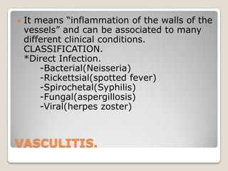
- 1. VASCULITIS. It means ―inflammation of the walls of the vessels‖ and can be associated to many different clinical conditions. CLASSIFICATION. *Direct Infection. -Bacterial(Neisseria) -Rickettsial(spotted fever) -Spirochetal(Syphilis) -Fungal(aspergillosis) -Viral(herpes zoster)
- 2. VASCULITIS. CLASSIFICATION.(cont.) *Immunologic. A. Immune complex- mediated. -Infection-induced(Hepatitis B,C) -Henoch-Schonlein purpura -SLE, etc. -Drug-induced -Cryoglobulinemia(Ig, IgM) -Serum sickness
- 3. VASCULITIS. CLASSIFICATION.(cont.) B. ANCA-mediated. -Wegener granulomatosis -Microscopic polyangeitis -Churg-Strauss syndrome C. Direct Ab-induced -Goodpasture syndrome(anti-GBM Abs) -Kawasaki disease(anti-endoth.Ab)
- 4. VASCULITIS. CLASSIFICATION.(cont.) D. Cell-mediated. -Organ-allograft rejection -Inflammatory bowel disease -Paraneoplastic vasculitis *Unknown. -Giant.cell arteritis -Takayasu arteritis -Polyarteritis nodosa
- 6. CLASSIFICATION TREE Vasculitis Large Blood Vessel • Temporal Arteritis • Takayasu Arteritis Medium Blood Vessel • Polyarteritis Nodosa • Kawasaki’s Disease Small Blood Vessel ANCAAssociated • Wegener’s Granulomatosis • Churg-Strauss Vasculitis • Microscopic Polyangiitis • Drug Induced Non-ANCAAssociated Immune Complex • Hypersensitivity Vasculitis • Cryoglobulinemic Vasculitis • CTD related Vasculitis • Henoch Schonlein Purpura • Behcet’s Miscellaneous • Paraneoplastic Vasculitis • Inflammatory Bowel Disease
- 9. GIANT CELL (TEMPORAL) ARTERITIS. A vasculitis of unknown etiology occurring primarily in the elderly. Other terms commonly used include temporal arteritis, cranial arteritis and granulomatous arteritis. Is the MOST common form of systemic vasculitis in adults, acute or chronic, often granulomatous inflammation of large/small size. It affects temporal arteries, but also vertebral, ophtalmic (blindness) and aorta (aneurysm) Involved arteries have nodular thickening reduction of lumenthrombosis. Granulomatous inflammation in inner half of media with mononuclear cells+ giant cells.
- 18. GIANT CELL (TEMPORAL) ARTERITIS. CLINICAL: old patients with: ◦ fever, ◦ fatigue, ◦ loss of weight, ◦ with or w/o facial pain and headache.
- 19. GIANT CELL (TEMPORAL) ARTERITIS. Others, with more severe form and involvement of ophtalmic arterydiplopia or blindness of abrupt onset. Dx.: significantly elevated ESR + arterial biopsy.
- 21. TAKAYASU ARTERITIS. Takayasu’s arteritis is a chronic inflammatory disorder of unknown etiology primarily affecting the aorta and its major branches. Predominant in females below 40´s w/probable autoimmune mechanism, Arteritis may also affects pulmonary arteries in ½ of cases as well as coronary/renal arteries.
- 22. Aortic arch arteriogram in a patient with Takayasu‘s arteritis. Smooth tapered stenosis of bilateral common carotid arteries (upper arrows) and of the right subclavian artery (lower arrow) can be seen. There is poststenotic dilatation beyond the left common carotid narrowed segment. Total involvement is seen along the
- 25. TAKAYASU ARTERITIS.(cont.) MICROSCOPIC: mononuclear infiltration of adventiciamid-layer with involvement of vasa vasorum.
- 26. TAKAYASU ARTERITIS.(cont.) CLINICAL: ◦ reduced brachial pulse, ◦ difference in BP between R & L arm >10 mm Hg, ◦ Coldness + numbness of fingers, ◦ bruising above a subclavia and/or aorta, ◦ hypertension, visual defectsblindness.
- 28. POLYARTERITIS NODOSA Is a small/medium sized arteritis affecting multiple organs (skin, peripheral nerves, gut, kidney and heart. Age of onset is from childhoodlate adulthood (average 40´s) and it has been associated w/Hepatitis B,C or both (most common in injection drug abusers). Probably mediated by immune complexes (Igs + viral Ags)circulating and deposited in inflammed vessels.
- 34. POLYARTERITIS NODOSA CLINICAL: onset is gradual (wksmos.) and nonspecific: ◦ malaise, ◦ fever, ◦ weight loss, ◦ abdominal pain, ◦ melena, ◦ myalgias, ◦ muscular weakness
- 35. POLYARTERITIS NODOSA ◦ diastolic pressure >90 mm Hg, ◦ mononeuropathy or polyneuropathy ◦ palpable purpura, ◦ livedo reticularis, ◦ digital gangrene or tender nodules
- 36. POLYARTERITIS NODOSA ◦ Renal involvementhypertension. ◦ In GI tract abdominal angina (hemorrhage, perforation). ◦ In heart: myocarditis/myocardial infarction. ◦ Eye: scleritis.
- 37. POLYARTERITIS NODOSA Dx. ◦ Elevated BUN or creatinine ◦ Proven hepatitis B/C virus infection ◦ Angiographic signs of aneuryms/vascular occlusion ◦ Demonstration of granulocytes in small or medium sized vessels (biopsy).
- 38. BUERGER´S DISEASE Also known as Thromboangiitis obliterans, is characterized by segmental thrombosis + acute/chronic inflammation of medium and small arteries MOSTLY tibial and radial arteries, and secondarily involvement of veins and nerves of limbs. Apparently heavy cigarette-smokers are MOST frequently affected (endoth. cells hypersensitivity?)
- 39. BUERGER´S DISEASE Microscopically: acute + chronic inflammation in arterial walls + thrombosisorganization/recanalization. Also, thrombosis contains microabscesses + granulomatous inflammationextension to veins/nerves. Late complication: chronic ulceration of toes or fingersgangrene.
- 43. BUERGER´S DISEASE CLINICAL: Claudication in feet and/or hands sometimes centrally radiated. Also, numbness and/or tingling in limbs + Raynaud´s phenomenon. Skin ulcerations + gangrene of digits
- 44. KAWASAKI DISEASE
- 45. KAWASAKI DISEASE (Mucocutaneous lymph node syndrome). Is an arteritis that frequently affects coronary arteries USUALLY in children/infants (about 80% are < 4 yrs old) Associated w/mucocutaneous lymph node syndrome of acute and self-limited evolution: ◦ fever, conjunctivitis, sore throat, ◦ generalized rash, redding of palms/soles, peeling of fingers/ toes and mucopurulent cervical lymphadenopathy. It is epidemic in Japan, Hawaii and US
- 47. KAWASAKI DISEASE Dx.: ◦ Leukocytosis (PMN´s), thrombocytosis. ◦ Some children have ANCA+ in plasma and ◦ Aprox.1/5 of patients involvement of coronary arteries/myocardium aneurysm/myocarditis that usually resolves spontaneously(mortality 2%).
- 52. Behçet’s Syndrome A systemic vasculitis of unknown cause with mucocutaneous and frequent ocular and musculoskeletal involvement. A marked geographic distribution is characterized by highest prevalence in Turkey, Iran and Japan.
- 53. Behçet’s Syndrome: Clinical features Recurrent oral and/or genital aphthous ulceration. Chronic relapsing uveitis leading to blindness in 10% of all cases. A variety of skin manifestations, including the ‗pathergy‘ phenomenon. Musculoskeletal, neurologic, major artery and vein involvement. An undulating course that generally abates in intensity with the passage of time.
- 55. MICROSCOPIC POLYANGIITIS It affects arterioles, capillaries and venules (smaller than PAN) and the leions are in the same stage. Typically presents as ―palpable purpura‖ (skin), but also with mucous membranes, lungs, brain, heart, GI tract, kidneys, muscle, PNS.
- 60. MICROSCOPIC POLYANGIITIS CLINICAL: ◦ Necrotizing GN (90%)hematuria ◦ Lungshemoptysis ◦ GI tractabdominal pain ◦ Jointsarthralgias ◦ Weight loss(>70%) ◦ Nerve damage(60%)numbness/tingling in limbs. ◦ Chronically: muscle wasting ◦ Skin: purpura(>60%) in legs, feet, buttocks
- 63. MICROSCOPIC POLYANGIITIS About 70% of patients may have a clear relation with a recent immunologic reaction to an Ag (drugs, microrganisms, tumor Ags,etc) having + pANCAS. MPA is quite similar histologically to PAN microscopic changes EXCEPT that muscular/large arteries are usually spared. It reminds Wegener vasculitis BUT w/o granulomatosis.
- 64. CHURG-STRAUSS SYNDROME Asthma is the cardinal clinical feature of CSS (8- 10 yrs BEFORE symptoms of vasculitis) with 8-10 yrs of evolutionrespiratory failure. Eosinophilia(>10% in CBC) Allergy Mono/polyneuropathy(ocular symptoms) Lung hemorrhage, pleural effusion, pulmonary infiltrates Changes in paranasal sinuses Eosinophilic granulomatosis vasculitis/perivasc. Abdominal cramps/heart failure(myocarditis)
- 70. History of Wegener’s In 1936, Wegener first described a distinct syndrome in three patients found to have necrotizing granulomas involving the upper and lower respiratory tract. In 1954, seven more patients described, resulting in definite criteria
- 71. Criteria for Classification Nasal or oral inflammation ◦ Development of painful or painless oral ulcers or purulent or bloody nasal discharge Abnormal chest radiograph ◦ Chest radiograph showing the presence of nodules, fixed infiltrates, or cavities Abnormal Urinary sediment ◦ Microhematuria (>5 red blood cells per high power field) or red cell casts in urine sediment Granulomatous inflammation on biopsy ◦ Histologic changes showing granulomatous inflammation within the wall of an artery or in the perivascular or extravascular area (artery or arteriole) * For purposes of classification, a patient shall be said to have Wegener's granulomatosis if at least 2 of these 4 criteria are present. The presence of any 2 or more criteria yields a sensitivity of 88.2% and a specificity of 92.0%
- 72. Classic Symptoms Upper respiratory tract ◦ sinuses ◦ nose ◦ ears ◦ trachea Lungs Kidneys
- 74. Upper Respiratory Tract Ear Ear infections that are slow to resolve. Recurrent otitis media. Decrease in hearing.
- 75. Upper Respiratory Tract Nose Nasal crusting Frequent nosebleeds Erosion and perforation of the nasal septum. ◦ The bridge of the nose can collapse resulting in a ―saddle–nose deformity‖.
- 76. Upper Respiratory Tract Sinuses/Trachea Sinuses ◦ Chronic sinus inflammation Trachea ◦ subglottic stenosis
- 77. Lungs Nodules Alveolar opacities Pleural opacities Diffuse hazy opacities (which may reflect alveolar hemorrhage)
- 80. Kidney Glomerulonephritis w/ associated hematuria and proteinuria Can lead to renal failure if not treated aggressively Active urine sediment: red blood cell casts
- 81. RBC casts
- 82. Skin ―palpable purpura‖ most common Raynaud‘s phenomenon—due to inadequate blood flow to fingers and toes Ulcers
- 83. Miscellaneous Joints ◦ Arthritis can occur, with joint swelling and pain Nerves ◦ Peripheral nerve involvement leads to numbness, tingling, shooting pains in the extremities, and sometimes to weakness in a foot, hand, arm, or leg Meninges Prostate gland Genito–urinary tract Constitutional symptoms of fatigue, low– grade fever, and weight loss
- 84. Incidence of symptoms Symptom At Onset Total ENT 75% 95% Lung 50 85 Joints 30 70 Fever 25 50 Kidney 20 75 Cough 20 50 Eye 15 50 Skin 15 45 Weight Loss 10 35 Nervous System (Central/Peripheral) 0 10/15 One-third of patients may be without symptoms at onset of disease
- 85. Pathogenesis ANCA ANCAs may be not only markers for Wegener's granulomatosis and related disorders, but they may also be actors in pathogenesis Neutrophils exposed to cytokines such as TNF, express PR3 & MPO (the targets for ANCAs) Adding ANCAs to these cytokine-primed neutrophils causes them to generate oxygen radicals and release enzymes capable of damaging blood vessels.
- 91. Diagnosis Nasal or oral inflammation ◦ Development of painful or painless oral ulcers or purulent or bloody nasal discharge Abnormal chest radiograph ◦ Chest radiograph showing the presence of nodules, fixed infiltrates, or cavities Abnormal urinary sediment ◦ Microhematuria (>5 red blood cells per high power field) or red cell casts in urine sediment Granulomatous inflammation on biopsy ◦ Histologic changes showing granulomatous inflammation within the wall of an artery or in the perivascular or extravascular area (artery or arteriole) Criteria for Classification
- 92. Management Vasculitides are often serious and sometimes fatal - require prompt recognition and therapy Treatment – helpful - particularly in the acute phase During maintenance therapy - adverse effects - superimposed infections
- 93. Management cont. Early deaths – due to active disease Late deaths – may be due to the complications of therapy The risk-versus-benefit ratio of any therapeutic approach should be weighed carefully
- 94. Management cont. Glucocorticoides and/or immunosuppressive therapy should be instituted immediately in diseases where irreversible organ system dysfunction and high morbidity and mortality have been clearly established Aggressive therapy should be avoided for vasculitic manifestations that rarely results in irreversible organ system dysfunction and that usually do not respond to such therapy
- 95. Management cont. Treatment of aggressive small vessel vasculitis 1. Induction of remission 2. Maintenance of remission 3. Treatment of relapse Induction therapy (to 3 months after remission, usually 6 months from diagnosis) Cyclophosphamide 2.0mg/kg/day (maximum 200mg/day) Age > 60years, reduce dose by 25% > 75years, by 50% Prednisolone 1mg/kg/day (maximum 80mg/day) reduced weekly to 25mg/day by 8 weeks and then more slowly to 10mg/day by 6 months
- 96. Management cont. In severe, life threatening disease (pulmonary haemorrhage, severe crescentic glomerulonephritis with creatinine > 500μmol/L) consider, ◦ Plasma exchange, 7-10 treatments over 14 days or ◦ Three pulses of methylprednisolone, 15mg/kg/day for 3 days
- 97. Management cont. Maintenance therapy (to 18-24 months, longer if clinically indicated) Azathioprine, 2.0mg/kg/day (maximum 200mg/day) Age > 60yrs, reduce dose by 25% > 75yrs, by 50% Relapse therapy Major relapse: return to induction therapy Minor relapse: increase dose of corticosteroid
- 98. Management cont. Stop cyclophosphamide or azathioprine if WBC < 4x10⁹/L. Restart with a dose reduced by at least 25mg when WBC > 4x10⁹/L on 2 consecutive tests.
- 99. Management cont. Most of other systemic vasculitides need at least corticosteroids Usual dose is 1mg/kg at the induction Gradual tapering during the remission