Basic ENT Hx & PE
•
40 gefällt mir•12,232 views

This document outlines the key components of performing a thorough head and neck examination in otolaryngology. It describes how to take a detailed patient history, including the HPI, and examines the various structures of the head and neck through inspection, palpation, and other examination techniques. Common abnormal findings and ENT diseases are also reviewed. Performing a systematic and well-documented head and neck exam is important for properly assessing patients' ENT complaints.

Empfohlen
Weitere ähnliche Inhalte
Was ist angesagt?
Was ist angesagt? (20)
Andere mochten auch
Andere mochten auch (20)
Ähnlich wie Basic ENT Hx & PE
Ähnlich wie Basic ENT Hx & PE (20)
Mehr von Notre Dame De Chartres Hospital
Mehr von Notre Dame De Chartres Hospital (20)
Kürzlich hochgeladen
Kürzlich hochgeladen (20)
Basic ENT Hx & PE
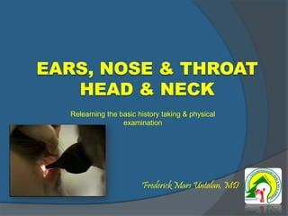
- 1. Frederick Mars Untalan, MD Relearning the basic history taking & physical examination
- 3. OUTLINE Know what to ask Know what to see Common ENT diseases
- 4. REMEMBER. . . . . . a thorough assessment begins with the HISTORY!
- 5. HISTORY of PRESENT ILLNESS (HPI or Symptom Analysis) Location and Radiation Timing: Onset, Frequency & Duration Quality and/or Characteristics Quantity and/or Severity Setting and/or Situation Aggravating Factors Alleviating Factors Associated Factors/Manifestations Underlying Concern and/or Perception
- 6. EQUIPMENT NEEDED Latex Gloves Light Source Cotton Cup with water (optional) Measuring tape (possible)
- 7. GENERAL CONSIDERATIONS The head and neck exam is not fixed in sequence. Different parts of the exam may be included and/or excluded depending on the history and the purpose of the exam.
- 9. Health History Determine presence/absence of age- and gender-specific diseases of the head and neck Common chief complaints Neck pain or stiff neck Hoarseness; nasal discharge or obstruction Neck mass Headache or facial pain Head injury ; otalgia; dysphagia; ear discharge
- 10. General Approach to Head and Neck Assessment Greet patient, explain assessment techniques Environment Quiet Warm Private Adequate lighting Upright sitting position Compare right and left sides Systematic approach
- 11. Where the Head Ends and Neck Begins Plane between the external occipital protuberance and inferior surface of the mandible Neck Anterior triangle is bordered by Mandible (above), Cervical midline(laterally) and Sternomastoid (anteriorly
- 12. Where the Head Ends and Neck Begins Neck – Posterior triangle is bordered by Clavicle (below), trapezius (posteriorly) and Sternomastoid (anteriorly)
- 13. Special Bony Areas External Occipial Protuberance (notch in occipital area Mastoid Process (behind Ear) Zygomatic Arch (Cheekbone) Orbit (eye socket) Maxilla (upper jaw) Mandible (lower Jaw)
- 14. Assessment of the Face Inspection Shape Symmetry Normal findings – Symmetrical features – Palpebral fissures equal – Nasolabial folds present bilaterally – Shape can be oval, round, or slightly square INSPECT Size, shape, and symmetry. Note placement of features , expression, movements and Skin characteristics. PALPATE Facial bones
- 15. ABNORMALS
- 16. Assessment of the Face Abnormal findings Deformed or absent structures Asymmetry More or less pronounced facial features Diseases which may alter facial features: Bell’s palsy, Down syndrome, Graves’ disease, Myxedema, Cachexia, Cushing’s syndrome
- 17. Replacement of Nonfunctional Facial Muscles Microneurovascular free muscle transfer
- 18. Mandible Palpate and auscultate the TMJ when the client opens and closes the mouth Normal findings No discomfort, joint articulates smoothly without clicking or crepitus Abnormal findings Pain, tenderness, crepitus, clicking, or snapping sound
- 19. TEMPORMANDIBULAR JOINT (TMJ) Located anterior to tragus, bilaterally Assess Palpate with movement Auscultate: Bell Abnormals Tenderness, crepitus, clicking Bruit Pain with trismus
- 25. External Auditory Canal Foreign Body
- 26. Otitis Externa
- 27. Otowick in EAC
- 28. Pharynx
- 29. Esophagus - mid
- 31. Foreign Bodies
- 33. Vocal Nodules
- 36. Nasal Polyp
- 39. Middle Meatus-Maxillary Sinus Ostium Purulent Drainage Normal
- 40. SINUSES: Frontal & Maxillary Assess Inspect Palpate Percuss: Direct Transilluminati on: Darkened room Abnormals Swelling Tenderness Flatter sound Unequal light
- 41. CT Scan - Sinusitis
- 47. Dorsal Deficiency More likely with osteotome rather than sharp rasp Corrected with completion osteotomies or on- lay graft
- 48. Septal Hematoma
- 49. Cleft Anatomy
- 50. Cleft Anatomy - The Nose
- 51. Cleft Anatomy
- 52. Controversies
- 57. Cleft Palate - Bifid Uvula
- 58. Ranula
- 59. Squamous Cell Carcinoma of Tongue ExophyticUlcerative
- 61. Parotitis
- 65. THYROID Inspect (Tangential lighting when swallowing) Palpate Palpate trachea Posterior Approach Anterior Approach Auscultate (with Bell)
- 66. Enlarged Thyroid
- 67. PALPATE the THYROID Posterior Approach Displace to one side then palpate Swallow
- 68. Neck Masses
- 69. NECK
- 71. LYMPH NODES Landmarks Anterior Triangle ○ Mandible ○ Trachea ○ SCM muscle Posterior Triangle ○ Clavical ○ Trapezius muscle ○ SCM muscle
- 73. LYMPH NODES Preauricular Nodes
- 74. LOCATION of LYMPH NODES of the NECK Anterior Cervical Chain Overlying the sternocleidomastoid muscle Posterior Cervical Chain In the posterior triangle along the trapezius muscle Internal Jugular Chain Deep under the sternocleidomastoid muscle. Not normally palpated. Supraclavicular Just above & behind the clavicle at the sternocleidomastoid muscle
- 75. LYMPH NODES Anterior Cervical Nodes
- 76. LYMPH NODES Posterior Cervical Chain
- 77. LYMPH NODES Supraclavicular Nodes
- 78. DOCUMENTATION Head: Normocephalic, no lumps, no lesions, no tenderness. Hair medium texture, evenly distributed.
- 79. DOCUMENTATION Face: Symmetric, no drooping, no weakness, no involuntary movements. Temporal artery pulses palpable, no bruits. TMJ articulates smoothly without clicking, crepitus or pain.
- 81. DOCUMENTATION Neck: Supple with full ROM, no pain. Symmetric, no lymphadenopathy or masses, trachea midline. Thyroid not palpable. No bruits.
- 82. DOCUMENTATION Ears Inspection & otoscopy; tuning fork tests Nose Inspection, anterior & posterior rhinoscopy Oral cavity & oropharynx Indirect laryngoscopy to check the laryngeal inlet & hypopharynx
- 83. OUTLINE Know what to ask Know what to see Common ENT diseases
- 84. Frederick Mars Untalan, MD Relearning the basic history taking & physical examination