
Empfohlen
Weitere ähnliche Inhalte
Ähnlich wie anatomy_axial_skeleton_pearson.ppt
Ähnlich wie anatomy_axial_skeleton_pearson.ppt (20)
Kürzlich hochgeladen
Kürzlich hochgeladen (20)
anatomy_axial_skeleton_pearson.ppt
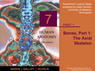
- 1. PowerPoint® Lecture Slides prepared by Leslie Hendon, University of Alabama, Birmingham HUMAN ANATOMY fifth edition MARIEB | MALLATT | WILHELM 7 Copyright © 2008 Pearson Education, Inc., publishing as Benjamin Cummings Bones, Part 1: The Axial Skeleton PART 1
- 2. Copyright © 2008 Pearson Education, Inc., publishing as Benjamin Cummings The Skeleton Consists of Bones, cartilage, joints, and ligaments Composed of 206 named bones grouped into two divisions Axial skeleton (80 bones) Appendicular skeleton (126 bones)
- 3. Copyright © 2008 Pearson Education, Inc., publishing as Benjamin Cummings The Axial Skeleton Formed from 80 named bones Consists of skull, vertebral column, and bony thorax Figure 7.1a
- 4. Copyright © 2008 Pearson Education, Inc., publishing as Benjamin Cummings The Axial Skeleton Figure 7.1b
- 5. Copyright © 2008 Pearson Education, Inc., publishing as Benjamin Cummings Bone Markings Projections that provide attachment for muscles and ligaments Projections that help form joints Depressions and openings for passage of nerves and blood vessels
- 6. Copyright © 2008 Pearson Education, Inc., publishing as Benjamin Cummings Figure 7.2a The Skull Formed by cranial and facial bones
- 7. Copyright © 2008 Pearson Education, Inc., publishing as Benjamin Cummings The Cranium The cranium serves to Enclose brain Provide attachment sites for some head and neck muscles
- 8. Copyright © 2008 Pearson Education, Inc., publishing as Benjamin Cummings The Face Facial bones serve to Form framework of the face Form cavities for the sense organs of sight, taste, and smell Provide openings for the passage of air and food Hold the teeth in place Anchor muscles of the face
- 9. Copyright © 2008 Pearson Education, Inc., publishing as Benjamin Cummings Overview of Skull Geography Facial bones form anterior aspect Cranium is divided into cranial vault and the base Internally, prominent bony ridges divide skull into distinct fossae
- 10. Copyright © 2008 Pearson Education, Inc., publishing as Benjamin Cummings Overview of Skull Geography The skull contains smaller cavities Middle and inner ear cavities – in lateral aspect of cranial base Nasal cavity – lies in and posterior to the nose Orbits – house the eyeballs Air-filled sinuses – occur in several bones around the nasal cavity
- 11. Copyright © 2008 Pearson Education, Inc., publishing as Benjamin Cummings Overview of Skull Geography The skull contains approximately 85 named openings Foramina, canals, and fissures Provide openings for important structures Spinal cord Blood vessels serving the brain 12 pairs of cranial nerves
- 12. Copyright © 2008 Pearson Education, Inc., publishing as Benjamin Cummings Cranial Bones Formed from eight large bones Paired bones include Temporal bones Parietal bones Unpaired bones include Frontal bone Occipital bone Sphenoid bone Ethmoid bone
- 13. Copyright © 2008 Pearson Education, Inc., publishing as Benjamin Cummings Frontal Bones Forms the forehead and roofs of the orbits Forms superciliary arches Internally, it contributes to the anterior cranial fossa Contains frontal sinuses
- 14. Copyright © 2008 Pearson Education, Inc., publishing as Benjamin Cummings Parietal Bones and Sutures Sutures of the cranium (continued) Sagittal suture – occurs where right and left parietal bones meet superiorly Lambdoid suture – occurs where the parietal bones meet the occipital bone posteriorly
- 15. Copyright © 2008 Pearson Education, Inc., publishing as Benjamin Cummings Sutural Bones Small bones that occur within sutures Irregular in shape, size, and location Not all people have sutural bones
- 16. Copyright © 2008 Pearson Education, Inc., publishing as Benjamin Cummings Occipital Bone Forms the posterior portion of the cranium and cranial base Articulates with the temporal bones and parietal bones Forms the posterior cranial fossa Foramen magnum located at its base
- 17. Copyright © 2008 Pearson Education, Inc., publishing as Benjamin Cummings Occipital Bone Features and structures Occipital condyles Hypoglossal foramen External occipital protuberunce Superior nuchal lines Inferior nuchal lines
- 18. Copyright © 2008 Pearson Education, Inc., publishing as Benjamin Cummings Inferior Aspect of the Skull Figure 7.4a
- 19. Copyright © 2008 Pearson Education, Inc., publishing as Benjamin Cummings Temporal Bones Lie inferior to parietal bones Form the inferolateral portion of the skull Term “temporal” Comes from Latin word for time Specific regions of temporal bone Squamous, temporal, petrous, and mastoid regions
- 20. Copyright © 2008 Pearson Education, Inc., publishing as Benjamin Cummings Lateral Aspect of the Skull Figure 7.3a
- 21. Copyright © 2008 Pearson Education, Inc., publishing as Benjamin Cummings The Temporal Bone Figure 7.5
- 22. Copyright © 2008 Pearson Education, Inc., publishing as Benjamin Cummings The Sphenoid Bone Spans the width of the cranial floor Resembles a butterfly or bat Consists of a body and three pairs of processes Contains five important openings
- 23. Copyright © 2008 Pearson Education, Inc., publishing as Benjamin Cummings The Sphenoid Bone Figure 7.6b
- 24. Copyright © 2008 Pearson Education, Inc., publishing as Benjamin Cummings The Ethmoid Bone Lies between nasal and sphenoid bones Forms most of the medial bony region between the nasal cavity and orbits
- 25. Copyright © 2008 Pearson Education, Inc., publishing as Benjamin Cummings Figure 7.7 The Ethmoid Bone
- 26. Copyright © 2008 Pearson Education, Inc., publishing as Benjamin Cummings Bones of the Skull Table 7.1 (1 of 2)
- 27. Copyright © 2008 Pearson Education, Inc., publishing as Benjamin Cummings Facial Bones Unpaired bones Mandible and vomer Paired bones Maxillae Zygomatic bones Nasal bones Lacrimal bones Palatine bones Inferior nasal conchae
- 28. Copyright © 2008 Pearson Education, Inc., publishing as Benjamin Cummings Mandible The lower jawbone is the largest and strongest facial bone Composed of two main parts Horizontal body Two upright rami
- 29. Copyright © 2008 Pearson Education, Inc., publishing as Benjamin Cummings Mandible Figure 7.8a
- 30. Copyright © 2008 Pearson Education, Inc., publishing as Benjamin Cummings Maxillary Bones Articulate with all other facial bones except the mandible Contain maxillary sinuses – largest paranasal sinuses Forms part of the inferior orbital fissure
- 31. Copyright © 2008 Pearson Education, Inc., publishing as Benjamin Cummings Maxillary Bones Figure 7.8b
- 32. Copyright © 2008 Pearson Education, Inc., publishing as Benjamin Cummings Maxillary Bones Figure 7.4a
- 33. Copyright © 2008 Pearson Education, Inc., publishing as Benjamin Cummings Other Bones of the Face Zygomatic bones Form lateral wall of orbits Nasal bones Form bridge of nose Lacrimal bones Located in the medial orbital walls Palatine bones Complete the posterior part of the hard palate
- 34. Copyright © 2008 Pearson Education, Inc., publishing as Benjamin Cummings Other Bones of the Face Vomer Forms the inferior part of the nasal septum Inferior nasal conchae Thin, curved bones that project medially form the lateral walls of the nasal cavity
- 35. Copyright © 2008 Pearson Education, Inc., publishing as Benjamin Cummings Bones of the Face Figure 7.2a
- 36. Copyright © 2008 Pearson Education, Inc., publishing as Benjamin Cummings Special Parts of the Skull Orbits Nasal cavity Paranasal sinuses Hyoid bone
- 37. Copyright © 2008 Pearson Education, Inc., publishing as Benjamin Cummings Nasal Cavity Figure 7.9a
- 38. Copyright © 2008 Pearson Education, Inc., publishing as Benjamin Cummings Nasal Septum Figure 7.9b
- 39. Copyright © 2008 Pearson Education, Inc., publishing as Benjamin Cummings Orbits Figure 7.10b
- 40. Copyright © 2008 Pearson Education, Inc., publishing as Benjamin Cummings Figure 7.12 The Hyoid Bone Lies inferior to the mandible The only bone with no direct articulation with any other bone Acts as a movable base for the tongue
- 41. Copyright © 2008 Pearson Education, Inc., publishing as Benjamin Cummings The Vertebral Column Formed from 26 bones in the adult Transmits weight of trunk to the lower limbs Surrounds and protects the spinal cord
- 42. Copyright © 2008 Pearson Education, Inc., publishing as Benjamin Cummings The Vertebral Column Serves as attachment sites for muscles of the neck and back Held in place by ligaments Anterior and posterior longitudinal ligaments Ligamentum flavum
- 43. Copyright © 2008 Pearson Education, Inc., publishing as Benjamin Cummings The Vertebral Column Figure 7.13
- 44. Copyright © 2008 Pearson Education, Inc., publishing as Benjamin Cummings Intervertebral Discs Cushion-like pads between vertebrae Act as shock absorbers Compose about 25% of height of vertebral column Composed of Nucleus pulposus and annulus fibrosis
- 45. Copyright © 2008 Pearson Education, Inc., publishing as Benjamin Cummings Intervertebral Discs Nucleus pulposus The gelatinous inner sphere of intervertebral disc Enables spine to absorb compressive stresses
- 46. Copyright © 2008 Pearson Education, Inc., publishing as Benjamin Cummings Intervertebral Discs Annulus fibrosis An outer collar of ligaments and fibrocartilage Contains the nucleus pulposus Functions to bind vertebrae together, resist tension on the spine, and absorb compressive forces
- 47. Copyright © 2008 Pearson Education, Inc., publishing as Benjamin Cummings Ligaments and Intervertebral Discs Figure 7.14a
- 48. Copyright © 2008 Pearson Education, Inc., publishing as Benjamin Cummings Herniated Disc May be caused by trauma to the spine Aging is also a contributing factor Nucleus pulposes loses cushioning properties Anulus fibrosis weakens Figure 7.14c
- 49. Copyright © 2008 Pearson Education, Inc., publishing as Benjamin Cummings Regions and Normal Curvatures Vertebral column is about 70 cm (28 inches) Vertebral column is divided into five major regions Cervical vertebrae 7 vertebrae of the neck region Thoracic vertebrae 12 vertebrae of the thoracic region
- 50. Copyright © 2008 Pearson Education, Inc., publishing as Benjamin Cummings Regions and Normal Curvatures Vertebral column is divided into five major regions (continued) Lumbar vertebrae 5 vertebrae of the lower back Sacrum Inferior to lumbar vertebrae Articulates with coxal bones Coccyx Most inferior region of the vertebral column
- 51. Copyright © 2008 Pearson Education, Inc., publishing as Benjamin Cummings Regions and Normal Curvatures Four distinct curvatures give vertebral column an S-shape Cervical and lumbar curvature Are concave posteriorly Thoracic and sacral curvatures Are convex posteriorly Curvatures increase the resilience of the spine
- 52. Copyright © 2008 Pearson Education, Inc., publishing as Benjamin Cummings Regions and Normal Curvatures Figure 7.13 PLAY Spine (vertical)
- 53. Copyright © 2008 Pearson Education, Inc., publishing as Benjamin Cummings General Structure of Vertebrae PLAY Spine (horizontal) Figure 7.15
- 54. Copyright © 2008 Pearson Education, Inc., publishing as Benjamin Cummings Regions Vertebral Characteristics Specific regions of the spine perform specific functions Types of movement that occur between vertebrae Flexion and extension Lateral flexion Rotation in the long axis
- 55. Copyright © 2008 Pearson Education, Inc., publishing as Benjamin Cummings Cervical Vertebrae Seven cervical vertebrae (C1 – C7) – smallest and lightest vertebrae C3 – C7 are typical cervical vertebrae Body is wider laterally Spinous processes are short and bifid (except C7) Vertebral foramen are large and triangular Transverse processes contain transverse foramina Superior articular facets face superoposteriorly
- 56. Copyright © 2008 Pearson Education, Inc., publishing as Benjamin Cummings Cervical Vertebrae Table 7.2a
- 57. Copyright © 2008 Pearson Education, Inc., publishing as Benjamin Cummings Cervical Vertebrae Figure 7.17a
- 58. Copyright © 2008 Pearson Education, Inc., publishing as Benjamin Cummings The Atlas C1 is termed the atlas Lacks a body and spinous process Supports the skull Superior articular facets receive the occipital condyles Allows flexion and extension of neck Nodding the head “yes”
- 59. Copyright © 2008 Pearson Education, Inc., publishing as Benjamin Cummings The Atlas Figure 7.16a
- 60. Copyright © 2008 Pearson Education, Inc., publishing as Benjamin Cummings The Atlas Figure 7.16b
- 61. Copyright © 2008 Pearson Education, Inc., publishing as Benjamin Cummings The Axis Has a body and spinous process Dens (odontoid process) projects superiorly Formed from fusion of the body of the atlas with the axis Acts as a pivot for rotation of the atlas and skull Participates in rotating the head from side to side
- 62. Copyright © 2008 Pearson Education, Inc., publishing as Benjamin Cummings The Axis Figure 7.16c
- 63. Copyright © 2008 Pearson Education, Inc., publishing as Benjamin Cummings Thoracic Vertebrae (T1 – T12) All articulate with ribs Have heart-shaped bodies from the superior view Each side of the body of T1 – T10 bears demifacts for articulation with ribs T1 has a full facet for the first rib T10 – T12 only have a single facet
- 64. Copyright © 2008 Pearson Education, Inc., publishing as Benjamin Cummings Thoracic Vertebrae Table 7.2b
- 65. Copyright © 2008 Pearson Education, Inc., publishing as Benjamin Cummings Thoracic Vertebrae Spinous processes are long and point inferiorly Vertebral foramen are circular Transverse processes articulate with tubercles of ribs Superior articular facets point posteriorly Inferior articular processes point anteriorly Allows rotation and prevents flexion and extension
- 66. Copyright © 2008 Pearson Education, Inc., publishing as Benjamin Cummings Lumbar Vertebrae (L1 – L5) Bodies are thick and robust Transverse processes are thin and tapered Spinous processes are thick, blunt, and point posteriorly Vertebral foramina are triangular Superior and inferior articular facets directly medially Allows flexion and extension – rotation prevented
- 67. Copyright © 2008 Pearson Education, Inc., publishing as Benjamin Cummings Lumbar Vertebrae Table 7.2c
- 68. Copyright © 2008 Pearson Education, Inc., publishing as Benjamin Cummings Lumbar Vertebrae Figure 7.17c
- 69. Copyright © 2008 Pearson Education, Inc., publishing as Benjamin Cummings Sacrum (S1 – S5) Shapes the posterior wall of pelvis Formed from 5 fused vertebrae Superior surface articulates with L5 Inferiorly articulates with coccyx Sacral promontory Where the first sacral vertebrae bulges into pelvic cavity Center of gravity is 1 cm posterior to sacral promontory
- 70. Copyright © 2008 Pearson Education, Inc., publishing as Benjamin Cummings Sacrum Sacral foramina Ventral foramina Passage for ventral rami of sacral spinal nerves Dorsal foramina Passage for dorsal rami of sacral spinal nerves
- 71. Copyright © 2008 Pearson Education, Inc., publishing as Benjamin Cummings Sacrum Figure 7.18a, b
- 72. Copyright © 2008 Pearson Education, Inc., publishing as Benjamin Cummings Coccyx Is the “tailbone” Formed from 3 – 5 fused vertebrae Offers only slight support to pelvic organs
- 73. Copyright © 2008 Pearson Education, Inc., publishing as Benjamin Cummings Bony Thorax Forms the framework of the chest Components of the bony thorax Thoracic vertebrae – posteriorly Ribs – laterally Sternum and costal cartilage – anteriorly Protects thoracic organs Supports shoulder girdle and upper limbs Provides attachment sites for muscles
- 74. Copyright © 2008 Pearson Education, Inc., publishing as Benjamin Cummings The Bony Thorax Figure 7.19a
- 75. Copyright © 2008 Pearson Education, Inc., publishing as Benjamin Cummings The Bony Thorax Figure 7.19b
- 76. Copyright © 2008 Pearson Education, Inc., publishing as Benjamin Cummings Sternum Formed from three sections Manubrium – superior section Articulates with medial end of clavicles Body – bulk of sternum Sides are notched at articulations for costal cartilage of ribs 2–7 Xiphoid process – inferior end of sternum Ossifies around age 40
- 77. Copyright © 2008 Pearson Education, Inc., publishing as Benjamin Cummings Sternum Anatomical landmarks Jugular notch Central indentation at superior border of the manubrium Sternal angle A horizontal ridge where the manubrium joins the body
- 78. Copyright © 2008 Pearson Education, Inc., publishing as Benjamin Cummings Ribs All ribs attach to vertebral column posteriorly True ribs - superior seven pairs of ribs Attach to sternum by costal cartilage False ribs – inferior five pairs of ribs Ribs 11–12 are known as floating ribs
- 79. Copyright © 2008 Pearson Education, Inc., publishing as Benjamin Cummings Ribs Figure 7.20a
- 80. Copyright © 2008 Pearson Education, Inc., publishing as Benjamin Cummings Ribs Figure 7.20b
- 81. Copyright © 2008 Pearson Education, Inc., publishing as Benjamin Cummings Disorders of the Axial Skeleton Abnormal spinal curvatures Scoliosis – an abnormal lateral curvature Kyphosis – an exaggerated thoracic curvature Lordosis – an accentuated lumbar curvature – “swayback” Stenosis of the lumbar spine A narrowing of the vertebral canal
- 82. Copyright © 2008 Pearson Education, Inc., publishing as Benjamin Cummings The Axial Skeleton Throughout Life Membrane bones begin to ossify in second month of development Bone tissue grows outward from ossification centers Fontanels Unossified remnants of membranes
- 83. Copyright © 2008 Pearson Education, Inc., publishing as Benjamin Cummings Fontanels Figure 7.21a
- 84. Copyright © 2008 Pearson Education, Inc., publishing as Benjamin Cummings Fontanels Figure 7.21b
- 85. Copyright © 2008 Pearson Education, Inc., publishing as Benjamin Cummings The Axial Skeleton Throughout Life Many bones of the face and skull form by intramembranous ossification Endochondral bones of the skull Occipital bone Sphenoid Ethmoid bones Parts of the temporal bone
- 86. Copyright © 2008 Pearson Education, Inc., publishing as Benjamin Cummings The Axial Skeleton Throughout Life Curvatures of the vertebral column Primary curvatures – thoracic and sacral curvatures An infant's spine is C-shaped at birth Secondary curvatures – cervical and lumbar curvatures Develop when a baby begins to walk Redistributes weight of the upper body over the lower limbs
- 87. Copyright © 2008 Pearson Education, Inc., publishing as Benjamin Cummings The Axial Skeleton Throughout Life Aging of the axial skeleton Water content of the intervertebral discs decreases By age 55, loss of a few centimeters in height is common Thorax becomes more rigid Bones lose mass with age