Stereotactic Radiosurgery (SRS)
•Als PPTX, PDF herunterladen•
14 gefällt mir•5,302 views

Radiosurgery is a discipline that utilizes externally generated ionizing radiation in certain cases to inactivate or eradicate a defined target(s) in the head or spine without the need to make an incision. Its uses in Neurosurgery is immense.

Empfohlen
Weitere ähnliche Inhalte
Was ist angesagt?
Was ist angesagt? (20)
Ähnlich wie Stereotactic Radiosurgery (SRS)
Ähnlich wie Stereotactic Radiosurgery (SRS) (20)
Mehr von suresh Bishokarma
Mehr von suresh Bishokarma (20)
Kürzlich hochgeladen
Kürzlich hochgeladen (20)
Stereotactic Radiosurgery (SRS)
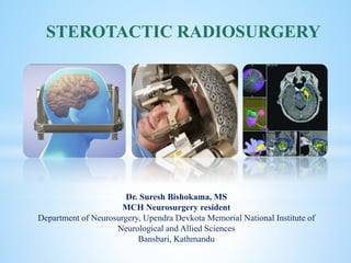
- 1. cka Dr. Suresh Bishokama, MS MCH Neurosurgery resident Department of Neurosurgery, Upendra Devkota Memorial National Institute of Neurological and Allied Sciences Bansbari, Kathmandu STEROTACTIC RADIOSURGERY
- 2. Radiation therapy is the use of high-energy photons or charged particles that takes advantage of the ionizing radiation portion of the electromagnetic spectrum to induce specific biologic changes for the treatment of various pathologic entities including tumors and vascular lesions. Radiosurgery, as defined by the American Association of Neurologic Surgeons (AANS) is “a distinct discipline that utilizes externally generated ionizing radiation in certain cases to inactivate or eradicate a defined target(s) in the head or spine without the need to make an incision.” DEFINITION
- 3. Ionizing radiation contains enough energy to result in the removal of electrons from their atoms, and thus leads to the creation of reactive species that cause damage to cells. DNA replicative failure: The most critical form of damage is DNA strand break. Double strand breaks are more difficult for cells to repair, and the repair process can generate aberrant chromosomes that result in mitotic catastrophe, or mutations that result in reduced replicative fitness. Vascular endothelium dysfunction: Endothelial cell apoptosis, endothelial cell death may lead to direct hypoxic necrosis of the tumor or may secrete signaling molecules that result in tumor cdl death. However, other studies have shown that at even higher MECHANISM OF THERAPEUTIC RADIATION
- 4. Clinical radiation dose, measured in grays (Gy), represents the energy deposited by ionization in material per unit mass of the material. Maximizing the dose of radiation to the target and minimizing the dose to normal tissue is the central goal of radiosurgery. DOSING
- 5. 1 Gy=100 rads Cells are most sensitive to radiation during the G2/M phase of the cell cycle and resistant during the late S phase and G1. FACTS
- 6. S N TUMOR SRS DOSE (Gy) TRADITION XRT DOSE 1 MENINGIOMA 12-16 Gy 55-60 Gy 2 LGG NA 54Gy infraction of 1.8Gy 3 EPEDYMOMA NA 59.4Gy 4 MEDULLOBLASTOMA NA 35-40Gy whole CSI+10-15Gy tumor bed and spinal boost, Fractionated over 6-7wks 5 VESTIBULAR SCHWANNOMA 12.5-14Gy MENINGIOMA 55-60Gy 7 PCNSL 40-50Gy 1.8-3 Gy daily fraction 8 CRANIOPHARYNGIOMA 8 Gy 54 Gy in 30 fractions 9 PITUITARY ADENOMA 40-50 Gy over 4-6weeks NA 10 DAVF 16-20Gy NA 11 AVM 15-25Gy NA 12 PINEAL TUMOR NA 55Gy in 1.8-Gy daily fractions, with 40Gy to the ventricular system and an additional 15 Gy to the tumor bed 13 TECTAL GLIOMA <14Gy 45-55Gy DOSAGE
- 7. SRS typically consists of a single high-dose treatment of radiation delivered using a precise localization system. Conventional radiotherapy typically involves lower doses delivered in daily fractions to reduce the effects of radiation on normal tissue. Radiation delivery system Fractionation VS radiosurgery (SRS)
- 8. 1. Re-oxygenation of hypoxic areas, resulting in increased sensitivity of malignant cells that were previously hypoxic. 2. Re-assortment of cells in the cell cycle, as cells are most sensitive to radiation during the G2/M phase of the cell cycle and resistant during the late S phase and G1. Allows resistant phases of the cell cycle to move to more sensitive phases of the cell cycle during subsequent fractions. (Late S phase and G1 G2/M). 3. DNA repair to occur, which favors normal cells that retain the full complement of DNA repair proteins. 4. Re-population of tumor cells during the therapy. FRACTIONATION INDUCES 4R OF RADIOBIOLOGY: “OARP”
- 9. Differential sensitivity of tissues to fractionation (/ ratio): which is a radio biologic concept based on a model of radiation response. Tissues with a high / ratio respond quickly to radiotherapy and are sensitive to smaller fraction sizes. / RATIO
- 10. 1. Photon beam radiosurgery: (X-ray/Gamma) 2. Proton beam radiosurgery: (Helium or Proton) TYPES OF RADIOSURGERY
- 11. Photon particle: no mass: speed of light: carries the energy present in all electromagnetic radiation, including microwaves, visible light, ultraviolet light, and x-rays. Photon is the most common form of ionizing radiation used in radiotherapy. Mechanism: Photons interact with matter predominantly via the photoelectric effect. Ionizing radiation results in the ejection of electrons from the atoms and scattering of the photon. The scattered photon has a change in its energy. Photons interact with a molecule of water, resulting in the production of superoxide, hydroxyl radicals, and other reactive oxygen species, which damage the DNA and result in replicative failure. Radiotherapy is believed to be more effective in the presence of oxygen, and it is thought that hypoxic areas of tumors may be less sensitive to the effects of ionizing radiation. PHOTON BEAM RADIOSURGERY
- 12. Three main categories (based on sources of radiation) are: Gamma knife Linear accelerator based, and Heavy charged particle radiosurgery. PHOTON BEAM RADIOSURGERY
- 13. The Gamma Knife is specifically designed for cranial and upper cervical lesions, and is best suited for smaller lesions (<3 cm diameter). The unit, as currently constructed, contains 201 fixed sources of 60Co (Half-life:5.3 years) distributed in a hemisphere, each of which is a thin rod with the long axis oriented along the radius of the sphere, converging on a single point, called the treatment isocenter. Because there are no moving parts during treatment, there is a high degree of setup accuracy. The GK cobalt-60 sources decay to nickel-60 with the release of two energies of gamma rays at 1.117 and 1.33 MeV, which are the main treatment energies. GAMMA KNIFE RADIOSURGERY
- 14. Linear accelerator-based radiotherapy (LINAC) system generates photon x-rays by accelerating electrons to a high speed using microwave energy to deliver SRS onto the target. LINAC produces 6-MeV x-rays. Fixed to the LINAC platform: true stereotactic frame, noninvasive facemask, and dental mold . The x-ray source and beam collimation is mounted on a rotating gantry, creating a fixed isocenter in space. Beam convergence is achieved via rotating arcs with the isocenter fixated on the target. Cyberknife is a miniaturized linac that is mounted on a robotic arm with 6 degrees of rotational freedom (rather than an isocentrically-mounted linac). The linac and robotic arm combination can deliver multiple small beamlets of radiation from many different angles to produce a conformal dose plan. LINEAR ACCELERATOR-BASED RADIOSURGERY (LINACS)
- 15. Proton a nucleus of hydrogen atom are positively charged, subatomic particles of ~ 1 atomic mass unit (protons, helium, and carbon, to name a few) produced by stripping electrons from molecular hydrogen gas, before being accelerated to therapeutic energies in a synchrotron or cyclotron. As a particle travels through the medium it loses its energy in a myriad of these collisions and finally comes to a full stop. Because more energy is lost the slower the particle goes, a useful feature arises-a peak right at the end of the particle's travel ("Bragg peak"). HEAVY CHARGED PARTICLE RADIOSURGERY (PROTONS OR HELIUM IONS)
- 16. Cyclotron is used to generate high charged particle for use in radiosurgery. Proton production starts by stripping the electron from molecular hydrogen gas, and the resulting protons are then accelerated to a therapeutic energy level using alternating magnetic fields in a cyclotron or synchrotron. Unlike high-energy photons (gamma and x-rays), which deposit the majority of their energy upon entrance into tissue and continue to deposit decreasing amount of energy as they travel through the body, heavy charged particle beams have a shorter, bounded range of penetration wherein particles sharply increase energy deposition near the terminal depth of penetration (Bragg peak effect). Particle radiosurgery achieves a well-localized volume of high dose radiation by taking advantage of cross firing of a number of beams as well as the Bragg peak. Due to the expense and increased complexity of heavy charged particle SRS, this therapy is only available in a few centers in the world.
- 17. Absence of dose beyond the target and the decrease in dose proximally. To cover lesions, which extend in depth, multiple Bragg peaks, originating from protons with different initial energies, are superimposed to create a spread-out Bragg peak (SOBP). ADVANTAGES OF PROTONS OVER PHOTONS
- 18. Physical uncertainty and cost. Given the sharp dose fall-off, it is critical to be able to calculate and deliver proton dose precisely. Large accelerators are needed to generate proton (football field size); compact technology is under development) DISADVANTAGE OF PROTONS OVER PHOTONS
- 19. 1. VASCULAR LESIONS ○ AVMs (including dural arteriovenous fistulas) ○ Cavernous malformations 2. TUMORS ○ Metastases ○ Vestibular schwannomas ○ Meningiomas ○ Pituitary adenomas ○ Gliomas ○ Others: craniopharyngioma, pineal tumors. INDICATIONS OF RADIOSURGERY 3. FUNCTIONAL DISORDERS ○ Trigeminal neuralgia ○ Intractable chronic pain: thalamotomy ○ Movement disorders: pallidotomy for Parkinson’s disease or thalamotomy for tremor ○ Psychiatric diseases (e.g. obsessive compulsive disorder) ○ Epilepsies 4. SPINE: PLIF, TLIF: to shave the end plates. SRS is useful for well-circumscribed lesions less than approximately 3 cm diameter (in general)
- 20. Compressive tumors of the spinal cord, brainstem or optic structures: (Even with the sharp fall of radiation dose, there remains radiation delivered within a few millimeters of the margins of the isocenter). This, together with post-radiation swelling, might create significant risk of neurologic injury. CONTRAINDICATIONS
- 21. Treatment unlikely to result in functional improvement or clinically meaningful disease stabilization, not otherwise achievable. Patients with wide-spread cerebral or extra-cranial metastases with limited life expectancy unlikely to gain clinical benefit within their remaining life. Patients with poor performance status (Karnofsky Performance Status less than 40 or ECOG Performance greater than 3) Essential tremor, coverage should be limited to the patient who cannot be controlled with medication, has major systemic disease or coagulopathy, and who is unwilling or unsuited for invasive surgical procedure. Coverage should further be limited to unilateral thalamotomy. SRS is not considered medically necessary
- 22. 1. Cranial nerves: Optic nerve cant tolerate >8Gy radiation within 2mm. 2. Damage to small nutrient vessels and Schwann cells or oligodendroglia are the possible mechanisms of radiation injury to cranial nerves. 3. Special sensory nerves (optic, vestibulocochlear) are the most radiosensitive 4. SRS treatment may also have a deleterious effect in structures sensitive to swelling, such as brainstem. 5. Additionally critical radiation sensitive structures include: optic vitreous, nerve, and chiasm, brain stem, pituitary gland, and cochlea. RED FLAG ZONE
- 23. The treatment procedure includes placement of stereotactic frame (in framed-based SRS), obtaining stereotactic images, target definition, treatment planning and execution of treatment. TREATMENT PROCEDURE
- 24. Methylprednisolone 40 mg IV and phenobarbital 90 mg IV immediately after the radiation dose to patients with tumors or AVMs to reduce these adverse effects PREMEDICATION
- 25. 1. Position stabilization (attachment of a frame or frameless). 2. Imaging for localization (CT, MRI, angiography, PET, etc.). 3. Computer-assisted tumor localization (i.e., “image guidance”). 4. Treatment planning – number of isocenters; number, placement and length of arcs or angles; number of 5. beams, beam size and weight, etc. 6. Isodose distributions, dosage prescription and calculation. 7. Setup and accuracy verification testing. 8. Simulation of prescribed arcs or fixed portals. 9. Radiation treatment delivery. COMPONENT OF SRS PROCEDURES
- 26. 1. SRS is best accepted for the treatment of small to moderate-sized (< 3 cm) AVMs that are deep or 2. Border on eloquent brain and have a “compact” (i.e. sharply demarcated) nidus. 3. The radiation induces endothelial cell damage, smooth muscle cell proliferation, thickening of the vascular wall, and ultimately obliteration of the lumen over a period of 2-3 years (latency period). AVFs pose high risk of hemorrhage. SRS is of no benefit for venous angiomas. SRS for cavernous malformations remains controversial. ESPECIAL CONSIDERATION AVM AND VASCULAR LESIONS
- 27. ● Total tumor number ≤ 10 ● Total tumor volume ≤ 15 cm3 ● Single tumor volume is <10 cm3, and ● No leptomeningeal disease present. BRAIN METASTASES:
- 28. Indications are: poor surgical candidates (due to poor medical condition and/ or advanced age, some use >65 or 70 years as a cutoff), patient refusing surgery, bilateral VS. VESTIBULAR SCHWANNOMA
- 29. SRS is generally not indicated as a primary treatment for infiltrating tumors, e.g. gliomas INFILTRATING TUMORS
- 30. 1. Focal deficits, seizures, or headache. 2. Radiation necrosis and permanent deficits ( Mechanism: glial cell damage, breakdown of blood brain barrier or early venous thrombosis 3. Premature venous thrombosis or occlusion before obliteration of AVM nidus can produce venous hyperemia or intracranial hemorrhage. 4. Vasculopathy 5. Cranial nerve deficits (Incidence is higher with tumors of CPA or skull base): Mechanism is due to damage of small nutrient vessels and Schwann cells or oligodendroglia) 6. Radiation induced tumors: Astrocytoma DRAWBACKS OF SRS
- 31. NATIONAL INSTITUTE OF NEUROLOGICAL AND ALLIED SCIENCES, BANSBARI, KATHMANDU STEROTACTIC RADIOSURGERY Dr. Suresh Bishokama, MS MCH Neurosurgery resident Department of Neurosurgery, Upendra Devkota Memorial National Institute of Neurological and Allied Sciences Bansbari, Kathmandu drsureshbk@gmail.com
Hinweis der Redaktion
- Radiosurgery is a discipline that utilizes externally generated ionizing radiation in certain cases to inactivate or eradicate a defined target(s) in the head or spine without the need to make an incision.