SlideShare ist ein Scribd-Unternehmen logo
1 von 47
cka
Dr. Suresh Bishokama, MS
MCH Neurosurgery resident
Department of Neurosurgery, Upendra Devkota Memorial National Institute of
Neurological and Allied Sciences
Bansbari, Kathmandu
MEDULLOBLASTOMA
 Cushing Report: 1930
 Coined Medulloblastoma: Bailey and Cushing
 1983: PNET
HISTORY
 Medulloblastoma is the most common malignant brain tumor of childhood (WHO IV) and
comprises up to 18-30% of all pediatric brain tumors.
 Medulloblastomas are typically midline cerebellar lesions arising from the vermis in the
inferior medullary velum; however, in older children and adults they can arise from the
cerebellar hemispheres.
 Medulloblastomas are related to primitive neuroectodermal tumors (PNETs) and occur
exclusively in the posterior fossa.
INTRODUCTION
 Overall medulloblastomas account for 12-25 % of all pediatric CNS tumors, and 30-40%
of pediatric posterior fossa tumors.
 They are also seen in adults but only account for 0.4-1.0% of adult brain tumor.
 M:F ratio of 2:1, although this is only true of group 3 and 4 tumors
 They usually present in childhood with 77% of cases before the age of 19.
 The median age of diagnosis is 9 years.
 When diagnosed in adulthood, they typically present in the 3rd and 4th decades and are
more likely to arise in atypical locations (see below).
 When they present in adulthood, there is often a better prognosis. (Younger the poorer)
EPIDEMIOLOGY
 N Nitroso compound
 Vegetables are protective
 JC virus
 Genetic conditions: Li-Fraumani, Gorlin, Turcot
 P53, IGF, c-MYC and N-MYC oncogenic transcription factor, Apoptotic pathway: Bcl-2
ASSOCIATION
 Medulloblastoma to arise from the external granule layer of the developing cerebellum
and others have proposed that the cell of origin arises from a subventricular progenitor
zone.
PATHOGENESIS
 WNT is a ligand for the transmembrane protein FRIZZLED and major cytoplasmic
component of the WNT pathway is APC/GSK-3B/AXIN complex.
 In absence of WNT this complex phosphorylates B catenin, thereby targeting it for
degradation. B catenin in nucleus promotes cellular proliferation.
WNT PATHWAY
Sonig Hedge Hog (SHH)
 Sonig Hedge Hog (SHH) is a diffusible mitogenic protein that binds to the
transmembrane receptor protein (PATCHED 1-PTCH1).
 In the absence of SHH, PTCH1 binds with SMO.
 This complex inhibit glioma associated oncogene homolog (GLI) and hence cellular
proliferation.
 However, when PTCH1 binds with SHH, inhibitor effect of SMO-PTCH1 complex are
reversed leading to activation of GLI and enhanced oncogenesis.
 WNT tumours should be identified by two of the following markers:
 Nuclear ß-catenin accumulation (IHC)
 Monosomy 6 (whole chromosome loss) by FISH
 CTNNB1 mutation
 WNT pattern by DNA methylation or by gene expression profiling
 SHH, Group 3 and Group 4
 IHC
 c-myc amplification (FISH)
 Genome-wide methylation
 Classic : (90%): Undifferentiated cells with hyperchromatic nuclei, (Homer
wright rosette suggesting neuronal differentiation, Blue tumor
 Desmoplastic : (6%): Classic + Glomeruli (pale island of collagen): Least
aggressive
 Nodularity MBEN : Diffuse variant in <3 years child: good prognosis
 Large cell Anaplastic : (4%): early seed, high mitosis: poor prognosis
HISTOLOGICAL SUBTYPES
Homer-Wright rosettes composed of neoplastic cells concentrically arranged around fibrillary processes
 Diffuse masses of small, undifferentiated oval or round cells.
 Some medulloblastomas show neuronal, glial and other differentiation.
 Nuclear moulding due to compactness inside cell.
 Neuronal differentiation is manifested by neuropil and rosette formation.
 Rosettes are groups of tumor cells arranged in a circle around a fibrillary center.
 Mature neurons may also be found infrequently. Glial differentiation in some tumors is reflected by
GFAP-positive cells. There may also be differentiation along oligodendroglial or ependymal lines.
CLASSIC MEDULLOBLASTOMA
 Pale nodules are composed uniform round to spindle shaped neuronal-appearing
cells which are not as active mitotically as the surrounding darker tumor.
 They are present within a reticulin-poor fine fibrillary background
DESMOPLASTIC MB
 Large cell anaplastic medulloblastoma composed of sheets of highly atypical cells.
 A focus of necrosis is present just above the center of the image.
 The tumor cells have irregular hyperchromatic nuclei with nuclear molding and little cytoplasm
ANAPLASTIC MB
 Medulloblastoma can spread along cerebrospinal fluid (CSF) pathways and also rarely to
systemic sites (bone, lung).
 Leptomeningeal dissemination constitutes the most common pattern of recurrence for
Group 3 and Group 4 patients, and usually leads to a quick progressive clinical decline
despite therapy.
New type: Medulloblastoma NOS.
CLINICOPATHOLOGICAL AND MOLECULAR
CHARACTERISTICS OF MB SUBGROUPS
CLINICOPATHOLOGICALAND MOLECULAR CHARACTERISTICS OF MB SUBGROUP
CHARACTER WNT 10% SHH: 25% GP3: 25% GP4: 40%; MC
GENDER M=F M=F M>F M>F
PEAK AGE Children Children Infants Children
OCCURRENCE
Arise from foramen of
luschka (CP angle)
Laterally cerebellar
hemisphere, and vermis
4th ventricle 4th ventricle
ENHANCEMENT
Intensely enhancing like
ependymoma but diffusion
restriction
Intermediate Intermediate Usually non-enhancing
HISTOLOGY Classic, rarely LCA
Desmoplastic/MBEN,
Classic, LCA
Classic, LCA Classic, LCA
GENE MUTATED
CTNNB1, TP53 CTNNB1, TP53,
3q gain, 9p deletion
MYC amp, OTX2,
SMARCA4
KDM6A, MYCN
SNCAIP
duplication
IHC
B-catenin +(including
nuclear and
cytoplasmic)
CTNNB1 mutation,
GAB1 (-)
Cytoplasmic B-Catenin+;
GLI amplification; SMO
mutation;
MYCN+
GAB1+, YAB1+, PTCH1+,
SMO+, P53±
MYCN+++;
YAB/GAB-
Minimal
amplification;
CDK6+;
YAB/GAB (-)
METASTASIS Low Extremely low High Frequent
PROGNOSIS
Best prognosis Intermediate Poor Intermediate
Molecular group /
Immunoreactivity
GAB1 ß-catenin YAP1
WNT Negative Nuclear +
Cytoplasmic +
Nuclear +
Cytoplasmic
SHH Cytoplasmic Cytoplasmic + Nuclear +
Cytoplasmic
Non-SHH/WNT Negative Cytoplasmic - Negative
MOLECULAR SUBGROUPS AND
IMMUNOREACTIVITY
One of the techniques that can be implemented widely across the globe is IHC-
based sub classification
IMMUNOREACTIVITY/
MOLECULAR GROUP
WNT SHH Non-SHH/WNT
ß-catenin (Nuclear) + - -
GAB-1 - + -
YAP1 + + -
p53 ±
Molecular subgroups and immunoreactivity
Surrogate IHC
Additional for Group 3: c-myc amp (FISH)
Additional for WNT: Monosomy Chr 6
(FISH)
 Homer-Wright rosettes composed of neoplastic cells concentrically arranged around
fibrillary processes are a common histological feature.
 Isochromosome17q was confirmed as the most common genetic alteration in
medulloblastomas, found in 28% of specimens.
 Synaptophysin, Neuron enolase, neurofilament protein, tubulin
 Metastases may have different immunohistology different from the actual primary.
PATHOLOGY
 Short time before diagnosis (<3months)
 Morning headache, vomiting, lethargic
 Features of raised ICP including papilloedema and diplopia (CN VI)
 Truncal and appendicular ataxia, nystagmus, bulbar and facial palsy (brainstem),
 Head tilt (tonsil descent in FM)
 Macrocrania (infant),
 Drop mets (Back pain, urinary retention and leg weakness): ~10-35% of MB seeds at the
time of dx and Extraneural mets occurs in 5% (sometime promoted by shunt: 10-20%;
overestimated ???)
CLINICAL PRESENTATION
 Classic features of a medulloblastoma on computed tomographic (CT) scan are
increased density on the non-contrast scan, midline location, well-defined margins, and
dense, homogeneous enhancement with injection of contrast, surrounding vasogenic
edema.
 Calcification (22%) and cyst formation (59%)
 The hyperdensity on plain CT scan, seen in medulloblastoma and in some ependymomas,
is secondary to the high cellularity of these tumors that have scanty cytoplasm and areas
of desmoplasia.
WORK UP
 MRI: There is a high degree of variability of MR appearances of medulloblastoma.
 T1 sequences are usually iso-hypointense to white matter and hyperintense on T2
sequences.
 Tumor enhancement can be both homogeneous or heterogeneous.
 Similar to ependymoma, approximately14% of medulloblastomas may show foraminal
extension.
 Banana sign (4th vent drapes around medulloblastoma unlike ependymoma where tumor
encroach 4th vent)
 HCP: 85-90%
MRI BRAIN WITH CONTRAST
T1W-MRI
T2W-MRI
FLAIR MAGNITUDE
SWI
DWI-MRI
ADC
Gd-MRI
FEATURES MEDULLOBLASTOMA EPENDYMOMAS PILOCYTIC ASTRO HEMANGIOBLASTOMA
Origin Roof of 4th V Floor Cerebellar Cerebellar
Clinical features
Age
Acute
5-15
Chronic
3-8
Chronic
5-15
Chronic
40-60
VHL: Young
NE-CT Hyperdense (cellularity) Hypo Hypo Hypo
Heterogenicity Less More Solid cystic Solid cystic
T1W-MRI Hypo Hypo Hypo Hyper (also cyst)
T2W-MRI Iso to Hyper Hyper Hyper Hyper, flow void
Mural Nodule None None May be present May be present
Enhancement Most (E); Non (gr 4) to
intense (WNT)
Intense Solid enhances Intense
Perilesional edema - - - +
Calcification Less More None Less
Hemorrhage Less More Rare Less
Extension into
cisternal spaces
Less More Nil Less
Diffusion restriction Yes No No No
MEDULLOBLASTOMA VS EPENDYMOMA
 MR spectroscopy may also be distinctive
 SHH
little or no taurine
low creatine
 Group 3 or 4
Taurine peak
high creatine
MRS
 MRI SPINE to assess the severity of diseases (Mets)
 Leptomeningeal seeding on MRI is found in approximately 33% of patients.
 The spinal canal is the most common location of seeding; however, the supratentorial
compartment may also be involved.
 Nodular or diffuse enhancement along the leptomeninges, nerve roots in the spinal canal,
or cranial nerves in the CPA are common findings of CSF seeding.
MRI SPINE
MRI and CSF cytometry may miss 14% of seeding
CSF cytometry
MODIFIED CHANG CLASSIFICATION
TREATMENT
1. Establish histological diagnosis
2. Maximally resect tumor mass
3. Relieve hydrocephalus
AIMS OF SURGERY
MEDULLOBLASTOMA
SURGICAL RESECTION
HCP TREATED
>3 YRS <3 YEARS
STANDARD RISK POOR RISK CHEMOTHERAPY
FOLLOW UP OR DELAY XRT OR
LOCAL FIELD XRT
PROTON
CSI OR REDUCED DOSE CSI+
CHEMO
CSI+ ADJ CHEMO (CCNU,
CISPLATIN, VINCRISTINE OR
CHEMO ON RESEARCH
PROTOCOL
FOLLOW
RISK STRATIFICATION
1. Age (less than <3 or >3 years); Presence of disseminated disease (M1-4); Extent of surgical
resection (<1.5 or >1.5cm residual) and Histological differentiation
(5 years PFS among group: 77% vs 35-50%)
Better outcome: Girls; WNT:good/ LCA/non SHH/WNT: Poor
MEDULLOBLASTOMA
FIG: PROPOSED ALGORITHM FOR TREATMENT OF PATIENT WITH MEDULLOBLATOMA
XRT: OPTIMAL DOSE
35-40Gy to whole CSI +
10-15 Gy boost to tumor
bed (PF) and to any spinal
mets, all fractionated over
6-7 weeks
Reduce dose by 20-25% or Chemo
Chemotherapy improves 5-yr PFS survival (87% vs 33%)
Low dose XRT 25 Gy
 Currently risk stratification is based on age (less than 3 years of age), presence of
disseminated disease (M1-4, and extent of surgical resection (<1.5 or >1.5cm residual)
and histological differentiation along glial, ependymal or neuronal line. (5 yrs PFS
among group: 77% vs 53%)
 In cases in which there is greater than 1.5 cm of residual tumor, a repeat procedure
should be considered, if safe and anatomically feasible, to place the patient in the best
prognostic category.
 Medulloblastoma is a radiosensitive tumor and incorporation of radiotherapy has become
a standard of care in treatment of children older than 3 years of age.
RISK STRATIFICATION
PROGNOSTIC FACTORS
Low-risk patients.
Nuclear accumulation of CTNNB1*
CTNNB1* mutations
Monosomy stratifies
*WNT subgroup markers
High-risk patients:
MYC/MYCN amplication and
isochromosome 17q
 It is generally agreed now that most patients do not require pre-operative shunting.
However, 20-35% may need post-operative shunt.
 Shunt risk of peritoneal dissemination: 10-20%.
 Pre-operative shunt: Demerit: Tumoral hemorrhage, seeding, upward herniation.
 If the drain continues to drain at the height of 20cm, hydrocephalus is shown to progress
or pseudomeningocele develops.
SHUNT OR NOT
TREATMENT STRATEGY
MECHANISM OF ACTION OF CHEMOTHERAPY
DRUG MOA SIDE EFFECT
CISPLATIN Induces cellular apoptosis by cross-linking DNA Ototoxicity and nephrotoxicity, dys-electrolytemia
CYCLOPHOSPHAMIDE Alkylating agent Hemorrhagic cystitis, infertility
LOMUSTINE Alkylating agent Myelosuppression, myeloid leukemia
VINCRISTINE Microtubule inhibitor that prevents cell division by
binding to the tubulin component of microtubules and
leading to metaphase arrest
Peripheral neuropathy. Muscle weakness, SEVERE
CONSTIPATION
 Radiation therapy (XRT) should begin ~30 days following definitive surgery.
 Historically, the entirety of the cerebellum was radiated.
 Therapy is delivered in daily fractions of 1.8 Gy to a final dose of 54 Gy - 59.4 Gy.
 Craniospinal irradiation (CSI) is delivered to the entire brain and spine and given
concurrently with primary site radiation for the first 13 days of therapy, to a total dose of
23.4 Gy (1.8x 13days)
RADIOTHERAPY
Posterior fossa ®: 1.8 Gy for 1 months (54-59.4 Gy)
CSI ®: 1.8 Gy for 13 days (23.4 Gy)
MEDULLOBLASTOMA
 The current standard of care for average risk medulloblastoma among >3 yrs child is
gross total resection, postoperative craniospinal radiation of 23.4 Gy with a tumor bed
irradiation t of 54 Gy, followed by 12 months of chemotherapy.
 Cyclophosphamide, vincristine, cisplatin and peripheral stem cell rescue. (5 years EFS
~70%)
 Child <3 years: chemotherapy has been used to delay radiation therapy until nervous
system matures or reduced (20-25% of total) dose of XRT.
 Administration of focal radiation to tumor bed is insufficient to control medulloblastoma,
even if there is no evidence of disseminated disease at diagnosis.
 Consequently, irradiation of entire neuraxis is critical in the treatment of
medulloblastoma.
CURRENT STANDARD OF CARE
 Cognitive decline, endocrine insufficiency, hearing loss, growth retardation, vascular
complications, leukoencephalopathy and secondary malignancies.
SIDE EFFECTS OF RADIATION
 Proton therapy: Bragg peak: capacity to deliver targeted radiation while sparing normal
structure. Target avoiding spinal growth plate, auditory canal, pituitary fossa.
NOVEL IRRADIATION PROMISE
MRI within 48 hours to minimize detection of non-tumoral post-operative changes
Postoperative surveillance imaging of the brain and spine in patients with medulloblastoma
is routinely employed at many institutions with 3–6 month intervals during the first 5 years
following initial diagnosis in the hope of detecting recurrent disease earlier.
.
POST OP IMAGING
 Post op neurological morbidity: 25-57%
 Cerebral mutism (25%); Dysarthria persist after mutism recover.
 Recurrence: Collin’s law to define period of recurrence (age at diagnosis + 9 months;
 Autologous stem cell rescue to deliver chemotherapy at dose that may provide better
response was tried limited by hematologic toxicity.
OUTCOME
 5 years survival:
 M0: 70%
 M1: 57%
 M ≥2: 40%
 Prognosis is most strongly influenced by molecular subtype
 WNT: very good
 SHH: infants good, others intermediate
 group 3: poor
 group 4: intermediate
 Outcome is good among girls
 Recurrence occurs in duration of 4-20 months.
 Survival after recurrence: 5months (poor)
PROGNOSIS
 Expression of the c-erbB-2 (HER2/neu) oncogene is useful in the staging of
medulloblastomas.
 Increased c-erbB-2 expression reflects an increase in the proliferative activity of
a tumor (widely used in breast cancer staging).
 No CSF metastases, complete surgical resection and negative c-erbB-2
expression: 5-year-survival 100%.
 No CSF metastases, complete surgical resection and positive c-erbB-2
expression: 5-year-survival 54%.
 CSF metastases and/or incomplete surgical resection: 5-year-survival 20%.
c-erbB-2 (HER2/neu) oncogene expression
 Protein phosphatase inhibitor Norcantharidin for WNT: + nuclear B- catenin.
 Smoothened (SMO) inhibitors: Vismodegib and Sonidegib targets Shh pathway
 Suppress MYC-related pathway activity.
FUTURE
 GOOD
1. WNT
2. B Catenin
3. Classic, Desmoplastic
CONFOUNDERS OF OUTCOME
 BAD
1. GAB1, YAB1
2. TP53
3. Large/Anaplastic
4. MYC amplification
Thank you

NATIONAL INSTITUTE OF NEUROLOGICAL AND ALLIED SCIENCES, BANSBARI, KATHMANDU
MEDULLOBLASTOMA
UPENDRA DEVKOTA MEMORIAL NATIONAL INSTITUTE OF NEUROLOGICALAND ALLIED SCIENCES,
BANSBARI, KATHMANDU

Weitere ähnliche Inhalte

Was ist angesagt?

Was ist angesagt? (20)

Craniopharyngiomas
Craniopharyngiomas Craniopharyngiomas
Craniopharyngiomas
 
Brain metastasis
Brain metastasis Brain metastasis
Brain metastasis
 
Diagnosis, Treatment & Management of Medulloblastoma
Diagnosis, Treatment & Management of Medulloblastoma Diagnosis, Treatment & Management of Medulloblastoma
Diagnosis, Treatment & Management of Medulloblastoma
 
Pediatric brain tumors Dr. Muhammad Bin Zulfiqar
Pediatric brain tumors Dr. Muhammad Bin Zulfiqar Pediatric brain tumors Dr. Muhammad Bin Zulfiqar
Pediatric brain tumors Dr. Muhammad Bin Zulfiqar
 
Anatomy of sellar suprasellar region
Anatomy of sellar suprasellar regionAnatomy of sellar suprasellar region
Anatomy of sellar suprasellar region
 
Pineal gland tumors
Pineal gland tumorsPineal gland tumors
Pineal gland tumors
 
Brain stem gliomas
Brain stem gliomasBrain stem gliomas
Brain stem gliomas
 
Brain stem glioma
Brain stem gliomaBrain stem glioma
Brain stem glioma
 
Medulloblastomas
MedulloblastomasMedulloblastomas
Medulloblastomas
 
Meningioma
MeningiomaMeningioma
Meningioma
 
Primary cns lymphoma ppt
Primary cns lymphoma pptPrimary cns lymphoma ppt
Primary cns lymphoma ppt
 
WHO BRAIN TUMOR CLASSIFICATION 5th EDITION
WHO BRAIN TUMOR CLASSIFICATION 5th EDITIONWHO BRAIN TUMOR CLASSIFICATION 5th EDITION
WHO BRAIN TUMOR CLASSIFICATION 5th EDITION
 
CNS infections in HIV
CNS infections in HIVCNS infections in HIV
CNS infections in HIV
 
Rhabdomyosarcoma
RhabdomyosarcomaRhabdomyosarcoma
Rhabdomyosarcoma
 
Cancer of unknown primary: Knowing the unknown
Cancer of unknown primary: Knowing the unknownCancer of unknown primary: Knowing the unknown
Cancer of unknown primary: Knowing the unknown
 
Gliomas - Brain Tumor
Gliomas - Brain TumorGliomas - Brain Tumor
Gliomas - Brain Tumor
 
Sellar, Suprasellar and Pineal tumor final pk .ppt
Sellar, Suprasellar and Pineal tumor final pk .pptSellar, Suprasellar and Pineal tumor final pk .ppt
Sellar, Suprasellar and Pineal tumor final pk .ppt
 
Glioblastoma Multiforme
Glioblastoma MultiformeGlioblastoma Multiforme
Glioblastoma Multiforme
 
EANO GUIDELINES FOR MANAGEMENT OF MENINGIOMA
EANO GUIDELINES FOR MANAGEMENT OF MENINGIOMAEANO GUIDELINES FOR MANAGEMENT OF MENINGIOMA
EANO GUIDELINES FOR MANAGEMENT OF MENINGIOMA
 
Meningioma of brain
Meningioma of brainMeningioma of brain
Meningioma of brain
 

Ähnlich wie MEDULLOBLASTOMA

Essentials of gliomas
Essentials of gliomas Essentials of gliomas
Essentials of gliomas
NeurologyKota
 

Ähnlich wie MEDULLOBLASTOMA (20)

Harbor UCLA Neuro-Radiology Case 6
Harbor UCLA Neuro-Radiology Case 6Harbor UCLA Neuro-Radiology Case 6
Harbor UCLA Neuro-Radiology Case 6
 
Primitive Neuroectodermal Tumor.pptx
Primitive Neuroectodermal Tumor.pptxPrimitive Neuroectodermal Tumor.pptx
Primitive Neuroectodermal Tumor.pptx
 
Harbor UCLA Neuro-Radiology Case 8
Harbor UCLA Neuro-Radiology Case 8Harbor UCLA Neuro-Radiology Case 8
Harbor UCLA Neuro-Radiology Case 8
 
Management of gliomas
Management of gliomasManagement of gliomas
Management of gliomas
 
Essentials of gliomas
Essentials of gliomas Essentials of gliomas
Essentials of gliomas
 
Neuroblastoma urology rotation
Neuroblastoma urology rotation Neuroblastoma urology rotation
Neuroblastoma urology rotation
 
Precocious puberty - Imaging
Precocious puberty - ImagingPrecocious puberty - Imaging
Precocious puberty - Imaging
 
Precocious puberty
Precocious pubertyPrecocious puberty
Precocious puberty
 
Medulloblastoma
MedulloblastomaMedulloblastoma
Medulloblastoma
 
Harbor UCLA Neuro-Radiology Case 6
Harbor UCLA Neuro-Radiology Case 6Harbor UCLA Neuro-Radiology Case 6
Harbor UCLA Neuro-Radiology Case 6
 
Spinal cord lesions and its radiological imaging finding.
Spinal cord lesions and its radiological imaging finding.Spinal cord lesions and its radiological imaging finding.
Spinal cord lesions and its radiological imaging finding.
 
Glioblastoma multiforme
Glioblastoma multiformeGlioblastoma multiforme
Glioblastoma multiforme
 
Neuroblastoma and nephroblastoma
Neuroblastoma and nephroblastoma Neuroblastoma and nephroblastoma
Neuroblastoma and nephroblastoma
 
Medulloblastoma - A Closer Look
Medulloblastoma - A Closer LookMedulloblastoma - A Closer Look
Medulloblastoma - A Closer Look
 
Embryonal brain tumours
Embryonal  brain tumours Embryonal  brain tumours
Embryonal brain tumours
 
Brain tumours
Brain tumoursBrain tumours
Brain tumours
 
high grade glioma.pptx
high grade glioma.pptxhigh grade glioma.pptx
high grade glioma.pptx
 
A Case of CNS Tumour
A Case of CNS TumourA Case of CNS Tumour
A Case of CNS Tumour
 
PEDIATRIC EMBRYONAL TUMOURS - UPDATE
PEDIATRIC EMBRYONAL TUMOURS - UPDATE PEDIATRIC EMBRYONAL TUMOURS - UPDATE
PEDIATRIC EMBRYONAL TUMOURS - UPDATE
 
Embryonal tumors in children
Embryonal tumors in childrenEmbryonal tumors in children
Embryonal tumors in children
 

Mehr von suresh Bishokarma

Posterior lumbar fusion vs Lumbar interbody fusion Evidence based.pptx
Posterior lumbar fusion vs Lumbar interbody fusion Evidence based.pptxPosterior lumbar fusion vs Lumbar interbody fusion Evidence based.pptx
Posterior lumbar fusion vs Lumbar interbody fusion Evidence based.pptx
suresh Bishokarma
 

Mehr von suresh Bishokarma (20)

Water dynamic of UBE Unilateral Biportal Endoscopy.pptx
Water dynamic of UBE Unilateral Biportal Endoscopy.pptxWater dynamic of UBE Unilateral Biportal Endoscopy.pptx
Water dynamic of UBE Unilateral Biportal Endoscopy.pptx
 
Posterior lumbar fusion vs Lumbar interbody fusion Evidence based.pptx
Posterior lumbar fusion vs Lumbar interbody fusion Evidence based.pptxPosterior lumbar fusion vs Lumbar interbody fusion Evidence based.pptx
Posterior lumbar fusion vs Lumbar interbody fusion Evidence based.pptx
 
Brain Abscess - Algorithmic approach
Brain Abscess - Algorithmic approachBrain Abscess - Algorithmic approach
Brain Abscess - Algorithmic approach
 
NEUROSURGICAL TENETS OF PITUITARY GLAND
NEUROSURGICAL TENETS OF PITUITARY GLANDNEUROSURGICAL TENETS OF PITUITARY GLAND
NEUROSURGICAL TENETS OF PITUITARY GLAND
 
Third ventricular surgical approaches
Third ventricular surgical approachesThird ventricular surgical approaches
Third ventricular surgical approaches
 
BRAIN ARTERIOVENOUS MALFORMATION
BRAIN ARTERIOVENOUS MALFORMATIONBRAIN ARTERIOVENOUS MALFORMATION
BRAIN ARTERIOVENOUS MALFORMATION
 
BRAIN ABSCESS: ALGORITHM SIMPLIFIED
BRAIN ABSCESS: ALGORITHM SIMPLIFIEDBRAIN ABSCESS: ALGORITHM SIMPLIFIED
BRAIN ABSCESS: ALGORITHM SIMPLIFIED
 
Screw trajectories and ergonomics in spine surgery
Screw trajectories and ergonomics in spine surgeryScrew trajectories and ergonomics in spine surgery
Screw trajectories and ergonomics in spine surgery
 
Stereotactic Radiosurgery (SRS)
Stereotactic Radiosurgery (SRS)Stereotactic Radiosurgery (SRS)
Stereotactic Radiosurgery (SRS)
 
Foramen Magnum Meningioma
Foramen Magnum MeningiomaForamen Magnum Meningioma
Foramen Magnum Meningioma
 
Brain metastasis - Simplified
Brain metastasis - SimplifiedBrain metastasis - Simplified
Brain metastasis - Simplified
 
Dandy Walker syndrome
Dandy Walker syndromeDandy Walker syndrome
Dandy Walker syndrome
 
Cranioplasty complications
Cranioplasty complicationsCranioplasty complications
Cranioplasty complications
 
Caroticocavernous fistula CCF
Caroticocavernous fistula CCFCaroticocavernous fistula CCF
Caroticocavernous fistula CCF
 
Choroid plexus
Choroid plexus Choroid plexus
Choroid plexus
 
Potts spine- TB spine.
Potts spine- TB spine.Potts spine- TB spine.
Potts spine- TB spine.
 
Lilliquist Membrane
Lilliquist MembraneLilliquist Membrane
Lilliquist Membrane
 
Head Injury AKA Traumatic Brain Injury
Head Injury AKA Traumatic Brain InjuryHead Injury AKA Traumatic Brain Injury
Head Injury AKA Traumatic Brain Injury
 
Cerebellar haematoma
Cerebellar haematomaCerebellar haematoma
Cerebellar haematoma
 
Cerebellum surgical anatomy
Cerebellum surgical anatomyCerebellum surgical anatomy
Cerebellum surgical anatomy
 

Kürzlich hochgeladen

Call Girls Bhubaneswar Just Call 9907093804 Top Class Call Girl Service Avail...
Call Girls Bhubaneswar Just Call 9907093804 Top Class Call Girl Service Avail...Call Girls Bhubaneswar Just Call 9907093804 Top Class Call Girl Service Avail...
Call Girls Bhubaneswar Just Call 9907093804 Top Class Call Girl Service Avail...
Dipal Arora
 
Russian Escorts Girls Nehru Place ZINATHI 🔝9711199012 ☪ 24/7 Call Girls Delhi
Russian Escorts Girls  Nehru Place ZINATHI 🔝9711199012 ☪ 24/7 Call Girls DelhiRussian Escorts Girls  Nehru Place ZINATHI 🔝9711199012 ☪ 24/7 Call Girls Delhi
Russian Escorts Girls Nehru Place ZINATHI 🔝9711199012 ☪ 24/7 Call Girls Delhi
AlinaDevecerski
 

Kürzlich hochgeladen (20)

Call Girls Ooty Just Call 8250077686 Top Class Call Girl Service Available
Call Girls Ooty Just Call 8250077686 Top Class Call Girl Service AvailableCall Girls Ooty Just Call 8250077686 Top Class Call Girl Service Available
Call Girls Ooty Just Call 8250077686 Top Class Call Girl Service Available
 
Best Rate (Guwahati ) Call Girls Guwahati ⟟ 8617370543 ⟟ High Class Call Girl...
Best Rate (Guwahati ) Call Girls Guwahati ⟟ 8617370543 ⟟ High Class Call Girl...Best Rate (Guwahati ) Call Girls Guwahati ⟟ 8617370543 ⟟ High Class Call Girl...
Best Rate (Guwahati ) Call Girls Guwahati ⟟ 8617370543 ⟟ High Class Call Girl...
 
Night 7k to 12k Chennai City Center Call Girls 👉👉 7427069034⭐⭐ 100% Genuine E...
Night 7k to 12k Chennai City Center Call Girls 👉👉 7427069034⭐⭐ 100% Genuine E...Night 7k to 12k Chennai City Center Call Girls 👉👉 7427069034⭐⭐ 100% Genuine E...
Night 7k to 12k Chennai City Center Call Girls 👉👉 7427069034⭐⭐ 100% Genuine E...
 
Top Rated Bangalore Call Girls Richmond Circle ⟟ 9332606886 ⟟ Call Me For Ge...
Top Rated Bangalore Call Girls Richmond Circle ⟟  9332606886 ⟟ Call Me For Ge...Top Rated Bangalore Call Girls Richmond Circle ⟟  9332606886 ⟟ Call Me For Ge...
Top Rated Bangalore Call Girls Richmond Circle ⟟ 9332606886 ⟟ Call Me For Ge...
 
Call Girls Bhubaneswar Just Call 9907093804 Top Class Call Girl Service Avail...
Call Girls Bhubaneswar Just Call 9907093804 Top Class Call Girl Service Avail...Call Girls Bhubaneswar Just Call 9907093804 Top Class Call Girl Service Avail...
Call Girls Bhubaneswar Just Call 9907093804 Top Class Call Girl Service Avail...
 
Call Girls Haridwar Just Call 8250077686 Top Class Call Girl Service Available
Call Girls Haridwar Just Call 8250077686 Top Class Call Girl Service AvailableCall Girls Haridwar Just Call 8250077686 Top Class Call Girl Service Available
Call Girls Haridwar Just Call 8250077686 Top Class Call Girl Service Available
 
Best Rate (Hyderabad) Call Girls Jahanuma ⟟ 8250192130 ⟟ High Class Call Girl...
Best Rate (Hyderabad) Call Girls Jahanuma ⟟ 8250192130 ⟟ High Class Call Girl...Best Rate (Hyderabad) Call Girls Jahanuma ⟟ 8250192130 ⟟ High Class Call Girl...
Best Rate (Hyderabad) Call Girls Jahanuma ⟟ 8250192130 ⟟ High Class Call Girl...
 
Russian Escorts Girls Nehru Place ZINATHI 🔝9711199012 ☪ 24/7 Call Girls Delhi
Russian Escorts Girls  Nehru Place ZINATHI 🔝9711199012 ☪ 24/7 Call Girls DelhiRussian Escorts Girls  Nehru Place ZINATHI 🔝9711199012 ☪ 24/7 Call Girls Delhi
Russian Escorts Girls Nehru Place ZINATHI 🔝9711199012 ☪ 24/7 Call Girls Delhi
 
Call Girls Kochi Just Call 8250077686 Top Class Call Girl Service Available
Call Girls Kochi Just Call 8250077686 Top Class Call Girl Service AvailableCall Girls Kochi Just Call 8250077686 Top Class Call Girl Service Available
Call Girls Kochi Just Call 8250077686 Top Class Call Girl Service Available
 
VIP Service Call Girls Sindhi Colony 📳 7877925207 For 18+ VIP Call Girl At Th...
VIP Service Call Girls Sindhi Colony 📳 7877925207 For 18+ VIP Call Girl At Th...VIP Service Call Girls Sindhi Colony 📳 7877925207 For 18+ VIP Call Girl At Th...
VIP Service Call Girls Sindhi Colony 📳 7877925207 For 18+ VIP Call Girl At Th...
 
Bangalore Call Girls Nelamangala Number 9332606886 Meetin With Bangalore Esc...
Bangalore Call Girls Nelamangala Number 9332606886  Meetin With Bangalore Esc...Bangalore Call Girls Nelamangala Number 9332606886  Meetin With Bangalore Esc...
Bangalore Call Girls Nelamangala Number 9332606886 Meetin With Bangalore Esc...
 
Top Rated Bangalore Call Girls Ramamurthy Nagar ⟟ 9332606886 ⟟ Call Me For G...
Top Rated Bangalore Call Girls Ramamurthy Nagar ⟟  9332606886 ⟟ Call Me For G...Top Rated Bangalore Call Girls Ramamurthy Nagar ⟟  9332606886 ⟟ Call Me For G...
Top Rated Bangalore Call Girls Ramamurthy Nagar ⟟ 9332606886 ⟟ Call Me For G...
 
Best Rate (Patna ) Call Girls Patna ⟟ 8617370543 ⟟ High Class Call Girl In 5 ...
Best Rate (Patna ) Call Girls Patna ⟟ 8617370543 ⟟ High Class Call Girl In 5 ...Best Rate (Patna ) Call Girls Patna ⟟ 8617370543 ⟟ High Class Call Girl In 5 ...
Best Rate (Patna ) Call Girls Patna ⟟ 8617370543 ⟟ High Class Call Girl In 5 ...
 
Call Girls Cuttack Just Call 9907093804 Top Class Call Girl Service Available
Call Girls Cuttack Just Call 9907093804 Top Class Call Girl Service AvailableCall Girls Cuttack Just Call 9907093804 Top Class Call Girl Service Available
Call Girls Cuttack Just Call 9907093804 Top Class Call Girl Service Available
 
Call Girls Bangalore Just Call 8250077686 Top Class Call Girl Service Available
Call Girls Bangalore Just Call 8250077686 Top Class Call Girl Service AvailableCall Girls Bangalore Just Call 8250077686 Top Class Call Girl Service Available
Call Girls Bangalore Just Call 8250077686 Top Class Call Girl Service Available
 
VIP Hyderabad Call Girls Bahadurpally 7877925207 ₹5000 To 25K With AC Room 💚😋
VIP Hyderabad Call Girls Bahadurpally 7877925207 ₹5000 To 25K With AC Room 💚😋VIP Hyderabad Call Girls Bahadurpally 7877925207 ₹5000 To 25K With AC Room 💚😋
VIP Hyderabad Call Girls Bahadurpally 7877925207 ₹5000 To 25K With AC Room 💚😋
 
Pondicherry Call Girls Book Now 9630942363 Top Class Pondicherry Escort Servi...
Pondicherry Call Girls Book Now 9630942363 Top Class Pondicherry Escort Servi...Pondicherry Call Girls Book Now 9630942363 Top Class Pondicherry Escort Servi...
Pondicherry Call Girls Book Now 9630942363 Top Class Pondicherry Escort Servi...
 
Book Paid Powai Call Girls Mumbai 𖠋 9930245274 𖠋Low Budget Full Independent H...
Book Paid Powai Call Girls Mumbai 𖠋 9930245274 𖠋Low Budget Full Independent H...Book Paid Powai Call Girls Mumbai 𖠋 9930245274 𖠋Low Budget Full Independent H...
Book Paid Powai Call Girls Mumbai 𖠋 9930245274 𖠋Low Budget Full Independent H...
 
Call Girls Jabalpur Just Call 8250077686 Top Class Call Girl Service Available
Call Girls Jabalpur Just Call 8250077686 Top Class Call Girl Service AvailableCall Girls Jabalpur Just Call 8250077686 Top Class Call Girl Service Available
Call Girls Jabalpur Just Call 8250077686 Top Class Call Girl Service Available
 
Call Girls Gwalior Just Call 8617370543 Top Class Call Girl Service Available
Call Girls Gwalior Just Call 8617370543 Top Class Call Girl Service AvailableCall Girls Gwalior Just Call 8617370543 Top Class Call Girl Service Available
Call Girls Gwalior Just Call 8617370543 Top Class Call Girl Service Available
 

MEDULLOBLASTOMA

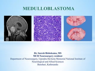
  • 1. cka Dr. Suresh Bishokama, MS MCH Neurosurgery resident Department of Neurosurgery, Upendra Devkota Memorial National Institute of Neurological and Allied Sciences Bansbari, Kathmandu MEDULLOBLASTOMA
  • 2.  Cushing Report: 1930  Coined Medulloblastoma: Bailey and Cushing  1983: PNET HISTORY
  • 3.  Medulloblastoma is the most common malignant brain tumor of childhood (WHO IV) and comprises up to 18-30% of all pediatric brain tumors.  Medulloblastomas are typically midline cerebellar lesions arising from the vermis in the inferior medullary velum; however, in older children and adults they can arise from the cerebellar hemispheres.  Medulloblastomas are related to primitive neuroectodermal tumors (PNETs) and occur exclusively in the posterior fossa. INTRODUCTION
  • 4.  Overall medulloblastomas account for 12-25 % of all pediatric CNS tumors, and 30-40% of pediatric posterior fossa tumors.  They are also seen in adults but only account for 0.4-1.0% of adult brain tumor.  M:F ratio of 2:1, although this is only true of group 3 and 4 tumors  They usually present in childhood with 77% of cases before the age of 19.  The median age of diagnosis is 9 years.  When diagnosed in adulthood, they typically present in the 3rd and 4th decades and are more likely to arise in atypical locations (see below).  When they present in adulthood, there is often a better prognosis. (Younger the poorer) EPIDEMIOLOGY
  • 5.  N Nitroso compound  Vegetables are protective  JC virus  Genetic conditions: Li-Fraumani, Gorlin, Turcot  P53, IGF, c-MYC and N-MYC oncogenic transcription factor, Apoptotic pathway: Bcl-2 ASSOCIATION
  • 6.  Medulloblastoma to arise from the external granule layer of the developing cerebellum and others have proposed that the cell of origin arises from a subventricular progenitor zone. PATHOGENESIS
  • 7.  WNT is a ligand for the transmembrane protein FRIZZLED and major cytoplasmic component of the WNT pathway is APC/GSK-3B/AXIN complex.  In absence of WNT this complex phosphorylates B catenin, thereby targeting it for degradation. B catenin in nucleus promotes cellular proliferation. WNT PATHWAY
  • 8. Sonig Hedge Hog (SHH)  Sonig Hedge Hog (SHH) is a diffusible mitogenic protein that binds to the transmembrane receptor protein (PATCHED 1-PTCH1).  In the absence of SHH, PTCH1 binds with SMO.  This complex inhibit glioma associated oncogene homolog (GLI) and hence cellular proliferation.  However, when PTCH1 binds with SHH, inhibitor effect of SMO-PTCH1 complex are reversed leading to activation of GLI and enhanced oncogenesis.
  • 9.  WNT tumours should be identified by two of the following markers:  Nuclear ß-catenin accumulation (IHC)  Monosomy 6 (whole chromosome loss) by FISH  CTNNB1 mutation  WNT pattern by DNA methylation or by gene expression profiling  SHH, Group 3 and Group 4  IHC  c-myc amplification (FISH)  Genome-wide methylation
  • 10.  Classic : (90%): Undifferentiated cells with hyperchromatic nuclei, (Homer wright rosette suggesting neuronal differentiation, Blue tumor  Desmoplastic : (6%): Classic + Glomeruli (pale island of collagen): Least aggressive  Nodularity MBEN : Diffuse variant in <3 years child: good prognosis  Large cell Anaplastic : (4%): early seed, high mitosis: poor prognosis HISTOLOGICAL SUBTYPES Homer-Wright rosettes composed of neoplastic cells concentrically arranged around fibrillary processes
  • 11.  Diffuse masses of small, undifferentiated oval or round cells.  Some medulloblastomas show neuronal, glial and other differentiation.  Nuclear moulding due to compactness inside cell.  Neuronal differentiation is manifested by neuropil and rosette formation.  Rosettes are groups of tumor cells arranged in a circle around a fibrillary center.  Mature neurons may also be found infrequently. Glial differentiation in some tumors is reflected by GFAP-positive cells. There may also be differentiation along oligodendroglial or ependymal lines. CLASSIC MEDULLOBLASTOMA
  • 12.  Pale nodules are composed uniform round to spindle shaped neuronal-appearing cells which are not as active mitotically as the surrounding darker tumor.  They are present within a reticulin-poor fine fibrillary background DESMOPLASTIC MB
  • 13.  Large cell anaplastic medulloblastoma composed of sheets of highly atypical cells.  A focus of necrosis is present just above the center of the image.  The tumor cells have irregular hyperchromatic nuclei with nuclear molding and little cytoplasm ANAPLASTIC MB
  • 14.  Medulloblastoma can spread along cerebrospinal fluid (CSF) pathways and also rarely to systemic sites (bone, lung).  Leptomeningeal dissemination constitutes the most common pattern of recurrence for Group 3 and Group 4 patients, and usually leads to a quick progressive clinical decline despite therapy.
  • 15. New type: Medulloblastoma NOS. CLINICOPATHOLOGICAL AND MOLECULAR CHARACTERISTICS OF MB SUBGROUPS CLINICOPATHOLOGICALAND MOLECULAR CHARACTERISTICS OF MB SUBGROUP CHARACTER WNT 10% SHH: 25% GP3: 25% GP4: 40%; MC GENDER M=F M=F M>F M>F PEAK AGE Children Children Infants Children OCCURRENCE Arise from foramen of luschka (CP angle) Laterally cerebellar hemisphere, and vermis 4th ventricle 4th ventricle ENHANCEMENT Intensely enhancing like ependymoma but diffusion restriction Intermediate Intermediate Usually non-enhancing HISTOLOGY Classic, rarely LCA Desmoplastic/MBEN, Classic, LCA Classic, LCA Classic, LCA GENE MUTATED CTNNB1, TP53 CTNNB1, TP53, 3q gain, 9p deletion MYC amp, OTX2, SMARCA4 KDM6A, MYCN SNCAIP duplication IHC B-catenin +(including nuclear and cytoplasmic) CTNNB1 mutation, GAB1 (-) Cytoplasmic B-Catenin+; GLI amplification; SMO mutation; MYCN+ GAB1+, YAB1+, PTCH1+, SMO+, P53± MYCN+++; YAB/GAB- Minimal amplification; CDK6+; YAB/GAB (-) METASTASIS Low Extremely low High Frequent PROGNOSIS Best prognosis Intermediate Poor Intermediate
  • 16. Molecular group / Immunoreactivity GAB1 ß-catenin YAP1 WNT Negative Nuclear + Cytoplasmic + Nuclear + Cytoplasmic SHH Cytoplasmic Cytoplasmic + Nuclear + Cytoplasmic Non-SHH/WNT Negative Cytoplasmic - Negative MOLECULAR SUBGROUPS AND IMMUNOREACTIVITY One of the techniques that can be implemented widely across the globe is IHC- based sub classification
  • 17. IMMUNOREACTIVITY/ MOLECULAR GROUP WNT SHH Non-SHH/WNT ß-catenin (Nuclear) + - - GAB-1 - + - YAP1 + + - p53 ± Molecular subgroups and immunoreactivity Surrogate IHC Additional for Group 3: c-myc amp (FISH) Additional for WNT: Monosomy Chr 6 (FISH)
  • 18.  Homer-Wright rosettes composed of neoplastic cells concentrically arranged around fibrillary processes are a common histological feature.  Isochromosome17q was confirmed as the most common genetic alteration in medulloblastomas, found in 28% of specimens.  Synaptophysin, Neuron enolase, neurofilament protein, tubulin  Metastases may have different immunohistology different from the actual primary. PATHOLOGY
  • 19.  Short time before diagnosis (<3months)  Morning headache, vomiting, lethargic  Features of raised ICP including papilloedema and diplopia (CN VI)  Truncal and appendicular ataxia, nystagmus, bulbar and facial palsy (brainstem),  Head tilt (tonsil descent in FM)  Macrocrania (infant),  Drop mets (Back pain, urinary retention and leg weakness): ~10-35% of MB seeds at the time of dx and Extraneural mets occurs in 5% (sometime promoted by shunt: 10-20%; overestimated ???) CLINICAL PRESENTATION
  • 20.  Classic features of a medulloblastoma on computed tomographic (CT) scan are increased density on the non-contrast scan, midline location, well-defined margins, and dense, homogeneous enhancement with injection of contrast, surrounding vasogenic edema.  Calcification (22%) and cyst formation (59%)  The hyperdensity on plain CT scan, seen in medulloblastoma and in some ependymomas, is secondary to the high cellularity of these tumors that have scanty cytoplasm and areas of desmoplasia. WORK UP
  • 21.  MRI: There is a high degree of variability of MR appearances of medulloblastoma.  T1 sequences are usually iso-hypointense to white matter and hyperintense on T2 sequences.  Tumor enhancement can be both homogeneous or heterogeneous.  Similar to ependymoma, approximately14% of medulloblastomas may show foraminal extension.  Banana sign (4th vent drapes around medulloblastoma unlike ependymoma where tumor encroach 4th vent)  HCP: 85-90% MRI BRAIN WITH CONTRAST
  • 26. FEATURES MEDULLOBLASTOMA EPENDYMOMAS PILOCYTIC ASTRO HEMANGIOBLASTOMA Origin Roof of 4th V Floor Cerebellar Cerebellar Clinical features Age Acute 5-15 Chronic 3-8 Chronic 5-15 Chronic 40-60 VHL: Young NE-CT Hyperdense (cellularity) Hypo Hypo Hypo Heterogenicity Less More Solid cystic Solid cystic T1W-MRI Hypo Hypo Hypo Hyper (also cyst) T2W-MRI Iso to Hyper Hyper Hyper Hyper, flow void Mural Nodule None None May be present May be present Enhancement Most (E); Non (gr 4) to intense (WNT) Intense Solid enhances Intense Perilesional edema - - - + Calcification Less More None Less Hemorrhage Less More Rare Less Extension into cisternal spaces Less More Nil Less Diffusion restriction Yes No No No MEDULLOBLASTOMA VS EPENDYMOMA
  • 27.  MR spectroscopy may also be distinctive  SHH little or no taurine low creatine  Group 3 or 4 Taurine peak high creatine MRS
  • 28.  MRI SPINE to assess the severity of diseases (Mets)  Leptomeningeal seeding on MRI is found in approximately 33% of patients.  The spinal canal is the most common location of seeding; however, the supratentorial compartment may also be involved.  Nodular or diffuse enhancement along the leptomeninges, nerve roots in the spinal canal, or cranial nerves in the CPA are common findings of CSF seeding. MRI SPINE
  • 29. MRI and CSF cytometry may miss 14% of seeding CSF cytometry
  • 32. 1. Establish histological diagnosis 2. Maximally resect tumor mass 3. Relieve hydrocephalus AIMS OF SURGERY
  • 33. MEDULLOBLASTOMA SURGICAL RESECTION HCP TREATED >3 YRS <3 YEARS STANDARD RISK POOR RISK CHEMOTHERAPY FOLLOW UP OR DELAY XRT OR LOCAL FIELD XRT PROTON CSI OR REDUCED DOSE CSI+ CHEMO CSI+ ADJ CHEMO (CCNU, CISPLATIN, VINCRISTINE OR CHEMO ON RESEARCH PROTOCOL FOLLOW RISK STRATIFICATION 1. Age (less than <3 or >3 years); Presence of disseminated disease (M1-4); Extent of surgical resection (<1.5 or >1.5cm residual) and Histological differentiation (5 years PFS among group: 77% vs 35-50%) Better outcome: Girls; WNT:good/ LCA/non SHH/WNT: Poor MEDULLOBLASTOMA FIG: PROPOSED ALGORITHM FOR TREATMENT OF PATIENT WITH MEDULLOBLATOMA XRT: OPTIMAL DOSE 35-40Gy to whole CSI + 10-15 Gy boost to tumor bed (PF) and to any spinal mets, all fractionated over 6-7 weeks Reduce dose by 20-25% or Chemo Chemotherapy improves 5-yr PFS survival (87% vs 33%) Low dose XRT 25 Gy
  • 34.  Currently risk stratification is based on age (less than 3 years of age), presence of disseminated disease (M1-4, and extent of surgical resection (<1.5 or >1.5cm residual) and histological differentiation along glial, ependymal or neuronal line. (5 yrs PFS among group: 77% vs 53%)  In cases in which there is greater than 1.5 cm of residual tumor, a repeat procedure should be considered, if safe and anatomically feasible, to place the patient in the best prognostic category.  Medulloblastoma is a radiosensitive tumor and incorporation of radiotherapy has become a standard of care in treatment of children older than 3 years of age. RISK STRATIFICATION PROGNOSTIC FACTORS Low-risk patients. Nuclear accumulation of CTNNB1* CTNNB1* mutations Monosomy stratifies *WNT subgroup markers High-risk patients: MYC/MYCN amplication and isochromosome 17q
  • 35.  It is generally agreed now that most patients do not require pre-operative shunting. However, 20-35% may need post-operative shunt.  Shunt risk of peritoneal dissemination: 10-20%.  Pre-operative shunt: Demerit: Tumoral hemorrhage, seeding, upward herniation.  If the drain continues to drain at the height of 20cm, hydrocephalus is shown to progress or pseudomeningocele develops. SHUNT OR NOT
  • 36. TREATMENT STRATEGY MECHANISM OF ACTION OF CHEMOTHERAPY DRUG MOA SIDE EFFECT CISPLATIN Induces cellular apoptosis by cross-linking DNA Ototoxicity and nephrotoxicity, dys-electrolytemia CYCLOPHOSPHAMIDE Alkylating agent Hemorrhagic cystitis, infertility LOMUSTINE Alkylating agent Myelosuppression, myeloid leukemia VINCRISTINE Microtubule inhibitor that prevents cell division by binding to the tubulin component of microtubules and leading to metaphase arrest Peripheral neuropathy. Muscle weakness, SEVERE CONSTIPATION
  • 37.  Radiation therapy (XRT) should begin ~30 days following definitive surgery.  Historically, the entirety of the cerebellum was radiated.  Therapy is delivered in daily fractions of 1.8 Gy to a final dose of 54 Gy - 59.4 Gy.  Craniospinal irradiation (CSI) is delivered to the entire brain and spine and given concurrently with primary site radiation for the first 13 days of therapy, to a total dose of 23.4 Gy (1.8x 13days) RADIOTHERAPY Posterior fossa ®: 1.8 Gy for 1 months (54-59.4 Gy) CSI ®: 1.8 Gy for 13 days (23.4 Gy) MEDULLOBLASTOMA
  • 38.  The current standard of care for average risk medulloblastoma among >3 yrs child is gross total resection, postoperative craniospinal radiation of 23.4 Gy with a tumor bed irradiation t of 54 Gy, followed by 12 months of chemotherapy.  Cyclophosphamide, vincristine, cisplatin and peripheral stem cell rescue. (5 years EFS ~70%)  Child <3 years: chemotherapy has been used to delay radiation therapy until nervous system matures or reduced (20-25% of total) dose of XRT.  Administration of focal radiation to tumor bed is insufficient to control medulloblastoma, even if there is no evidence of disseminated disease at diagnosis.  Consequently, irradiation of entire neuraxis is critical in the treatment of medulloblastoma. CURRENT STANDARD OF CARE
  • 39.  Cognitive decline, endocrine insufficiency, hearing loss, growth retardation, vascular complications, leukoencephalopathy and secondary malignancies. SIDE EFFECTS OF RADIATION
  • 40.  Proton therapy: Bragg peak: capacity to deliver targeted radiation while sparing normal structure. Target avoiding spinal growth plate, auditory canal, pituitary fossa. NOVEL IRRADIATION PROMISE
  • 41. MRI within 48 hours to minimize detection of non-tumoral post-operative changes Postoperative surveillance imaging of the brain and spine in patients with medulloblastoma is routinely employed at many institutions with 3–6 month intervals during the first 5 years following initial diagnosis in the hope of detecting recurrent disease earlier. . POST OP IMAGING
  • 42.  Post op neurological morbidity: 25-57%  Cerebral mutism (25%); Dysarthria persist after mutism recover.  Recurrence: Collin’s law to define period of recurrence (age at diagnosis + 9 months;  Autologous stem cell rescue to deliver chemotherapy at dose that may provide better response was tried limited by hematologic toxicity. OUTCOME
  • 43.  5 years survival:  M0: 70%  M1: 57%  M ≥2: 40%  Prognosis is most strongly influenced by molecular subtype  WNT: very good  SHH: infants good, others intermediate  group 3: poor  group 4: intermediate  Outcome is good among girls  Recurrence occurs in duration of 4-20 months.  Survival after recurrence: 5months (poor) PROGNOSIS
  • 44.  Expression of the c-erbB-2 (HER2/neu) oncogene is useful in the staging of medulloblastomas.  Increased c-erbB-2 expression reflects an increase in the proliferative activity of a tumor (widely used in breast cancer staging).  No CSF metastases, complete surgical resection and negative c-erbB-2 expression: 5-year-survival 100%.  No CSF metastases, complete surgical resection and positive c-erbB-2 expression: 5-year-survival 54%.  CSF metastases and/or incomplete surgical resection: 5-year-survival 20%. c-erbB-2 (HER2/neu) oncogene expression
  • 45.  Protein phosphatase inhibitor Norcantharidin for WNT: + nuclear B- catenin.  Smoothened (SMO) inhibitors: Vismodegib and Sonidegib targets Shh pathway  Suppress MYC-related pathway activity. FUTURE
  • 46.  GOOD 1. WNT 2. B Catenin 3. Classic, Desmoplastic CONFOUNDERS OF OUTCOME  BAD 1. GAB1, YAB1 2. TP53 3. Large/Anaplastic 4. MYC amplification
  • 47. Thank you  NATIONAL INSTITUTE OF NEUROLOGICAL AND ALLIED SCIENCES, BANSBARI, KATHMANDU MEDULLOBLASTOMA UPENDRA DEVKOTA MEMORIAL NATIONAL INSTITUTE OF NEUROLOGICALAND ALLIED SCIENCES, BANSBARI, KATHMANDU