SlideShare ist ein Scribd-Unternehmen logo
1 von 56
Bone And Joint Infection In Children
Dr Sunetra Roy
DCH, DNB (PEDIATRICS)
Introduction
 Bone infections in children are relatively common and
important because of their potential to cause permanent
disability.
 Early recognition and prompt institution of appropriate
medical and surgical therapy minimize permanent
damage.
 The risk is greatest if the physis (the growth plate of
bone) is damaged.
Epidemiology
 The median age 6 yr.∼
 Incidence = 1:5,000   
 Boys > Girls
 No predilection for any Race (Except Sickle cell disease – Blacks)
 In Previously healthy children- mostly hematogenous.
 Minor closed trauma - common preceding event ( 30% cases)∼
 Infection of bones can follow penetrating injuries or open
fractures.
 Following orthopedic surgery.
 Impaired host defenses increases the risk of skeletal infection.
Etiology of Osteomyelitis and Septic Arthritis
Infant 0-2 months Staphylococcus aureus
Streptococcus agalactiae
Gram-negative enteric bacteria
Candida
<5 y Staphylococcus aureus
Streptococcus pyogenes
Streptococcus pneumoniae
Kingella kingae
Haemophilus influenzae type b
>5 y S. aureus
S. Pyogenes
Adolescent Neisseria gonorrhoeae
MOST COMMON CLINICAL
ASSOCIATION
MICROORGANISM
Frequent microorganism in any type of
osteomyelitis
Staph. aureus
Common in nosocomial infection Enterobacteriaceae, Pseudomonas
aeruginosa, candida spp.
Foreign body–associated infection Coagulase-negative staphylococci or
other skin flora, atypical mycobacteria
Decubitus ulcers Streptococci and/or anaerobic bacteria
Populations in which tuberculosis is
prevalent
Mycobacterium tuberculosis
Exposure to kittens Bartonella henselae
Immunocompromised patients Aspergillus spp., Candida albicans, or
Mycobacteria spp.
Microorganisms Isolated fromPatients with Osteomyelitis
Pathogenesis
In the metaphysis, nutrient arteries branch into
non-anastomosing capillaries under the
physis make a sharp loop before entering
venous sinusoids draining into the marrow
( Hair-pin Ends)
Blood flow, sluggish and provides an ideal
environment for bacterial seeding
Relative paucity of phagocytic cells in this
area
Pathogenesis- In neonates and young infants
 Transphyseal blood vessels connect
the metaphysis and epiphysis
 commonly pus from the metaphysis
enters the joint space
 Result in abnormal growth and bone
or joint deformity
Joint involvement takes place when
the metaphysis is intra- articular as in
hip, ankle, shoulder, and elbow joint
or when subperiosteal pus ruptures
into the joint space
Pathogenesis- In child >18 months
 Spread through the cortex.
 Porous metaphyseal cortex provides easy assess for the exit
of exuate or pus
 Elevates the periosteum (thick periosteum loosly adherent to
the cortex)
 Subperiosteal pus collection(further impairing blood supply to
the cortex and metaphysis)
 Enough periosteal destruction soft tissue abcess.→
Pathogenesis…In later childhood and adolescence
The growth plate closes, hematogenous osteomyelitis more often
begins in the diaphysis and can spread to the entire
intramedullary canal.
Closed plate is relativley resistant to spread of infection.
 The periosteum becomes more adherent, favoring pus to
decompress through the periosteum
 The fluid formed seeks the path of least resistance from the
metaphysis
Clinical Features
 Age - greater in infants and toddlers than in older
children
 Sex - boys > girls, usually 2 : 1
 Signs and symptoms are highly dependent on the age of
the patient
 The initial s/s are often subtle
 The hallmark of osteomyelitis is fever plus localised bony
tenderness
 Neonates may exhibit pseudoparalysis or pain with movement
of the affected extremity- Diaper changes
 Half of neonates do not have fever and may not appear ill
Clinical Features…
 Signs and symptoms
 Older infants and children are more likely to have fever,
pain, and localizing signs such as edema, erythema,
and warmth
 With involvement of the lower extremities, limp or refusal to
walk
 Careful Physical examination may reveal focal bony
tenderness.
Distribution of Affected Bones in Acute Hematogenous
Osteomyelitis
• Usually only a single site
of bone or joint
involvement, the exception
is osteomyelitis in
neonates.
• Several bones are
infected in <10% of cases;
BONE %
Femur 23-28
Tibia 20-24
Humerus 5-13
Radius 5-6
Phalanx 3-5
Pelvis 4-8
Calcaneus 4-8
Ulna 4-8
Metatarsal ∼2
Vertebrae ∼2
Sacrum ∼2
Clavicle ∼2
Skull ∼1
Diagnosis
 Children with acute bone pain and systemic signs of
sepsis should be considered to have acute hematogenous
Osteomyelitis until proved otherwise.
 Diagnosis may be established if a patient fulfills two of
the following criteria:
1. Bone aspiration yield pus
2. Bacterial culture of bone or blood positive
3. Presence of the classical s/s of acute osteomyelitis
4. Radiographic changes typical for osteomyelitis.
LABORATORY FINDINGS
 No specific laboratory tests.
 Elevations in peripheral WBC, ESR, and CRP –
 ESR-Nonspecific acute phase reactant
 Increased 48-72 hrs
 Increased in 90% of cases
 Not affected by antibiotic tx
 CRP- Increased in 98% of cases
 Blood culture-
 Positive in 30-50%
 Decreased with antibiotic
 48 hours to get most organisms
Radiology
 Plain xray
 Sensitivity 43-75%
 Specificity 75-83%
 Soft tissue swelling 48hrs
 Periosteal reaction 5-7days
 Osteolysis 7-14 days of infection(need 30-50% bone loss)
 Magnetic Rsonance Imaging (MRI)
 Computed Tomography (CT)
 Radionuclide Study
Plain radiography
 Bone changes - not seen for at least 10-14 days after the
onset of infection.
• Plain radiographs-normal in
the first 7 to 10 days
• Soft tissue swelling with loss
of the normal soft tissue
planes is seen before bone
changes become apparent
•Periosteal elevation or thickening -
new bone formation, pus, or
reactive edema from adjacent soft
tissue infection
Radionuclide imaging…
 Radionuclide imaging can be valuable in suspected bone
infections especially if multiple foci are suspected
 Bone scans in acute haematogenous osteomyelitis have a
sensitivity of 84% to 100% and a specificity of 40% to
96%.
(N Susan Stott, Journal of Orthopaedic Surgery; Review
article: Paediatric bone and joint infection)
 Can detect osteomyelitis within 24–48 hr after onset
of symptoms
 The sensitivity in neonates is much lower owing to
poor bone mineralization
 A) Plain radiographs of the ankle demonstrate deep soft tissue swelling inferior to
the medial malleolus. B) A technetium 99m bone scan shows increased uptake in
the distal tibia in the blood flow and bone uptake (four hour) phases.
Early Osteomyelitis, with pain and fever
Bone Scan…
Tc99
24-48hrs +ve
Decreased uptake
in early phase d/t
increased pressure
Ultrasound
 can detect features of osteomyelitis several days earlier Xray
(predominately in children).
 Acute osteomyelitis is recognized by elevation of the periosteum
by a hypoechoic layer
 Soft tissue abscesses related to chronic osteomyelitis are
identified as hypoechoic or anechoic fluid collections, which may
extend around the bony contours.
 Finally, US guided aspiration of joints
MRI
 have comparable positive predictive value to radionuclide
imaging in acute osteomyelitis
 particularly useful in the evaluation of vertebral osteomyelitis
and diskitis
Bone Aspiration
 A bacteriologic diagnosis is made by culturing the
involved bone or pus, once a clinical diagnosis of acute
osteomyelitis is established.
 Useful to determine whether an abscess is present.
 The organism detection rate is increased to 75% to 80%
by aspiration of the affected bone
 K. kingae may need to be identified by polymerase chain
reaction
 Also useful in determining the future course of therapy
of the child
Differential Diagnosis
 Trauma
 Cellulitis
 Myositis and pyomyositis
 Leukemia
 Neuroblastoma with bone involvement
 Primary bone tumors (Ewing sarcoma)
 Septic arthritis
Classification-
Acute hematogenous Osteomyelitis
Subacute hematogenous Osteomyelitis
Chronic Multifocal Osteomyelitis
Parameters
Subacute Acute
WBC Frequently normal Frequently elevated
ESR Frequently elevated Frequently elevated
Blood Cultures Rarely Positive 50% Positive
Bone Cultures 60% Positive 90% Positive
Localization Diaphysis, metaphysis,
epiphysis, cross physis
Metaphysis
Pain Mild to Moderate Severe
Systemic Illness No Fever, malaise
Loss of function No or minimal Marked
Prior antibiotics 30%-40% Occasional
Initial radiograph Frequently abnormal Bone normal
Brodie’s abscess, central oval lucency surrounded
By reactive sclerosis usually within or close to the
Metaphysis and the lesion may extend across the
physis.
Garré sclerosing osteomyelitis, or chronic nonsuppurative sclerosing
osteomyelitis, is a form of chronic osteomyelitis. Mild inflammation and infection
lead to subperiosteal bone deposition. It is frequently asymptomatic. The
characteristic radiographic appearance is an area of periosteal proliferation
surrounded by successive layers of condensed cortical bone (arrows), described
as an onion skin appearance.
TREATMENT OF OSTEOMYELITIS
Duration of antibiotic therapy
 Most infections- eg S. aureus, minimal duration is 21-28 days, if
prompt resolution of signs and symptoms (within 5-7 days)
and the CRP and ESR have normalized;
 a total of 4-6 wk of therapy may be required.
 For group A streptococcus, S. pneumoniae, or H. influenzae
type b, treatment duration maybe shorter.
 A total of 7-10 postoperative days of treatment is adequate
for Pseudomonas osteochondritis when thorough curettage of
infected tissue has been performed.
 Immunocompromised patients, mycobacterial or fungal
infection -require prolonged courses of therapy.
Indication of Surgical Treatment
 Abscess collection (Subperiosteal , soft tissue, or
intramedullary)
 Patient is not responding to appropriate antibiotic
therapy after a negative bone aspiration
(If a child with acute hematogenous osteomyelitis does not show
symptomatic improvement with decrease in swelling and
tenderness after 36-48 hrs of appropriate antibiotic treatment, the
bone should be aspirated again and consideration given to surgical
drainage)
 In subacute Osteomyelitis when debridement is
necessary (granulation tissue within the cavity even
though no pus)
 Radiographic lesion, sequestrum
Complications
 Bone abscess
 Bacteremia
 Fracture
 Loosening of the prosthetic implant
 Overlying soft-tissue cellulitis
 Draining soft-tissue sinus tracts
 Growth disturbance
Prognosis
 Improvement in signs and symptoms is rapid when timely
intervened.
 Failure to improve or worsening by 72 hr requires review of
antibiotic therapy, the need for surgical intervention, or the
correctness of the diagnosis.
 Recurrence of disease and development of chronic infection after
treatment occur in <10% of patients.
Septic Arthritis
 An infection of a synovial joint which may occur in all age
groups in children
 Has a specific infantile form affecting the infant from birth
to the first year of life
 Relatively uncommon
Septic Arthritis
 More common in infants and toddlers than in older
children
 Half of all cases occur by 2 yr of age and three fourths of all
cases occur by 5 yr of age
 Immuno-compromised hosts may have a higher incidence
ETIOLOGY
Pathogenesis
 Results from hematogenous seeding of the synovial space
 Organisms enter the joint space by direct inoculation or
extension from a contiguous focus.
 The synovial membrane (rich vascular supply and lacks a
basement membrane) providing an ideal environment for
hematogenous seeding.
 Cytokines stimulate chemotaxis of neutrophils into the joint
space, proteolytic enzymes and elastases are released by
neutrophils cartilage damage
 Increased pressure in joint space from can compromise the
vascular supply and induce pressure necrosis of the cartilage.
Pathogenesis….
 May be associated with acute osteomyelitis esp in the
proximal femur
 Routes of Infection-
 Usually hematogenous
 Trauma (penetrating injuries)
 Post procedure (arthroscopy, prosthetic joint surgery, intra-
articular steroid injection, and orthopedic surgery)
Pathology
 Acute inflammatory reaction.
 Degradation of the articular cartilage begins within 8
hours of infection.
 In neonates, metaphyseal osteomyelitis is often
associated with septic arthritis due to the presence of
transphyseal blood vessels that disappear by age 6 to 12
months
 Sixty to 100% of neonates with septic arthritis will
have adjacent osteomyelitis
CLINICAL MANIFESTATIONS
 Hot, swollen joint painful on any movement indicates a septic
arthritis until proved otherwise
 In neonate, present as a pseudo-paralysis of one limb
 Erythema and edema of the skin and soft tissue overlying
the site of infection are seen earlier in suppurative
arthritis than in osteomyelitis
 Joint kept at position of ease.
Distribution of Affected Joints in Acute
Suppurative Arthritis
BONE %
Knee ∼40
Hip 22-40
Ankle 4-13
Elbow 8-12
Wrist 1-4
Shoulder ∼3
Interphalangeal <1
Metatarsal <1
Sacroiliac <1
Acromioclavicular <1
Metacarpal <1
Toe ∼1
Investigations
 The investigations are similar to those required in
osteomyelitis
 Blood culture
 Aspiration of the joint fluid for Gram stain and culture,
definitive diagnostic technique and provides the optimal
specimen for culture to confirm the diagnosis
 Elevations of WBCs, ESR, and CRP are very sensitive for joint
infections but are nonspecific
 Synovial fluid analysis – Cell Counts >50,000-100,000 cells/mm3
generally
indicate an infectious process. Synovial fluid characteristics of septic
arthritis can suggest infection but are not sufficiently specific to exclude
infection.
Infected Fluid
 Appearance / Clarity: Turbid to very turbid (N- Clear and colorless)
 Viscosity: Greatly reduced (similar to water) (N-Very viscous)
 Leukocytes / cubic cm: 15,000- >200,000 (N : Average 63; Range: 13-180.)
usually >50,000 and >100,000 is virtually diagnostic
 Percent polymorphoneuclear: 50-100% (N-<25%)
90% almost always indicates infection
 Mucin clot: Poor to very poor (N-Good)
 Glucose level difference versus plasma: >40 mg/100ml less than plasma (N-
Within 10 mg/100ml of plasma concentration)
Differential Diagnosis
Depends on the joint or joints involved and the age of the
patient
 Hip
 toxic synovitis, Legg-Calvé-Perthes disease, slipped capital femoral
epiphysis, psoas abscess, and proximal femoral, pelvic, or vertebral
osteomyelitis as well as diskitis
 Knee
 distal femoral or proximal tibial osteomyelitis, pauciarticular
rheumatoid arthritis, and referred pain from the hip
 Others like trauma, cellulitis, pyomyositis, sickle cell disease,
hemophilia, and Henoch-Schönlein purpura, also Reactive
arthritis, Acute rheumatic fever (multiple joints involved)
Radiographic Evaluation
 Plain Xrays
 Widening of the joint capsule, soft-tissue edema, and obliteration of
normal fat lines
 Ultrasonography
 helpful in identifying effusions in deep joints such as the hip
 may serve as an aid in performing hip aspiration
 CT
 MRI
 Both are useful in confirming the presence of joint fluid in patients
with suspected osteoarthritis infections
 MRI may be useful in excluding adjacent osteomyelitis
 Radionuclide studies
 In suppurative arthritis, three-phase imaging with technetium-99
methylene diphosphonate shows symmetric uptake on both sides of
the joint, limited to the bony structures adjacent to the joint
TREATMENT
 Hip joint
 An infection of the hip joint is an emergency, as potential for
the development of avascular necrosis of the femoral head.
 Aspiratiion through Fluroscopy guided
For joints other than the hip, daily aspirations of synovial fluid
may be required
• If fluid continues to accumulate after 4–5 days, arthrotomy is
needed
APView of Left Hip showing avascularnecrosis left head of
femur
 In selected patients, obtaining a plain radiograph of the joint
before completing therapy can provide evidence (typically
periosteal new bone) of a previously unappreciated contiguous
site of osteomyelitis that would likely prolong antibiotic
treatment.
 Oral antibiotics can be used to complete therapy once the
patient is afebrile for 48-72 hr and is clearly improving
Monitoring of patient
 Failure to improve or worsening by 72 hr requires
 review of the appropriateness of the antibiotic therapy
 the need for surgical intervention
 the correctness of the diagnosis
 Failure of either of acute-phase reactants to follow the
usual course should raise concerns about the adequacy of
therapy
 Recurrence of disease and development of chronic
infection after treatment occur in <10% of patients.
THANK YOU

Weitere ähnliche Inhalte

Was ist angesagt?

Bone and joint infection in children
Bone and joint infection in childrenBone and joint infection in children
Bone and joint infection in children
Sunil Agrawal
 
Chronic osteomyelitis
Chronic osteomyelitisChronic osteomyelitis
Chronic osteomyelitis
group7usmkk
 
Approach to limping child
Approach to limping childApproach to limping child
Approach to limping child
Hardik Pawar
 

Was ist angesagt? (20)

Slipped capital femoral epiphysis
Slipped capital femoral epiphysisSlipped capital femoral epiphysis
Slipped capital femoral epiphysis
 
Unusual presentation of psoas abscess _a case
Unusual presentation of psoas abscess  _a caseUnusual presentation of psoas abscess  _a case
Unusual presentation of psoas abscess _a case
 
Acute osteomyelitis
Acute osteomyelitisAcute osteomyelitis
Acute osteomyelitis
 
Bone and joint infection in children
Bone and joint infection in childrenBone and joint infection in children
Bone and joint infection in children
 
Psoas abscess
Psoas abscessPsoas abscess
Psoas abscess
 
Septic arthritis in children
Septic arthritis in childrenSeptic arthritis in children
Septic arthritis in children
 
Acute and Chronic Osteomyelitis - Infection of Bone
Acute and Chronic Osteomyelitis - Infection of BoneAcute and Chronic Osteomyelitis - Infection of Bone
Acute and Chronic Osteomyelitis - Infection of Bone
 
Chronic osteomyelitis
Chronic osteomyelitisChronic osteomyelitis
Chronic osteomyelitis
 
Psoas abscess
Psoas abscessPsoas abscess
Psoas abscess
 
Infected nonunion tibia
Infected  nonunion tibiaInfected  nonunion tibia
Infected nonunion tibia
 
Bone and joint infections: Osteomyelitis, Septic Arthritis
Bone and joint infections: Osteomyelitis, Septic ArthritisBone and joint infections: Osteomyelitis, Septic Arthritis
Bone and joint infections: Osteomyelitis, Septic Arthritis
 
Acute osteomyelitis
Acute osteomyelitisAcute osteomyelitis
Acute osteomyelitis
 
Spondyloarthropathy
SpondyloarthropathySpondyloarthropathy
Spondyloarthropathy
 
Skeletal Tuberculosis
Skeletal TuberculosisSkeletal Tuberculosis
Skeletal Tuberculosis
 
Perthes Disease LCPD
Perthes Disease LCPDPerthes Disease LCPD
Perthes Disease LCPD
 
Acute infections of bones and joints
Acute infections of bones and jointsAcute infections of bones and joints
Acute infections of bones and joints
 
Tuberculosis of the hip
Tuberculosis of the hipTuberculosis of the hip
Tuberculosis of the hip
 
Juvenile idiopathic arthritis (JIA)
Juvenile idiopathic arthritis (JIA)Juvenile idiopathic arthritis (JIA)
Juvenile idiopathic arthritis (JIA)
 
Approach to limping child
Approach to limping childApproach to limping child
Approach to limping child
 
Inflamatory arthritis
Inflamatory arthritisInflamatory arthritis
Inflamatory arthritis
 

Ähnlich wie Bone infection in children ppt

Ähnlich wie Bone infection in children ppt (20)

Bone+joint infections
Bone+joint infectionsBone+joint infections
Bone+joint infections
 
Acute musculoskeletal infections in children
Acute musculoskeletal infections in childrenAcute musculoskeletal infections in children
Acute musculoskeletal infections in children
 
Osteomyelitis
OsteomyelitisOsteomyelitis
Osteomyelitis
 
Osteomyelitis
OsteomyelitisOsteomyelitis
Osteomyelitis
 
bone diseases.pptx
bone diseases.pptxbone diseases.pptx
bone diseases.pptx
 
Osteomyelitis
OsteomyelitisOsteomyelitis
Osteomyelitis
 
Osteomyelitis in Children
Osteomyelitis in ChildrenOsteomyelitis in Children
Osteomyelitis in Children
 
Principles of antibiotic use in management of osteomyelitis
Principles of antibiotic use in management of osteomyelitisPrinciples of antibiotic use in management of osteomyelitis
Principles of antibiotic use in management of osteomyelitis
 
Bone and Joint Infection
Bone and Joint InfectionBone and Joint Infection
Bone and Joint Infection
 
Osteomyelitis
OsteomyelitisOsteomyelitis
Osteomyelitis
 
Acute osteomyelitis
Acute osteomyelitisAcute osteomyelitis
Acute osteomyelitis
 
Acute osteomyelitis
Acute osteomyelitisAcute osteomyelitis
Acute osteomyelitis
 
Introduction to radiological diagnosis of osteomyelitis for undergraduates
Introduction to radiological diagnosis of osteomyelitis for undergraduatesIntroduction to radiological diagnosis of osteomyelitis for undergraduates
Introduction to radiological diagnosis of osteomyelitis for undergraduates
 
Septicarthritis
Septicarthritis Septicarthritis
Septicarthritis
 
Bone Infections
Bone InfectionsBone Infections
Bone Infections
 
Osteomyelitis.ppt
Osteomyelitis.pptOsteomyelitis.ppt
Osteomyelitis.ppt
 
Osteomyelitis
OsteomyelitisOsteomyelitis
Osteomyelitis
 
Osteosarcoma
Osteosarcoma Osteosarcoma
Osteosarcoma
 
Orthopedics 5th year, 2nd lecture (Dr. Omar Barawi)
Orthopedics 5th year, 2nd lecture (Dr. Omar Barawi)Orthopedics 5th year, 2nd lecture (Dr. Omar Barawi)
Orthopedics 5th year, 2nd lecture (Dr. Omar Barawi)
 
Osteomyelitis
OsteomyelitisOsteomyelitis
Osteomyelitis
 

Kürzlich hochgeladen

Russian Call Girls Lucknow Just Call 👉👉7877925207 Top Class Call Girl Service...
Russian Call Girls Lucknow Just Call 👉👉7877925207 Top Class Call Girl Service...Russian Call Girls Lucknow Just Call 👉👉7877925207 Top Class Call Girl Service...
Russian Call Girls Lucknow Just Call 👉👉7877925207 Top Class Call Girl Service...
adilkhan87451
 

Kürzlich hochgeladen (20)

Call Girls in Delhi Triveni Complex Escort Service(🔝))/WhatsApp 97111⇛47426
Call Girls in Delhi Triveni Complex Escort Service(🔝))/WhatsApp 97111⇛47426Call Girls in Delhi Triveni Complex Escort Service(🔝))/WhatsApp 97111⇛47426
Call Girls in Delhi Triveni Complex Escort Service(🔝))/WhatsApp 97111⇛47426
 
Premium Call Girls In Jaipur {8445551418} ❤️VVIP SEEMA Call Girl in Jaipur Ra...
Premium Call Girls In Jaipur {8445551418} ❤️VVIP SEEMA Call Girl in Jaipur Ra...Premium Call Girls In Jaipur {8445551418} ❤️VVIP SEEMA Call Girl in Jaipur Ra...
Premium Call Girls In Jaipur {8445551418} ❤️VVIP SEEMA Call Girl in Jaipur Ra...
 
Night 7k to 12k Navi Mumbai Call Girl Photo 👉 BOOK NOW 9833363713 👈 ♀️ night ...
Night 7k to 12k Navi Mumbai Call Girl Photo 👉 BOOK NOW 9833363713 👈 ♀️ night ...Night 7k to 12k Navi Mumbai Call Girl Photo 👉 BOOK NOW 9833363713 👈 ♀️ night ...
Night 7k to 12k Navi Mumbai Call Girl Photo 👉 BOOK NOW 9833363713 👈 ♀️ night ...
 
Call Girls Ahmedabad Just Call 9630942363 Top Class Call Girl Service Available
Call Girls Ahmedabad Just Call 9630942363 Top Class Call Girl Service AvailableCall Girls Ahmedabad Just Call 9630942363 Top Class Call Girl Service Available
Call Girls Ahmedabad Just Call 9630942363 Top Class Call Girl Service Available
 
Top Quality Call Girl Service Kalyanpur 6378878445 Available Call Girls Any Time
Top Quality Call Girl Service Kalyanpur 6378878445 Available Call Girls Any TimeTop Quality Call Girl Service Kalyanpur 6378878445 Available Call Girls Any Time
Top Quality Call Girl Service Kalyanpur 6378878445 Available Call Girls Any Time
 
Manyata Tech Park ( Call Girls ) Bangalore ✔ 6297143586 ✔ Hot Model With Sexy...
Manyata Tech Park ( Call Girls ) Bangalore ✔ 6297143586 ✔ Hot Model With Sexy...Manyata Tech Park ( Call Girls ) Bangalore ✔ 6297143586 ✔ Hot Model With Sexy...
Manyata Tech Park ( Call Girls ) Bangalore ✔ 6297143586 ✔ Hot Model With Sexy...
 
Most Beautiful Call Girl in Bangalore Contact on Whatsapp
Most Beautiful Call Girl in Bangalore Contact on WhatsappMost Beautiful Call Girl in Bangalore Contact on Whatsapp
Most Beautiful Call Girl in Bangalore Contact on Whatsapp
 
Models Call Girls In Hyderabad 9630942363 Hyderabad Call Girl & Hyderabad Esc...
Models Call Girls In Hyderabad 9630942363 Hyderabad Call Girl & Hyderabad Esc...Models Call Girls In Hyderabad 9630942363 Hyderabad Call Girl & Hyderabad Esc...
Models Call Girls In Hyderabad 9630942363 Hyderabad Call Girl & Hyderabad Esc...
 
Jogeshwari ! Call Girls Service Mumbai - 450+ Call Girl Cash Payment 90042684...
Jogeshwari ! Call Girls Service Mumbai - 450+ Call Girl Cash Payment 90042684...Jogeshwari ! Call Girls Service Mumbai - 450+ Call Girl Cash Payment 90042684...
Jogeshwari ! Call Girls Service Mumbai - 450+ Call Girl Cash Payment 90042684...
 
Model Call Girls In Chennai WhatsApp Booking 7427069034 call girl service 24 ...
Model Call Girls In Chennai WhatsApp Booking 7427069034 call girl service 24 ...Model Call Girls In Chennai WhatsApp Booking 7427069034 call girl service 24 ...
Model Call Girls In Chennai WhatsApp Booking 7427069034 call girl service 24 ...
 
Russian Call Girls Lucknow Just Call 👉👉7877925207 Top Class Call Girl Service...
Russian Call Girls Lucknow Just Call 👉👉7877925207 Top Class Call Girl Service...Russian Call Girls Lucknow Just Call 👉👉7877925207 Top Class Call Girl Service...
Russian Call Girls Lucknow Just Call 👉👉7877925207 Top Class Call Girl Service...
 
Saket * Call Girls in Delhi - Phone 9711199012 Escorts Service at 6k to 50k a...
Saket * Call Girls in Delhi - Phone 9711199012 Escorts Service at 6k to 50k a...Saket * Call Girls in Delhi - Phone 9711199012 Escorts Service at 6k to 50k a...
Saket * Call Girls in Delhi - Phone 9711199012 Escorts Service at 6k to 50k a...
 
Premium Bangalore Call Girls Jigani Dail 6378878445 Escort Service For Hot Ma...
Premium Bangalore Call Girls Jigani Dail 6378878445 Escort Service For Hot Ma...Premium Bangalore Call Girls Jigani Dail 6378878445 Escort Service For Hot Ma...
Premium Bangalore Call Girls Jigani Dail 6378878445 Escort Service For Hot Ma...
 
Call Girls Hosur Just Call 9630942363 Top Class Call Girl Service Available
Call Girls Hosur Just Call 9630942363 Top Class Call Girl Service AvailableCall Girls Hosur Just Call 9630942363 Top Class Call Girl Service Available
Call Girls Hosur Just Call 9630942363 Top Class Call Girl Service Available
 
Top Rated Hyderabad Call Girls Erragadda ⟟ 9332606886 ⟟ Call Me For Genuine ...
Top Rated  Hyderabad Call Girls Erragadda ⟟ 9332606886 ⟟ Call Me For Genuine ...Top Rated  Hyderabad Call Girls Erragadda ⟟ 9332606886 ⟟ Call Me For Genuine ...
Top Rated Hyderabad Call Girls Erragadda ⟟ 9332606886 ⟟ Call Me For Genuine ...
 
Call Girls Kolkata Kalikapur 💯Call Us 🔝 8005736733 🔝 💃 Top Class Call Girl Se...
Call Girls Kolkata Kalikapur 💯Call Us 🔝 8005736733 🔝 💃 Top Class Call Girl Se...Call Girls Kolkata Kalikapur 💯Call Us 🔝 8005736733 🔝 💃 Top Class Call Girl Se...
Call Girls Kolkata Kalikapur 💯Call Us 🔝 8005736733 🔝 💃 Top Class Call Girl Se...
 
The Most Attractive Hyderabad Call Girls Kothapet 𖠋 9332606886 𖠋 Will You Mis...
The Most Attractive Hyderabad Call Girls Kothapet 𖠋 9332606886 𖠋 Will You Mis...The Most Attractive Hyderabad Call Girls Kothapet 𖠋 9332606886 𖠋 Will You Mis...
The Most Attractive Hyderabad Call Girls Kothapet 𖠋 9332606886 𖠋 Will You Mis...
 
All Time Service Available Call Girls Marine Drive 📳 9820252231 For 18+ VIP C...
All Time Service Available Call Girls Marine Drive 📳 9820252231 For 18+ VIP C...All Time Service Available Call Girls Marine Drive 📳 9820252231 For 18+ VIP C...
All Time Service Available Call Girls Marine Drive 📳 9820252231 For 18+ VIP C...
 
8980367676 Call Girls In Ahmedabad Escort Service Available 24×7 In Ahmedabad
8980367676 Call Girls In Ahmedabad Escort Service Available 24×7 In Ahmedabad8980367676 Call Girls In Ahmedabad Escort Service Available 24×7 In Ahmedabad
8980367676 Call Girls In Ahmedabad Escort Service Available 24×7 In Ahmedabad
 
Trichy Call Girls Book Now 9630942363 Top Class Trichy Escort Service Available
Trichy Call Girls Book Now 9630942363 Top Class Trichy Escort Service AvailableTrichy Call Girls Book Now 9630942363 Top Class Trichy Escort Service Available
Trichy Call Girls Book Now 9630942363 Top Class Trichy Escort Service Available
 

Bone infection in children ppt

  • 1. Bone And Joint Infection In Children Dr Sunetra Roy DCH, DNB (PEDIATRICS)
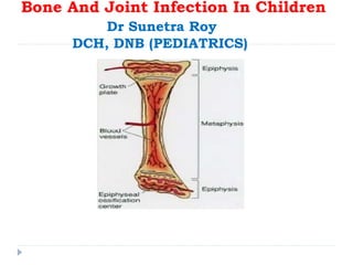
  • 2. Introduction  Bone infections in children are relatively common and important because of their potential to cause permanent disability.  Early recognition and prompt institution of appropriate medical and surgical therapy minimize permanent damage.  The risk is greatest if the physis (the growth plate of bone) is damaged.
  • 3.
  • 4. Epidemiology  The median age 6 yr.∼  Incidence = 1:5,000     Boys > Girls  No predilection for any Race (Except Sickle cell disease – Blacks)  In Previously healthy children- mostly hematogenous.  Minor closed trauma - common preceding event ( 30% cases)∼  Infection of bones can follow penetrating injuries or open fractures.  Following orthopedic surgery.  Impaired host defenses increases the risk of skeletal infection.
  • 5. Etiology of Osteomyelitis and Septic Arthritis Infant 0-2 months Staphylococcus aureus Streptococcus agalactiae Gram-negative enteric bacteria Candida <5 y Staphylococcus aureus Streptococcus pyogenes Streptococcus pneumoniae Kingella kingae Haemophilus influenzae type b >5 y S. aureus S. Pyogenes Adolescent Neisseria gonorrhoeae
  • 6. MOST COMMON CLINICAL ASSOCIATION MICROORGANISM Frequent microorganism in any type of osteomyelitis Staph. aureus Common in nosocomial infection Enterobacteriaceae, Pseudomonas aeruginosa, candida spp. Foreign body–associated infection Coagulase-negative staphylococci or other skin flora, atypical mycobacteria Decubitus ulcers Streptococci and/or anaerobic bacteria Populations in which tuberculosis is prevalent Mycobacterium tuberculosis Exposure to kittens Bartonella henselae Immunocompromised patients Aspergillus spp., Candida albicans, or Mycobacteria spp. Microorganisms Isolated fromPatients with Osteomyelitis
  • 7. Pathogenesis In the metaphysis, nutrient arteries branch into non-anastomosing capillaries under the physis make a sharp loop before entering venous sinusoids draining into the marrow ( Hair-pin Ends) Blood flow, sluggish and provides an ideal environment for bacterial seeding Relative paucity of phagocytic cells in this area
  • 8. Pathogenesis- In neonates and young infants  Transphyseal blood vessels connect the metaphysis and epiphysis  commonly pus from the metaphysis enters the joint space  Result in abnormal growth and bone or joint deformity Joint involvement takes place when the metaphysis is intra- articular as in hip, ankle, shoulder, and elbow joint or when subperiosteal pus ruptures into the joint space
  • 9. Pathogenesis- In child >18 months  Spread through the cortex.  Porous metaphyseal cortex provides easy assess for the exit of exuate or pus  Elevates the periosteum (thick periosteum loosly adherent to the cortex)  Subperiosteal pus collection(further impairing blood supply to the cortex and metaphysis)  Enough periosteal destruction soft tissue abcess.→
  • 10. Pathogenesis…In later childhood and adolescence The growth plate closes, hematogenous osteomyelitis more often begins in the diaphysis and can spread to the entire intramedullary canal. Closed plate is relativley resistant to spread of infection.  The periosteum becomes more adherent, favoring pus to decompress through the periosteum  The fluid formed seeks the path of least resistance from the metaphysis
  • 11.
  • 12. Clinical Features  Age - greater in infants and toddlers than in older children  Sex - boys > girls, usually 2 : 1  Signs and symptoms are highly dependent on the age of the patient  The initial s/s are often subtle  The hallmark of osteomyelitis is fever plus localised bony tenderness  Neonates may exhibit pseudoparalysis or pain with movement of the affected extremity- Diaper changes  Half of neonates do not have fever and may not appear ill
  • 13. Clinical Features…  Signs and symptoms  Older infants and children are more likely to have fever, pain, and localizing signs such as edema, erythema, and warmth  With involvement of the lower extremities, limp or refusal to walk  Careful Physical examination may reveal focal bony tenderness.
  • 14. Distribution of Affected Bones in Acute Hematogenous Osteomyelitis • Usually only a single site of bone or joint involvement, the exception is osteomyelitis in neonates. • Several bones are infected in <10% of cases; BONE % Femur 23-28 Tibia 20-24 Humerus 5-13 Radius 5-6 Phalanx 3-5 Pelvis 4-8 Calcaneus 4-8 Ulna 4-8 Metatarsal ∼2 Vertebrae ∼2 Sacrum ∼2 Clavicle ∼2 Skull ∼1
  • 15. Diagnosis  Children with acute bone pain and systemic signs of sepsis should be considered to have acute hematogenous Osteomyelitis until proved otherwise.  Diagnosis may be established if a patient fulfills two of the following criteria: 1. Bone aspiration yield pus 2. Bacterial culture of bone or blood positive 3. Presence of the classical s/s of acute osteomyelitis 4. Radiographic changes typical for osteomyelitis.
  • 16. LABORATORY FINDINGS  No specific laboratory tests.  Elevations in peripheral WBC, ESR, and CRP –  ESR-Nonspecific acute phase reactant  Increased 48-72 hrs  Increased in 90% of cases  Not affected by antibiotic tx  CRP- Increased in 98% of cases  Blood culture-  Positive in 30-50%  Decreased with antibiotic  48 hours to get most organisms
  • 17. Radiology  Plain xray  Sensitivity 43-75%  Specificity 75-83%  Soft tissue swelling 48hrs  Periosteal reaction 5-7days  Osteolysis 7-14 days of infection(need 30-50% bone loss)  Magnetic Rsonance Imaging (MRI)  Computed Tomography (CT)  Radionuclide Study
  • 18. Plain radiography  Bone changes - not seen for at least 10-14 days after the onset of infection. • Plain radiographs-normal in the first 7 to 10 days • Soft tissue swelling with loss of the normal soft tissue planes is seen before bone changes become apparent •Periosteal elevation or thickening - new bone formation, pus, or reactive edema from adjacent soft tissue infection
  • 19.
  • 20.
  • 21.
  • 22. Radionuclide imaging…  Radionuclide imaging can be valuable in suspected bone infections especially if multiple foci are suspected  Bone scans in acute haematogenous osteomyelitis have a sensitivity of 84% to 100% and a specificity of 40% to 96%. (N Susan Stott, Journal of Orthopaedic Surgery; Review article: Paediatric bone and joint infection)  Can detect osteomyelitis within 24–48 hr after onset of symptoms  The sensitivity in neonates is much lower owing to poor bone mineralization
  • 23.  A) Plain radiographs of the ankle demonstrate deep soft tissue swelling inferior to the medial malleolus. B) A technetium 99m bone scan shows increased uptake in the distal tibia in the blood flow and bone uptake (four hour) phases. Early Osteomyelitis, with pain and fever Bone Scan… Tc99 24-48hrs +ve Decreased uptake in early phase d/t increased pressure
  • 24. Ultrasound  can detect features of osteomyelitis several days earlier Xray (predominately in children).  Acute osteomyelitis is recognized by elevation of the periosteum by a hypoechoic layer  Soft tissue abscesses related to chronic osteomyelitis are identified as hypoechoic or anechoic fluid collections, which may extend around the bony contours.  Finally, US guided aspiration of joints MRI  have comparable positive predictive value to radionuclide imaging in acute osteomyelitis  particularly useful in the evaluation of vertebral osteomyelitis and diskitis
  • 25.
  • 26.
  • 27. Bone Aspiration  A bacteriologic diagnosis is made by culturing the involved bone or pus, once a clinical diagnosis of acute osteomyelitis is established.  Useful to determine whether an abscess is present.  The organism detection rate is increased to 75% to 80% by aspiration of the affected bone  K. kingae may need to be identified by polymerase chain reaction  Also useful in determining the future course of therapy of the child
  • 28. Differential Diagnosis  Trauma  Cellulitis  Myositis and pyomyositis  Leukemia  Neuroblastoma with bone involvement  Primary bone tumors (Ewing sarcoma)  Septic arthritis
  • 29. Classification- Acute hematogenous Osteomyelitis Subacute hematogenous Osteomyelitis Chronic Multifocal Osteomyelitis Parameters Subacute Acute WBC Frequently normal Frequently elevated ESR Frequently elevated Frequently elevated Blood Cultures Rarely Positive 50% Positive Bone Cultures 60% Positive 90% Positive Localization Diaphysis, metaphysis, epiphysis, cross physis Metaphysis Pain Mild to Moderate Severe Systemic Illness No Fever, malaise Loss of function No or minimal Marked Prior antibiotics 30%-40% Occasional Initial radiograph Frequently abnormal Bone normal
  • 30. Brodie’s abscess, central oval lucency surrounded By reactive sclerosis usually within or close to the Metaphysis and the lesion may extend across the physis.
  • 31.
  • 32. Garré sclerosing osteomyelitis, or chronic nonsuppurative sclerosing osteomyelitis, is a form of chronic osteomyelitis. Mild inflammation and infection lead to subperiosteal bone deposition. It is frequently asymptomatic. The characteristic radiographic appearance is an area of periosteal proliferation surrounded by successive layers of condensed cortical bone (arrows), described as an onion skin appearance.
  • 34. Duration of antibiotic therapy  Most infections- eg S. aureus, minimal duration is 21-28 days, if prompt resolution of signs and symptoms (within 5-7 days) and the CRP and ESR have normalized;  a total of 4-6 wk of therapy may be required.  For group A streptococcus, S. pneumoniae, or H. influenzae type b, treatment duration maybe shorter.  A total of 7-10 postoperative days of treatment is adequate for Pseudomonas osteochondritis when thorough curettage of infected tissue has been performed.  Immunocompromised patients, mycobacterial or fungal infection -require prolonged courses of therapy.
  • 35. Indication of Surgical Treatment  Abscess collection (Subperiosteal , soft tissue, or intramedullary)  Patient is not responding to appropriate antibiotic therapy after a negative bone aspiration (If a child with acute hematogenous osteomyelitis does not show symptomatic improvement with decrease in swelling and tenderness after 36-48 hrs of appropriate antibiotic treatment, the bone should be aspirated again and consideration given to surgical drainage)  In subacute Osteomyelitis when debridement is necessary (granulation tissue within the cavity even though no pus)  Radiographic lesion, sequestrum
  • 36. Complications  Bone abscess  Bacteremia  Fracture  Loosening of the prosthetic implant  Overlying soft-tissue cellulitis  Draining soft-tissue sinus tracts  Growth disturbance
  • 37. Prognosis  Improvement in signs and symptoms is rapid when timely intervened.  Failure to improve or worsening by 72 hr requires review of antibiotic therapy, the need for surgical intervention, or the correctness of the diagnosis.  Recurrence of disease and development of chronic infection after treatment occur in <10% of patients.
  • 38. Septic Arthritis  An infection of a synovial joint which may occur in all age groups in children  Has a specific infantile form affecting the infant from birth to the first year of life  Relatively uncommon
  • 39. Septic Arthritis  More common in infants and toddlers than in older children  Half of all cases occur by 2 yr of age and three fourths of all cases occur by 5 yr of age  Immuno-compromised hosts may have a higher incidence
  • 41. Pathogenesis  Results from hematogenous seeding of the synovial space  Organisms enter the joint space by direct inoculation or extension from a contiguous focus.  The synovial membrane (rich vascular supply and lacks a basement membrane) providing an ideal environment for hematogenous seeding.  Cytokines stimulate chemotaxis of neutrophils into the joint space, proteolytic enzymes and elastases are released by neutrophils cartilage damage  Increased pressure in joint space from can compromise the vascular supply and induce pressure necrosis of the cartilage.
  • 42. Pathogenesis….  May be associated with acute osteomyelitis esp in the proximal femur  Routes of Infection-  Usually hematogenous  Trauma (penetrating injuries)  Post procedure (arthroscopy, prosthetic joint surgery, intra- articular steroid injection, and orthopedic surgery)
  • 43. Pathology  Acute inflammatory reaction.  Degradation of the articular cartilage begins within 8 hours of infection.  In neonates, metaphyseal osteomyelitis is often associated with septic arthritis due to the presence of transphyseal blood vessels that disappear by age 6 to 12 months  Sixty to 100% of neonates with septic arthritis will have adjacent osteomyelitis
  • 44. CLINICAL MANIFESTATIONS  Hot, swollen joint painful on any movement indicates a septic arthritis until proved otherwise  In neonate, present as a pseudo-paralysis of one limb  Erythema and edema of the skin and soft tissue overlying the site of infection are seen earlier in suppurative arthritis than in osteomyelitis  Joint kept at position of ease.
  • 45. Distribution of Affected Joints in Acute Suppurative Arthritis BONE % Knee ∼40 Hip 22-40 Ankle 4-13 Elbow 8-12 Wrist 1-4 Shoulder ∼3 Interphalangeal <1 Metatarsal <1 Sacroiliac <1 Acromioclavicular <1 Metacarpal <1 Toe ∼1
  • 46. Investigations  The investigations are similar to those required in osteomyelitis  Blood culture  Aspiration of the joint fluid for Gram stain and culture, definitive diagnostic technique and provides the optimal specimen for culture to confirm the diagnosis  Elevations of WBCs, ESR, and CRP are very sensitive for joint infections but are nonspecific
  • 47.  Synovial fluid analysis – Cell Counts >50,000-100,000 cells/mm3 generally indicate an infectious process. Synovial fluid characteristics of septic arthritis can suggest infection but are not sufficiently specific to exclude infection. Infected Fluid  Appearance / Clarity: Turbid to very turbid (N- Clear and colorless)  Viscosity: Greatly reduced (similar to water) (N-Very viscous)  Leukocytes / cubic cm: 15,000- >200,000 (N : Average 63; Range: 13-180.) usually >50,000 and >100,000 is virtually diagnostic  Percent polymorphoneuclear: 50-100% (N-<25%) 90% almost always indicates infection  Mucin clot: Poor to very poor (N-Good)  Glucose level difference versus plasma: >40 mg/100ml less than plasma (N- Within 10 mg/100ml of plasma concentration)
  • 48. Differential Diagnosis Depends on the joint or joints involved and the age of the patient  Hip  toxic synovitis, Legg-Calvé-Perthes disease, slipped capital femoral epiphysis, psoas abscess, and proximal femoral, pelvic, or vertebral osteomyelitis as well as diskitis  Knee  distal femoral or proximal tibial osteomyelitis, pauciarticular rheumatoid arthritis, and referred pain from the hip  Others like trauma, cellulitis, pyomyositis, sickle cell disease, hemophilia, and Henoch-Schönlein purpura, also Reactive arthritis, Acute rheumatic fever (multiple joints involved)
  • 49.
  • 50. Radiographic Evaluation  Plain Xrays  Widening of the joint capsule, soft-tissue edema, and obliteration of normal fat lines  Ultrasonography  helpful in identifying effusions in deep joints such as the hip  may serve as an aid in performing hip aspiration  CT  MRI  Both are useful in confirming the presence of joint fluid in patients with suspected osteoarthritis infections  MRI may be useful in excluding adjacent osteomyelitis  Radionuclide studies  In suppurative arthritis, three-phase imaging with technetium-99 methylene diphosphonate shows symmetric uptake on both sides of the joint, limited to the bony structures adjacent to the joint
  • 52.  Hip joint  An infection of the hip joint is an emergency, as potential for the development of avascular necrosis of the femoral head.  Aspiratiion through Fluroscopy guided For joints other than the hip, daily aspirations of synovial fluid may be required • If fluid continues to accumulate after 4–5 days, arthrotomy is needed
  • 53. APView of Left Hip showing avascularnecrosis left head of femur
  • 54.  In selected patients, obtaining a plain radiograph of the joint before completing therapy can provide evidence (typically periosteal new bone) of a previously unappreciated contiguous site of osteomyelitis that would likely prolong antibiotic treatment.  Oral antibiotics can be used to complete therapy once the patient is afebrile for 48-72 hr and is clearly improving
  • 55. Monitoring of patient  Failure to improve or worsening by 72 hr requires  review of the appropriateness of the antibiotic therapy  the need for surgical intervention  the correctness of the diagnosis  Failure of either of acute-phase reactants to follow the usual course should raise concerns about the adequacy of therapy  Recurrence of disease and development of chronic infection after treatment occur in <10% of patients.

Hinweis der Redaktion

  1. the behavior of boys might predispose them to traumatic events.
  2. Local swelling and redness with osteomyelitis may mean that the infection has spread out of the metaphysis into the subperiosteal space
  3. The x-ray of the distal tibia demonstrates periosteal elevation (left-image arrowhead) and osteolysis (right-image arrowhead) findings consistent with osteomyelitis. The x-ray shown demonstrates osteomyelitis of the metacarpal head (arrow) secondary to a closed-fist injury (courtesy of David Effron). Radiographic evidence of bone destruction finally becomes apparent by 10 to 21 days.
  4. Osteomyelitis causes increased vascularity, inflammation, and increased osteoblastic activity, resulting in an increased concentration of 99mTc.
  5. Plain radiographs of the knee are unremarkable. B) T1 weighted MRI demonstrates marked decrease in signal intensity in the medial metaphysis and epiphysis of the distal femur. There is some involvement of the soft tissue. C) These findings are more pronounced in a T1 weighted MRI with gadolinium. Patients with sickle cell disease are at increased risk for bacterial infection, and osteomyelitis is the second most common infection. S aureus is still the most common microorganism responsible for infection, but Salmonella and Serratia species and Proteus mirabilis represent a disproportionate share compared with the general populace. The T1-weighted MRI shown demonstrates decreased signal within the metatarsal (arrow) in a patient with sickle cell disease due to osteomyelitis.  MRI is the best imaging study for osteomyelitis (yellow arrow), and may also reveal diskitis (blue arrow), abscess, and/or involvement of the cord (red arrow).
  6. Course of therapy – if no pus(cellulitis) – only antibiotics, if pus – surgical drainage required
  7. Radiographs show a central oval lucency surrounded by dense reactive sclerosis usually within or close to the metaphysis and the lesion may extend across the physis Chronic abscess resulting from incomplete resolution of acute osteomyelitis and isolation of the infection by sclerotic bone Brodie abscess A Brodie abscess is a subacute osteomyelitis that occurs in children and young adults from hematogenous spread. Disease onset is usually insidious with only mild symptoms, usually pain or ache, and no systemic response. The delay in disease progression is thought to be secondary to a strong host-pathogen response. It most commonly involves the distal tibia and appears as a rounded area of lucency (arrow). It may progress to a chronic localized abscess. Treatment with antibiotics or surgery usually results in a complete cure, but there is controversy over when to proceed with procedural intervention.
  8. Individualized depending on the organism isolated and clinical course. According to Maheshwori, Essential Pediatrics - &amp;lt;48 hr IV cefotax+ Amik for 2 weeks then Oral -&amp;gt;48hr – Surgical drainage
  9. Sequelae of skeletal infections might not become apparent for months or years long-term follow-up is necessary
  10. Proteolytic enzymes released from the synovial cells and chondrocytes also contribute to destruction of cartilage and synovium. Bacterial hyaluronidase breaks down the hyaluronic acid in the synovial fluid, making the fluid less viscous and diminishing its ability to lubricate and protect the joint cartilage. Synovial and cartilage destruction results from a combination of proteolytic enzymes and mechanical factors.
  11. manifested by glycosaminoglycan and collagen breakdown mediated via polymorphonuclear cells and cytokines secreted by the chondrocytes
  12. Joints of the lower extremity constitute 75% of all cases of suppurative arthritis. The elbow, wrist, and shoulder joints are involved in about 25% of cases, and small joints are uncommonly infected. Suppurative infections of the hip, shoulder, elbow, and ankle in older infants and children may be associated with an adjacent osteomyelitis of the proximal femur, proximal humerus, proximal radius, and distal tibia because the metaphysis extends intra-articularly
  13. Quantity: Average is 1.1cc. Range: 0.13 to 3.5 cc. Appearance / Clarity: Clear and colorless (one should be able to read a newspaper through the fluid) Viscosity: Very viscous Cell count: Average 63; Range: 13-180. Differential: Red blood cells: Zero Percent polymorphonuclear leukocytes: Average: 6.5%; Range 0-25 Lymphocytes: Average: 24.6%; Range 0-78 Monocytes: Average: 6.5%; Range 0-25 Mucin clot: Good Crystals : None Uric acid: Same as in plasma Glucose level difference versus plasma: Within 10 mg/100ml of plasma concentration