4. Function:
The Circulatory System is responsible for
transporting materials throughout the entire
body. It transports nutrients, water, and
oxygen to the body cells and carries away
wastes such as carbon dioxide that body cells
produce.
5. Circulatory system content
The circulatory System is divided into
three major parts:
1. The Heart
2. The Blood
3. The Blood Vessels
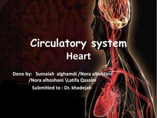
6. 1.Heart
• The heart is located near the anterior chest
wall , directly posterior to the sternum.
• The great veins arteries are connected to the
superior end of the heart at its base.
7. 1.Heart
• The heart surrounded the pericardial sac,
sits in the anterior portion of the
mediastinum.
• The mediastinum the region between the
two cavities , also contains the great
vessels , thymus ,esophagus and trachea.
8. 1.Heart
• The pericardium is lined by a delicate serous
membrane that can be divided into the
1. visceral pericardium which covers and adheres
closely to the outer surface of the heart.
2. parietal pericardium which lines the inner
surface of the pericardial sac , which surrounds
the heart.
• *The pericardial sac , or fibrous pericardium,
which consist of a dense network collagen fibers.
10. Anatomy of the heart
• The heart contains four muscular chambers,
two associated with each circuit.
1. The right atrium receives blood from the
systemic circuit and pass it to right ventricle
which pumps blood into the pulmonary
circuit.
2. The left atrium collects blood from the
pulmonary circuit and empties it into the left
ventricle , which pumps blood into the
systematic circuit.
11. Anatomy of the heart
• When the heartbeats, first the atria contract
, and then the ventricles contract.
• The two ventricle contract at the same time
and eject equal volumes of blood into the
pulmonary and systemic circuit.
13. The heart wall
The wall of the heart reveals three distinct layer:
1. The epicardium is the visceral pericardium
that covers the outer surface of the heart.
2.The myocardium or muscular wall of the heart
, forms the atria and ventricles. This layer
contains cardiac muscle tissue, blood vessels,
and nerves. The myocardium consists of con-
centric layers of cardiac muscle tissue.
14. The heart wall
3.The endocardium covers the inner surfaces
of the heart, including those of the heart
valves. This simple squamous epithelium is
continuous with the endothelium of the
attached great vessels.
16. Artioventricular and semilunar
valves:
• Although adjacent myocardial cells are joined
together mechanically and electrically by
intercalated discs, the atria and ventricles are
separated into two functional units by a sheet of
connective tissue – the fibrous skeleton
previously mentioned.
• Embedded within this sheet of tissue are one-
way atrioventricular (AV) valves.
• The AV valve located between the right atrium
and right ventricle has three flaps and is
therefore called the tricuspid valve .
17. Artioventricular and semilunar
valves:
• AV valve between the left atrium and left
ventricle has flaps and is this called the
bicuspid or alternative mitral valve.
• The AV valves allow blood to flow from the
atria ventricles, but they normally prevent the
backflow of blood the atria.
• Opening and closing of these valves occur as a
the ventricles are relaxed , the venous return
of blood to the causes the pressure in the atria
to exceed that in the ventricles .
18. Artioventricular and semilunar
valves:
• The AV valves therefore open, allowing blood to
enter the ventricle .As the ventricles contract, the
intraventricular pre rises above the pressure in the
atria and pushes the AV closed
22. THE CARDIAC CYCLE:
• Electrical activity coordinates the rhythmic contraction
and relaxation of the heart’s chambers.
• Most currents in the heart are less than a millionth of an
ampere, but they exert a powerful influence on the heart
muscle.
• The cardiac cycle consists of two phases
• DIASTOLE
[In this phase the heart’s ventricles are relaxed, is the longer
phase, taking up approximately two-thirds of the cycle]
• SYSTOLE
[the phase during which blood is
Ejected from the ventricles, takes up
the remaining one third]
23. During diastole:
• The sinus node generates an impulse that forces the two
atria to contract.
• In this phase, the tricuspid and mitral valves are open,
and blood is propelled from the atria into the relaxed
ventricles.
• By the end of diastole, the electric impulse reaches the
ventricles, causing them to contract.
24. During systole:
the contracting ventricles close the tricuspid and mitral
valves. Shortly afterward, the pressure of the blood inside the
ventricles rises sufficiently to force the pulmonary and aortic
valves to open, and blood is ejected into the pulmonary
artery and the aorta.
As the ventricles relax again, blood backs up from the
pulmonary artery and the aorta, closing down the pulmonary
and aortic valves.
The pressure in the relaxed ventricles is now lower than in
the atria, the tricuspid and mitral valves open again, and the
cardiac cycle starts anew.
This events in fact takes approximately a second.
25. The familiar double throb (lub dub) of the
beating heart corresponds to the two
sets of synchronized contractions that
occur during the cardiac cycle.
The throbbing sound we hear comes from:
Snapping of the valves,
Accompanying vibrations of other heart
structures
Turbulence produced by the flow of
blood.
27. Pulmonary and systemic circulations:
The circulatory system is extremely important in sustaining life.
It’s proper functioning is responsible for the delivery of oxygen
and nutrients to all cells, as well as the removal of carbon
dioxide, waste products, maintenance of optimum pH, and the
mobility of the elements, proteins and cells, of the immune
system.
Both atria contract at the same time and that both ventricles
contract at the same time.
The heart works as two pumps, one on the right and one on
the left that works simultaneously.
The right pump pumps the blood to the lungs or the pulmonary
circulation at the same time that the left pump pumps blood to
the rest of the body or the systemic circulation.
28. Systemic circulation
The part of the network that delivers
blood to all parts of the body except the
lungs
Pulmonary circulation:
The flow of blood through the lungs.
The pressure in Pulmonary circulation
system is only about one-sixth as great as
in the systemic circulation, and the walls
of pulmonary arteries and veins are
significantly thinner than the walls of
corresponding vessels in the rest of the
body.
In the pulmonary circulation, the roles of arteries
and veins are the opposite of what they are in the
systemic circulation, Blood in the arteries has less
oxygen, while blood in the veins is oxygen-rich.
29. Blood pathway in systemic and pulmonary
circulation
• Venous blood from systemic circulation (deoxygenated) enters the
right atrium through the superior and inferior vena cava. The right
atrium contracts and forces the blood through the tricuspid valve
(right atrioventricular valve) and into the right ventricles. The right
ventricles contract and force the blood through the pulmonary
semilunar valve into the pulmonary trunk and out the pulmonary
artery. The pulmonary arteries branch to transport blood to the
lung, where gas exchange occurs between the lung capillaries and
the air sacs (alveoli) of the lungs blood
releases carbon dioxide and receives
a new supply of oxygen.
The new blood is carried in the
pulmonary veins that take it to the
left atrium.
30. Continue…
• The left atrium then contracts and forces blood through the
left atrioventricular (bicuspid) or mitral valve into the left
ventricle. The left ventricle contracts forcing blood through
the aortic semilunar valve into the ascending aorta (a very
large, elastic artery). It then branches to arteries carrying
oxygen rich blood to all parts of the body.
• As a result of cellular respiration, the oxygen concentration
is lower and the carbon dioxide concentration is higher in
the tissues than in the capillary blood, blood that drain into
the systemic veins is thus partially depleted of oxygen and
increase in carbon dioxide content.
• These veins ultimately empty into two
large veins [the superior and interior
vena cavae] that return the oxygen
poor blood to the right atrium.
31. The path of blood
from the heart
(right ventricle)
through the lungs;
and back to the
heart (left atrium)
completes one
circuit: the
•The complete the
systemic
circulation
from the heart
(left ventricle),
through the organ
systems, and back
to the heart (right
atrium).
Summary
32. HEART RATE AND CARDIAC
OUTPUT
In an average adult, the pacemaker fires
approximately 70 impulses a minute at rest, which
means that in one minute the heart goes through a
full cardiac cycle 70 times.
The amount of blood pumped by the heart in one
minute is called the cardiac output.
When there is a need for an increased blood supply,
as during physical exertion, the heart most commonly
increases its output by beating faster—for example,
up to 140 or 150 beats per minute.
33. STROKE VOLUME:
• The cardiac output is determined not only by the
heart rate but also by the amount of blood the
ventricles eject or pump out with each
contraction,(This amount is called the stroke volume).
• A decrease in the stroke volume is one of the first
signs of a failing heart.
• cardiologists usually measure only the stroke volume
of the left ventricle, because it is the one that pumps
blood to all of the body’s organs except the lungs
34. 2. Blood
The blood is the substance that is constantly
flowing through our bodies. it is pumped by
heart.
• the blood travels through thousands of miles
of blood vessels right within the body.
• A young person has about a gallon of blood.
An adult has about 5 quarts.
• the blood is not just a red liquid but rather is
made up of liquids, solids and small amounts
of oxygen and carbon dioxide.
35. Blood content:
1- Red Blood Cells( erythrocytes)
• Red Blood Cells are responsible for
carrying oxygen and carbon dioxide.
• Red Blood Cells pick up oxygen in the lungs
and transport it to all the body cells. After
delivering the oxygen to the cells it gathers up
the carbon dioxide and transports carbon
dioxide back to the lungs where it is removed
from the body when we exhale(breath out).
There are about 5,000,000 Red Blood Cells in
ONE drop of blood.
36. Blood content:
2.White Blood Cells (leukocytes )
• White Blood Cells help the body fight off
germs. by attack and destroy germs when they
enter the body.
37. Blood content:
3.Platelets
• Platelets are blood cells that help stop bleeding. .,
fibers and other blood cells to help form a plug to
seal the broken blood vessel. When the platelet plug
is completely formed the wound stops bleeding.
38. Blood content:
4.Plasma
• Plasma is the liquid part of the blood.
Approximately half of your blood is made of
plasma. The plasma carries the blood cells and
other components throughout the body. Plasma
is made in the liver.
39. Blood content
5. hemoglobin
•Hemoglobin is the protein molecule in red blood cells
that carries oxygen from the lungs to the body's
tissues and returns carbon dioxide from the tissues
back to the lungs. Hemoglobin also plays an
important role in maintaining the shape of the red
blood cells.
40. Blood content…con
• Hemoglobin is made up
of four protein molecules
(globulin chains) that are
connected together. The
normal adult hemoglobin
(Hbg) molecule contains
two alpha-globulin chains
and two beta-globulin
chains.
41. Blood content…con
•Each globulin chain contains an important central
structure called the heme molecule.
•Heme molecule Embedded within the iron that is
vital in transporting oxygen and carbon dioxide in our
blood , and it’s responsible for the red color of blood.
42. Blood vessels
• Blood is carried in a closed system of vessels that
begins and ends at the heart.
• The three major types of vessels are arteries,
capillaries, and Veins.
• Arteries carry blood away from the heart (O2 rich,
except pulmonary).
• Veins carry blood toward the heart (O2 poor,
except pulmonary).
• Capillaries contact tissue cells and directly serve
cellular needs.
43.
44. Blood vessels wall
1. Tunica interna /intima- innermost: Endothelium
(simple squamous epithelium) of connected cells;
forms a smooth, flat , low friction surface.
• All vessels have this layer, and all but the teeny-
tiniest have a basement membrane associated
with the endothelium.
2. Tunica media-smooth muscle and elastic
connective tissue surrounding the interna,
Responsible for vasoconstriction and vasodilation.
46. Artery
• Their relatively thick, muscular walls make
arteries elastic and contractile.
• Elasticity permits the vessel diameter to
change passively in response to changes in
blood pressure.
• When stimulated, arterial smooth muscles
contract, constricting the artery—a process
called vasoconstriction.
47. Types of artery
1- Elastic arteries:
• Elastic arteries are also known as conducting
arteries because they carry large volumes of
blood away from the heart.
• The pulmonary trunk and aorta, as well as their
major branches.
• The walls of elastic arteries are extremely
resilient because
• The tunica media contains a high density of
elastic fibers and relatively few smooth muscle
cells.
• As a result, elastic arteries can tolerate the
pressure changes of the cardiac cycle.
48. 2- Muscular arteries:
• Muscular arteries, or medium-sized arteries, are
also known as distribution arteries because they
distribute blood to the body’s skeletal muscles and
internal organs.
• Most of the vessels of the arterial system are
muscular arteries.
• They are characterized by a thick tunica media. It
contains more smooth muscle cells than does the
tunica media of elastic arteries
• The external carotid arteries of the neck, the
brachial arteries of the arms, the mesenteric
arteries of the abdomen, and the femoral arteries
of the thighs are examples of muscular arteries.
49. 3-Arterioles:
• Arterioles have a poorly defined tunica externa.
• In the larger arterioles, the tunica media consists
of one or two layers of smooth muscle cells.
• In the smallest arterioles, the tunica media
contains scattered smooth muscle cells that do
not form a complete layer.
50.
51. capillary
• A capillary is an extremely small blood vessel
located within the tissues of the body, that
transports blood from arteries to veins.
• Capillaries are most abundant in tissues and
organs that are metabolically active, for example,
muscle tissues and the kidneys have a greater
amount of capillary networks than do connective
tissues.
• Capillary walls are thin and are composed of
endothelium, Oxygen, carbon dioxide, nutrients
and wastes are exchanged through the thin walls
of the capillaries.
52. Capillary types
1- Continuous- most common. Endothelial cells
connected by tight junctions, without large pores .
Small spaces between tight junctions allow fluid and
small solutes to pass into and out of blood.
2-Fenestrated- located in areas where lots of
exchange of relatively large particles take place, like
endocrine glands . Large pores in cells. Fenestrations
are large enough to allow polypeptides like
hormones .
3-Sinusoidal. Large spaces between endothelial cells
allow very large particles, including proteins and
RBC, to pass.
53.
54. Capillary Bed
• Capillaries function not as individual units, but rather,
as part of an interconnected network called a
capillary bed, or capillary plexus.
• A single arteriole generally gives rise to dozens of
capillaries.
• They empty into several venules, the smallest vessels
of the venous system.
• The entrance to each capillary is guarded by a
precapillary sphincter.
• Contraction of the smooth muscle cells narrow the
diameter of the capillary entrance, thereby reducing
the flow of blood.
• Relaxation of the sphincter dilate the opening,
allowing blood to enter the capillary faster.
55. • A capillary bed contains several direct
connections between arterioles and venules.
• The wall in the first part of such a passageway
contains smooth muscle that can change its
diameter, this segment is called a metarteriole.
• The rest of the passageway resembles a typical
capillary in structure and is called a thoroughfare
channel.
56. veins
• Veins collect blood from all tissues and organs
and return it to the heart.
• The walls of veins can be thinner than those
of corresponding arteries because the blood
pressure in veins is lower than that in
arteries.
• Even though their walls are thinner, in general
veins are larger in diameter than their
corresponding arteries.
57. Venules
Venules
• Are the smallest venous vessels.
• They collect blood from capillary beds, are the
smallest venous vessels.
• the smallest venules resemble expanded
capillaries.
Medium-Sized Veins
• Are comparable in size to muscular arteries.
• Their tunica media is thin and contains relatively
few smooth muscle cells.
• The thickest layer of a medium-sized vein is the
tunica externa, which contains longitudinal
bundles of elastic and collagen fibers.
58. Large Veins
• Large veins include the superior and inferior
venae cavae and their tributaries within the
abdominopelvic and thoracic cavities.
• All large veins have all three layers.
• The slender tunica media is surrounded by a
thick tunica externa composed of a mixture of
elastic and collagen fibers.
59.
60. Heart Disease
• Coronary Artery Disease
Heart disease is a result of plaque buildup in your
coronary arteries -- a condition called atherosclerosis --
that leads to blockages. The arteries, which start out
smooth and elastic, become narrow and rigid,
restricting blood flow to the heart. The heart becomes
starved of oxygen and the vital nutrients it needs to
pump properly
61. Heart Disease
• Abnormal Heart Rhythms
The heart is an amazing organ. It beats in a steady, even
rhythm, about 60 to 100 times each minute (that's
about 100,000 times each day!). But, sometimes your
heart gets out of rhythm. An irregular or abnormal
heartbeat is called an arrhythmia. An arrhythmia (also
called a dysrhythmia) can involve a change in the
rhythm, producing an uneven heartbeat, or a change in
the rate, causing a very slow or very fast heartbeat
62. Heart Disease
• Heart Failure
The term "heart failure" can be frightening. It does not
mean the heart has "failed" or stopped working. It
means the heart does not pump as well as it should.
This then leads to salt and water retention, causing
swelling and shortness of breath. The swelling and
shortness of breath are the primary symptoms of heart
failure.
• Congenital Heart Disease
Congenital heart disease is a type of defect in one or
more structures of the heart or blood vessels that
occurs before birth.
63. Heart Disease
• Cardiomyopathies
Cardiomyopathies are diseases of the heart muscle
itself. People with cardiomyopathies -- sometimes
called an enlarged heart -- have hearts that are
abnormally enlarged, thickened, and/or stiffened. As a
result, the heart's ability to pump blood is weakened.
64. Heart Disease
Pericarditis
Pericarditis is inflammation of the lining that surrounds
the heart. It is a rare condition that is often caused by
an infection.
Aorta Disease and Marfan Syndrome
The aorta is the large artery that leaves the heart and
provides oxygen-rich blood throughout the body. These
diseases and conditions can cause the aorta to dilate
(widen) or dissect (tear), increasing the risk for future
life-threatening events