R1KneeCyst_allow.pptx
•Als PPTX, PDF herunterladen•
0 gefällt mir•1 view
R1KneeCyst_allow.pptx
Melden
Teilen
Melden
Teilen

Empfohlen
Empfohlen
Weitere ähnliche Inhalte
Kürzlich hochgeladen
Kürzlich hochgeladen (20)
Khambhalia Escorts 8617370543 Khambhalia Call Girls Service
Khambhalia Escorts 8617370543 Khambhalia Call Girls Service
Just Call Vip call girls Farrukhabad Escorts ☎️8617370543 Two shot with one g...
Just Call Vip call girls Farrukhabad Escorts ☎️8617370543 Two shot with one g...
Russian Call Girls Lucknow Just Call 👉👉 📞 8617370543 Top Class Call Girl Serv...
Russian Call Girls Lucknow Just Call 👉👉 📞 8617370543 Top Class Call Girl Serv...
New Call Girls In Shamli 8617370543 Shamli Escorts Service
New Call Girls In Shamli 8617370543 Shamli Escorts Service
Nehru Nagar, Call Girls ☎️ ((#9711106444)), 💘 Full enjoy Low rate girl💘 Genui...
Nehru Nagar, Call Girls ☎️ ((#9711106444)), 💘 Full enjoy Low rate girl💘 Genui...
Orai call girls 📞 8617370543At Low Cost Cash Payment Booking
Orai call girls 📞 8617370543At Low Cost Cash Payment Booking
Call Girls Sultanpur Just Call 📞 8617370543 Top Class Call Girl Service Avail...
Call Girls Sultanpur Just Call 📞 8617370543 Top Class Call Girl Service Avail...
Russian Call Girls In Bhubaneswar 📱 Odisha 9777949614 Indore
Russian Call Girls In Bhubaneswar 📱 Odisha 9777949614 Indore
Call Girls Mehsana - 📞 8617370543 Our call girls are sure to provide you with...
Call Girls Mehsana - 📞 8617370543 Our call girls are sure to provide you with...
SB_ Dragons Riders of Berk_ Rough_ RiverPhan (2024)
SB_ Dragons Riders of Berk_ Rough_ RiverPhan (2024)
Kathmandu Escort❤ @Daminy@💞 50+ Call Girl PRofile in @Kathmandu New Housewife...
Kathmandu Escort❤ @Daminy@💞 50+ Call Girl PRofile in @Kathmandu New Housewife...
Jaunpur Escorts Service Girl ^ 9332606886, WhatsApp Anytime Jaunpur
Jaunpur Escorts Service Girl ^ 9332606886, WhatsApp Anytime Jaunpur
Theoretical Framework- Explanation with Flow Chart.docx
Theoretical Framework- Explanation with Flow Chart.docx
Call Girls Veraval Just Call 8617370543Top Class Call Girl Service Available
Call Girls Veraval Just Call 8617370543Top Class Call Girl Service Available
Call Girls In Goa, North Goa 👌9971646499👌Cash On delivery Goa Escorts Service
Call Girls In Goa, North Goa 👌9971646499👌Cash On delivery Goa Escorts Service
Empfohlen
More than Just Lines on a Map: Best Practices for U.S Bike Routes
This session highlights best practices and lessons learned for U.S. Bike Route System designation, as well as how and why these routes should be integrated into bicycle planning at the local and regional level.
Presenters:
Presenter: Kevin Luecke Toole Design Group
Co-Presenter: Virginia Sullivan Adventure Cycling AssociationMore than Just Lines on a Map: Best Practices for U.S Bike Routes
More than Just Lines on a Map: Best Practices for U.S Bike RoutesProject for Public Spaces & National Center for Biking and Walking
Empfohlen (20)
How to Prepare For a Successful Job Search for 2024
How to Prepare For a Successful Job Search for 2024
Social Media Marketing Trends 2024 // The Global Indie Insights
Social Media Marketing Trends 2024 // The Global Indie Insights
Trends In Paid Search: Navigating The Digital Landscape In 2024
Trends In Paid Search: Navigating The Digital Landscape In 2024
5 Public speaking tips from TED - Visualized summary
5 Public speaking tips from TED - Visualized summary
Google's Just Not That Into You: Understanding Core Updates & Search Intent
Google's Just Not That Into You: Understanding Core Updates & Search Intent
The six step guide to practical project management
The six step guide to practical project management
Beginners Guide to TikTok for Search - Rachel Pearson - We are Tilt __ Bright...
Beginners Guide to TikTok for Search - Rachel Pearson - We are Tilt __ Bright...
Unlocking the Power of ChatGPT and AI in Testing - A Real-World Look, present...
Unlocking the Power of ChatGPT and AI in Testing - A Real-World Look, present...
More than Just Lines on a Map: Best Practices for U.S Bike Routes
More than Just Lines on a Map: Best Practices for U.S Bike Routes
Ride the Storm: Navigating Through Unstable Periods / Katerina Rudko (Belka G...
Ride the Storm: Navigating Through Unstable Periods / Katerina Rudko (Belka G...
Good Stuff Happens in 1:1 Meetings: Why you need them and how to do them well
Good Stuff Happens in 1:1 Meetings: Why you need them and how to do them well
R1KneeCyst_allow.pptx
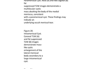
- 1. Parameniscal Cyst. Axial (A) and two sagittal (B) fat suppressed T2W images demonstrate a multilocular cystic mass abutting the body of the medial meniscus, consistent with a parameniscal cyst. These findings may indicate an underlying occult meniscal tear. Figure 20. Intrameniscal Cyst. Coronal T1W (A) and fat suppressed T2W (B) images demonstrate mass- like cystic enlargement of the lateral meniscal body secondary to a large intrameniscal cyst.