
Empfohlen
Empfohlen
Weitere ähnliche Inhalte
Ähnlich wie Obesity-and-Diabetes-Day-Presentation.pptx
Ähnlich wie Obesity-and-Diabetes-Day-Presentation.pptx (20)
Mehr von ssuser3fe82a
Kürzlich hochgeladen
Kürzlich hochgeladen (20)
Obesity-and-Diabetes-Day-Presentation.pptx
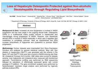
- 1. Loss of Hepatocyte Osteopontin Protected against Non-alcoholic Steatohepatitis through Regulating Lipid Biosynthesis Hui HAN 1, Romain Desert 1, Xiaodong Ge 1, Sukanta Das 1, Zhuolun Song 1, Dipti Athavale 1, Wei Chen 1, Harriet Gaskell 1, Daniel Lantvit 1, Grace Guzman 1 and Natalia Nieto 1 1 Department of Pathology, University of Illinois at Chicago, 840 S. Wood St., Suite 130 CSN, MC 847, Chicago, IL 60612, USA Background & Aims: increased de novo lipogenesis is involved in NASH progression and the main target in the ongoing clinical trials. Osteopontin (OPN) is a multifactorial matrix protein induced in hepatocytes and macrophages during progression of NASH. However, how hepatocyte derived OPN is involved in NASH especially in which way it regulates lipid metabolism is completely unknown. Here we hypothesized that OPN increase biosynthesis of triglyceride and cholesterol to promote NASH progression. Methodology: Human datasets were downloaded from Gene Expression Omnibus and analyzed by general statistical methods. Mice with Opn ablation in hepatocytes (OpnΔHep) was generated with the scheme in the figure. WT and OpnΔHep mice were fed a control or NASH-inducing diet (high fat, cholesterol, fructose) for 6 months. H&E slides were scored for disease severity. Liver triglyceride and cholesterol were measured with biochemical approach. Transcriptome profiling was performed by RNA sequencing followed by calculation of differentially expressed genes with Limma package in R platform. The gene enrichment analysis were performed by Networkanalyst. Upstream target analysis were performed by ingenuity pathway analysis (IPA). Abstract Opnfl/fl × AlbCre/+ OpnΔHep OPN NASH Feeding Histology Biochemistry Transcriptomic
- 2. 0% 50% 100% Low High OPN Expression is Associated to Development of Liver Steatosis during NASH progression (A) (B) ** 2 1 0 Steatosis Score OPN Expression OPN Expression Stage Steatosis OPN Expression Subject (%) * *** *** *** Figure 1: OPN Expression is Associated to Development of Liver Steatosis during NASH progression. (A) Relative OPN mRNA expression in human liver biopsies from GSE126848; (B) Relative OPN mRNA expression in non-fibrotic NASH patients from GSE151158 with different scores of steatosis; (C) Percent of NAFLD patients with corresponding steatosis score. Subjects were stratified by median SPP1 expression in GSE151158. *p<0.05, **p<0.01, ***p<0.0001, vs control, 0 or low. (C)
- 3. Ablation of OPN (OpnΔHep) from Hepaotcyte Decreased Progression of NASH Ctrl NASH Male Female Male Female WT OpnΔHep PV CV PV CV PV CV PV CV CV PV PV CV CV PV 200x 200x 200x 200x 200x 200x 200x 200x 400x 400x PV CV 400x 400x 200x 200x 200x WT OpnΔHep Ctrl NASH Ctrl NASH Steatosis 1.31±0.09 2.50±0.05## 2.00±0.07 1.36±0.05** Inflammation 1.15±0.05 2.00±0.10## 0.33±0.04* 1.09±0.05##** Ballooning 0.69±0.06 1.60±0.07## 0.67±0.08 1.45±0.05## NAS 3.15±0.15 6.10±0.19### 3.00±0.13 3.91±0.11** Figure 2: Ablation of OPN (OpnΔHep) from Hepatocyte Decreased Progression of NASH. Mice were fed for control or NASH diet for 6 months. (A) H&E staining. Red arrow, liver steatosis; blue arrow, inflammatory foci; green arrow, ballooning. (B) NASH activity score (NAS). H&E slides were scored by Kleiner's system. *p<0.05, **p<0.01, vs WT with same diet. #p<0.05, ##p<0.01, ###p<0.001, vs Ctrl diet with same genotype. n=10.
- 4. 0 2 4 6 8 10 12 (B) (A) Hepatic triglyceride (mg/g) 0 50 100 150 200 250 300 350 ### ### * 0 100 200 300 400 500 600 Hepatic cholesterol (mg/g) * ** ### Ctrl NASH Ctrl NASH WT OpnΔHep Ctrl NASH Ctrl NASH WT OpnΔHep Liver to b.w. Ratio (%) ### ## ** Ctrl NASH Ctrl NASH WT OpnΔHep ### (C) OpnΔHep Reduced Hepatic Triglyceride and Cholesterol Figure 3: OpnΔHep Reduced Hepatic Triglyceride and Cholesterol. Mice were fed for control or NASH diet for 6 months. (A) Liver to b.w. ratio. (B) Hepatic triglyceride. (C) Hepatic cholesterol. The hepatic lipids were normalized by protein concentration. *p<0.05, **p<0.01, vs WT with same diet. ##p<0.01, ###p<0.001, vs Ctrl diet with same genotype. n=10.
- 5. -6 -4 -2 0 2 0 3 4 2 1 -Log(p) Log2(Fold of Change) Metabolic Pathway (KEGG) Up-regulate Down-regulate (A) (C) Gene Set Description Hit/Size NES mmu00900 Terpenoid backbone biosynthesis 8/10 -2.4452 mmu00100 Steroid biosynthesis 12/13 -2.7275 mmu01100 Metabolic pathways 66/202 -2.8108 mmu00982 Drug metabolism 11/14 -2.2779 mmu00980 Metabolism of xenobiotics by cytochrome P450 10/11 -2.281 mmu04621 NOD-like receptor signaling pathway 11/12 2.3119 mmu00620 Pyruvate metabolism 6/11 -2.087 mmu01212 Fatty acid metabolism 9/22 -2.0997 mmu00061 Fatty acid biosynthesis 5/6 -1.9918 Fasn Scd1 Acaca Hmgcr Sqle WT OpnΔHep OpnΔHep Inhibited Fatty Acid and Cholesterol Biosynthesis Figure 4: OpnΔHep Inhibited Fatty Acid and Cholesterol Biosynthesis. Untreated mice were scarified at the age of two weeks. (A) Volcano plot for differentially expressed (DE) genes in total liver between OpnΔHep and WT. There are 131 DE genes (FDR<0.05, FC>1.5) belong to metabolic pathway (KEGG). (B) Heatmap for representative genes involving fatty acid and cholesterol biosynthesis. (C) Gene set enrichment analysis for the DE genes. The pathways with FDR<0.05 were listed. (B)
- 6. OpnΔHep Potentially Affected Activity of Lipid Sensing Nuclear Receptors Figure 5: OpnΔHep Potentially Affected Activity of Lipid Sensing Nuclear Receptors. DE genes were analyzed for potential upstream targets by ingenuity pathway analysis (Qiagen). The top inhibited transcription factors were SREBF1/2, NR1H3 (or Liver X Receptor), and NR1I2 (Pregnane X Receptor) Conclusions • Loss of hepatocyte OPN protects against NASH progression through inhibiting fatty acid and cholesterol biosynthesis • The protective effect is potentially due to lowered activities of LXR and SREBF1/2. • Whether OPN interact with lipid sensing nuclear receptors will be studied in the future. Nucleus Contact Hui HAN 909 S Wolcott AveChicago, IL 60612 COMB6140, Nieto’s Lab huihan@uic.edu