
Empfohlen
Weitere ähnliche Inhalte
Ähnlich wie precardium.pptx
Ähnlich wie precardium.pptx (20)
Kürzlich hochgeladen
Kürzlich hochgeladen (20)
precardium.pptx
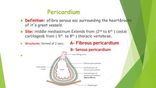
- 1. Pericardium Definition: afibro serous sac surrounding the heart&roots of it's great vessels. Site: middle mediastinum Extends from (2nd to 6th ) costal cartilages& from ( 5th to 8th ) thoracic vertebrae. Structures: formed of 2 sacs: A- Fibrous pericardium B- Serous pericardium
- 2. Fibrous pericardium Conical fibrous sac formed of fibrous tissue: It has: Base, Apex, ant, post& 2 lat surface. Base: bellow, attached to central tendon of diaphragm. Apex: above, up to sternal angle where it belend with the adventitia of great vessels & Pre tracheal fascia.
- 5. Serous pericardium Closed serous sac which is invaginated from above and behind by the heart & great vessels. Structures:formed of 2 layers - 1- parietal layer: - lines fibrous pericardium & continous with visceral layer at root of great vessels . 2- Visceral layer(epicardium): covers the heart.
- 7. Blood Supply Fibrous and parietal serous: Arterial blood supply from: - Internal mammary, musculophernic arteries& Descending thoracic aorta. Venous drainage:to internal mammary and azygose veins. - Visceral serous: as the heart.
- 8. Nerve Supply Fibrous and parietal serous: Phernic nerve (Sensitive to pain). Visceral layer: as heart (Autonomic→ insensetive to pain) but Sensitive to ischaemia .
- 9. sinuses of pericardium They are recesses inside the pericardal cavity lined by serous pericardium, they includes : 1- Transvers sinus 2- oblique sinuses.
- 12. Oblique sinus Ablined recess of the pericardal cavity that lies behind the base of the heart opened bellow to general pericardal cavity Boundaries: - Anteriorly: back of Lt atrium. - Post: fibrous pericardium - On left side: reflection of the serous pericardium on 2 left pulmonary veins. - On right Side: reflection of the serous pericardium on 2 right Pulmonary veins & 2 vena cava
- 14. هلل الحمد