
Empfohlen
Weitere ähnliche Inhalte
Was ist angesagt?
Was ist angesagt? (20)
Ähnlich wie 4.DISEASE OF HARD TISSUES OF TEETH.pptx
Ähnlich wie 4.DISEASE OF HARD TISSUES OF TEETH.pptx (20)
Kürzlich hochgeladen
Kürzlich hochgeladen (20)
4.DISEASE OF HARD TISSUES OF TEETH.pptx
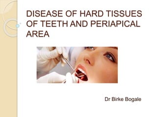
- 1. DISEASE OF HARD TISSUES OF TEETH AND PERIAPICAL AREA Dr Birke Bogale
- 2. Objectives Define Dental caries Explain Causes, Risk factors and Prevention of Dental caries Explain - Presentation - Diagnosis - Treatment of dental caries, diseases of the pulp and periapical tissue Identify different types of caries, diseases of the pulp and periapical tissue
- 3. DISEASE OF HARD TISSUES OF TEETH Dental Caries Also called tooth decay. It usually occurs in children and young adults, but can affect any person. It is a common cause of tooth loss in younger people.
- 4. Facts, WHO 2003 Dental diseases are the most prevalent chronic diseases worldwide, and a costly burden to health care services. Expensive treatment: 5 -10% of total health care expenditures in industrialized countries. Dental caries: High prevalence rate in most developing low-income countries more than 90% of caries is untreated. An estimated 5 billion people worldwide suffer from dental caries (tooth decay).
- 5. Etiology Common Bacteria: Streptococcus species ; S. Mutans Lactobacillus species Bacteria Actinomyces species Other Risk factors Teeth Anatomy Sugar Time Life style (Frequency of snacking, OH, infant feeding, etc.) Carbohydrates, sticky foods Systemic Ds. (Metabolic, eating do, Immunity, etc.) Worn dentures, appliances & fillings Fluoride D. Carie s
- 6. Bacteria: Normally present in the mouth. Convert all foods especially sugar and starch into acids. Bacteria, acid, food debris and saliva combine in the mouth to form a sticky substance called plaque that adheres to tooth surface. It is most prominent on molars, just above the gum line on all teeth and at the edges of fillings.
- 7. The acids in plaque dissolve the enamel surface of the tooth and create holes in the tooth (cavities). Cavities are usually painless until they grow very large and affect nerves or cause a tooth fracture. Untreated tooth decay also destroys the internal structures of the tooth (pulp) and ultimately causes the loss of the tooth.
- 8. Medical factors associated with increased caries risk Factors Risk increasing observation Gender Female > Male Age Children and adolescents are more prone Fluoride exposure No or less fluoride in drinking water Smoking Risk increases with intake Alcohol Risk increases with intake General Health Chronic illness and debilitating disease Medication Medication- w/c decrease salivary flow E.g.-Atropine -Antiepileptics -Pills /hormone
- 9. Clinical sites for caries initiation 1. Pit and fissures of enamel – most susceptible 2. Smooth surface of crown (proximal) – Second susceptible 3. Root surface (cervical area) – Third susceptible site 4. Sub gingival area
- 10. Presentation Highly variable. Risk factors and stages of development are similar. Initially it may appear as a small chalky area (Incipient caries), large cavitation. With continued acid attack the surface changes from being smooth to rough and may become stained.
- 11. Note: - Penetration of enamel caries into the dentino- enamel junction rapid lateral expansion DEJ has least resistance to caries attack. Progression and morphology of carious lesion is variable depending upon; -Site of origin -The condition of the mouth (oral hygiene).
- 12. Diagnosis Visual inspection: white spots or darker inactive lesions Probing: soft surface and cavity Radiographic exam. (Bitewing or OPG) : essential for interproximal caries, ◦ current radiographic techniques lack sensitivity in recognizing non-cavitating lesions or root surface caries. Newer techniques: quantitative light- induced fluorescence and fiber-optic trans illumination are currently under development and may improve caries detection.
- 13. * Rampant caries Acute and rapid infectious process, usually involving several teeth. It is usually occurred in children or infants. Causes: - Dietary habits Poor oral hygiene Systemic illness
- 14. *Secondary caries It is caries of filled teeth. It may occur due to defective margin of restoration and if caries is not completely removed before cavity preparation.
- 15. *Arrested Caries Under favorable conditions, a lesion become inactive, even regress. Clinically arrested dentine caries has a hard or leathery consistency and is darker in color than soft, yellow active decay. Arrested enamel caries can be stained dark brown.
- 16. Prevention Three main approaches are possible: 1. Tooth strengthening or protection 2. Reduction in the availability of microbial substrate. 3. Removal of plaque by physical or chemical means; practically: dietary advice, fluoride, Pit and fissure sealing regular tooth brushing. The relative value of these varies with age of the individual.
- 17. Saliva 1.Has buffering action (effect) against bacteria and act as intra-oral antacid due to its alkali PH at high flow rate 2.Has flushing effect – wash away bacteria and decrease plaque accumulation. 3.Produce antimicrobial products such as IgA, lysozyme, lactoferin, agglutins and lactoperioxidase 4.Has remeneralization effect, as it is supersaturated with calcium, phosphate and fluoride ions which give opportunity for remeneralization of enamel.
- 18. Treatment 1. Fillings : by removing the decayed tooth part with a drill and replacing with filling material. Aesthetic filling materials: GIC, Porcelain and composite resin More closely match the natural tooth appearance and preferred for front teeth. Non-aesthetic filling materials: Silver amalgam (alloy) and gold Considered to be stronger and are often
- 19. 2. Crowns or "caps" Used if tooth decay is extensive & limited tooth structure, which may cause weakened teeth. Large fillings and weak teeth increase the risk of the tooth breaking. The decayed or weakened area is removed and repaired. A crown is fitted over the remainder of the tooth. Crowns are often made of gold, porcelain or porcelain attached to metal. They are also used on fractured teeth due to trauma.
- 20. 3. Root canal treatment Recommended if the nerve in a tooth dies. The center of the tooth, including the nerve and blood vessel tissue (pulp), is removed along with decayed portions of the tooth. The root chamber and canals are filled with a sealing material. The tooth is filled and a crown may be placed over the tooth, if needed.
- 21. The development stages of dental caries a. Enamel caries: No pain b. Dentine caries: maybe sensitive to hot, cold and sweet foods/drinks and eating hard things; there may be pain c. Pulp involved: severe continuous or throbbing pain d. Abscess: deep acute pain which may disappear after a while.
- 23. Disease of the pulp and periapical Tissues I. Disease of the pulp (Pulpitis) Pulpitis - Inflammation of pulp Etiology: -Dental caries (primary cause) -Tooth fracture; expose dental pulp to oral fluids and bacterias -Chemical irritation to pulp; filling -Severe thermal change; common in large metallic restoration
- 24. Classification: 1. Pulp hyperemia (Focal Reversible Pulpitis) Earliest form of pulpitis Cause: -Deep caries -Large metallic restoration (especially with no adequate insulation) -Restoration with defective margin CF: -Sensitive to thermal changes, particularly to cold, but disappears upon removal of the stimulus or restoration of normal temperature.
- 25. 2. Reversible pulpitis CF:- Fleeting sensitivity / pain to hot, cold or sweet with immediate onset -pain usually is sharp and may be difficult to locate -Quickly subsides after removal of stimulus (pain <10 min) Rx:-Filling with sedative dressing (if needed) or permanent restoration with suitable pulp protection.
- 26. 3. Irreversible Pulpitis CF:-Spontaneous pain which may last several hours (>10 min), be worse at night and is pulsatile in nature -Pain elicited by hot and cold at first -Heat will be more significant and cold may actually ease symptoms in later stages . -Localization of pain may be difficult initially, - As the inflammation spread to the Periapical tissue, tooth will become more sensitive to pressure. -Tender to percussion Rx: -RCT, treatment of choice -Extraction
- 27. Diseases of periapical tissues Once infection has become established in the dental pulp, spread of the process can be in one direction through the root canals and into the periapical region. Progression of irreversible pulpitis ultimately leads to death of the pulp (pulpal necrosis). At this stage the patient may experience relief from pain. If neglected, the bacteria and the pulpal breakdown products leave the root canal system via the apical foramen lead to inflammatory changes and possibly severe pain.
- 28. Here a number of different tissue reactions may occur, depending on the circumstances The periapical lesions don’t represent individual and distinct entities, but rather that there is a subtle transformation from one type of lesion into another type in most cases. Note: - Sensitive to percussion is first evidence that infection has spread beyond the confines of the pulp.
- 29. 1. Apical periodontitis (Periapical granuloma) Granuloma :-Essentially localized mass of chronic granulation tissue formed in response to infection CF:-Sensitive to percussion, due to edema, hyperemia and inflammation of apical periodontal ligament. -Mild pain occasioned on biting or chewing solid foods -In some cases tooth feels slightly elongated in its socket and may actually be so. Rx: -Extraction -RCT, under certain condition with or without subsequent apicectomy
- 30. 2. Apical periodontal cyst Cyst – Pathological cavity lined by epithelium and is often fluid filled APC- is common and developed over long period of time. Cause:- Sequela of periapical granuloma - Necrosis of dental pulp CF:-The majority is asymptomatic and present no clinical evidence of their presence. -Seldom painful even sensitive to percussion. -Long standing may undergo acute exacerbation of inflammatory process and develop rapidly into an abscess may then proceed to a cellulites or draining fistula. Rx: -Extraction and curette of periapical tissue, carefully -RCT and apicectomy with inoculation in some
- 32. 3. Periapical Abscess (Dentoalveolar Abscess) An acute or chronic suppurative process of the dental periapical region. CF:-Tooth is extremely painful -Slightly extruded from its socket -Seldom severe systemic manifestation, regional lymphadenitis and fever may be present -Rapid extension to adjacent bone marrow spaces frequently occurs producing an actual osteomyelitis. Rx: -Drainage by either opening the pulp chamber or extraction -RCT, sometimes Note: - If not treated, leads to Orofacial soft tissue infections, osteomyelitis, bacteremia, etc.