SlideShare ist ein Scribd-Unternehmen logo
1 von 59
OCULAR SURFACE
SQUAMOUS
NEOPLASIA(OSSN)
INTRODUCTION
• The term Ocular Surface Squamous Neoplasia
[OSSN] presently refers to the entire spectrum
of dysplastic, pre-invasive and malignant
squamous lesions of the conjunctiva and
cornea
• We have suggested the use of the "umbrella“
term ocular surface epithelial dysplasia (OSED)
but now feel that ocular surface squamous
neoplasia (OSSN) is a better terminology.
• OCULAR SURFACE denotes involvement of the
conjunctiva or cornea;
• SQUAMOUS excludes other epithelial cells such
as basal cells and melanocytes
• and NEOPLASIA includes both dysplastic and
carcinomatous lesions.
Ocular Surface Squamous Neoplasia
Graham a. lee, MBBS, and Lawrence w. hirst, MD
Survey of ophthalmology volume 39, number 6. may-june
1995
The term Ocular Surface Squamous Neoplasia (OSSN) was
coined by Lee and Hirst which has three grades :-
I. BENIGN DYSPLASIA
• Papilloma
• Pseudoepitheliomatous hyperplasia
• Benign hereditary intraepithelial dyskeratosis
II. PREINVASIVE OSSN
• Conjunctival/corneal carcinoma in situ
III. INVASIVE OSSN
• Squamous carcinoma
• Mucoepidermoid carcinoma
EPIDEMIOLOGY
• Third most common ocular tumour after
melanoma and lymphoma
• Common in Caucasians
• Common in older age group(6-7 decade)
Males are affected more commonly than females
• Earlier onset in areas in latitude 30 degree from
equator, declines considerably with increase in
latitude
• Pts with HIV and Xeroderma pigmentosum
present earlier
All young patients with OSSN should be screened
for HIV.
ETIOLOGY
• LIMBAL TRANSITION ZONE/STEM CELL
THEORY
• Stem cells in the limbal epithelial crypts are
the likely originators of this disease, and may
take on cancer stem cell properties.
Effects of solar ultraviolet radiation
• UVB radiation causes direct DNA damage by
crosslinking adjacent bases to form cyclobutane
pyrimidine dimers (CPDs) and 6-4
photoproducts (6-4 PPs)
• Specific CC/TT dimer transitions of the p53
tumour-suppressor gene have been observed in
OSSN lesions
• 29% increase in incidence of SCC per unit
increase in exposure to ambient solar ultraviolet
radiation
• Other eye diseases thought to be related to
UV-B exposure also have a high association
with OSSN.
• pterygium
• pingueculum
• climatic droplet keratopathy
• cataract
• corneal degeneration
Reactivation of latent HPV infection.
• UV radiation also reactivates latent viruses
such as HPV 16
• HPV's E7 protein keeps infected cells in a
proliferative state
• while E6 inhibits cell cycle arrest of DNA-
damaged cells.
Failure of DNA repair mechanisms
• An increased incidence of OSSN has been
reported in conditions like Xeroderma
Pigmentosa, where the DNA repair
mechanism is defective
Immunosuppression
• UV radiation
• HIV and
• vitamin A deficiency weakens the tumour
surveillance system and allows DNA damaged
cells to proliferate into tumours.
• Vitamin A deficiency interferes with ocular
surface integrity creating micro-abrasions
through which HPV may invade the conjunctival
basement membrane and epithelial cells.
Miscellaneous
• Chronic exposure keratopathy secondary to long
• standing facial nerve palsy may be associated
with OSSN
• exposure to petroleum products
• heavy cigarette smoking
• chemicals such as trifluridine and arsenicals
• ocular surface injury
• Chronic inflammatory conditions-pemphigoid,
atopic eczema, chronic blepharoconjunctivitis
Clinical Features
• Patients with OSSN may be asymptomatic or
present with chronic redness and irritation of
the eye.
• Visual acuity is not affected unless there is
extensive corneal involvement
• OSSN may grow within weeks to years; in
most cases, the history is of several months
• The lesions are described as being slightly
elevated, variably shaped, relatively sharply
demarcated from the surrounding normal
tissues, accompanied by feeder blood vessels
and colored from pearly gray to reddish gray
depending on the vascularity of the tumor
• They most commonly straddle the nasal or
temporal limbus between the palpebral
fissures
• The following appearances may be seen.
• a Gelatinous mass with superficial vessels
b White leukoplakic plaque that covers the
lesion
c Papillomatous lesion with corkscrew-like
surface blood vessels
• In clinical practice, gelatinous type is the
commonest.
• The gelatinous lesion can again be
• circumscribed, which are most common
• nodular variety , which has a propensity for
rapid growth
• diffuse variety, the least common, which can
masquerade as chronic conjunctivitis
• The corneal side of the lesion may be seen as
an opalesence of the epithelium, slightly
raised in comparison to adjacent normal
epithelium the edges of which are usually
sharply defined.
• It is best appreciated by retroillumination.
• The lesion is usually nonpigmented though
pigmented OSSN may also occur.
• OSSN may sometimes mimic a pterygium or a
pinguecula. It has also been known to occur in
a pre-existing pinguecula or pterygium.
• It is nearly impossible to categorise OSSN as
benign or malignant based on clinical
appearance alone.
• Larger lesions that are fixated to the
underlying tissue are usually malignant.
• Staining with fluorescein sodium or Rose
Bengal helps in the diagnosis by showing up
the papillary or granular nature of the lesion
and by delineating its extent
• Anterior segment OCT may be done to find
out the extent of deep involvement as well as
intraocular and angle invasion
• Intraocular invasion is rare ,heralded by onset
of iritis, glaucoma, scleral thinning or retinal
detachment.
• Regional and systemic metastases are also
uncommon.
• Common sites of metastasis include
preauricular,submandibular and cervical
lymph nodes, parotid gland, lungs, and bone.
DIFFERENTIAL DIAGNOSIS
• Pterygium
• papilloma
• pingueculum,
• nevus.
• Malignant melanoma
• pyogenic granuloma
• pseudo-epitheliomatous hyperplasia
• Corneal pannus from any cause
• Mooren's ulcer,
• fatty degeneration of the cornea and epithelial
dystrophy of the cornea
• Papilloma may occur anywhere on the
conjunctiva, may be sessile or pedunculated,
has a punctate vascular pattern, occurs in
younger patients,but ultimately may only be
difterentiated by histologic examination
• Malignant melanoma has a regular smooth
surface, lacks gelatinous or leukoplakic surface
disturbances and may become ulcerated
• Benign nevi tend to occur in the interpalpebral
zone, from the limbus to the caruncle, to
occur in younger patients, and to have
distinctive cysts on slit-lamp examination
HISTOPATHOLOGY
• DYSPLASIA:
• Mild - less than a third thickness occupied by atypical
cells
• Moderate - three quarters thickness occupied by
atypical cells
• Severe -- nearly full thickness occupied by atypical cells
• CARCINOMA IN SITU: as above with loss of the normal
surface layer
• INVASIVE SQUAMOUS CELL CARCINOMA: as above
when the basement membrane of the basal epithelial
layer has been breached and invasion of the substantia
propria has occurred.
• Spindle cell, Mucoepidermoid and Adenoid-
Squamous are three aggressive variants
TREATMENT
• SURGICAL EXCISION
• video
TOPICAL CHEMOTHERAPY
• Topical chemotherapy may be used
preoperatively, intraoperatively or
postoperatively.
Its preoperative use is seen in cases with
extensive lesions where it helps to reduce the
tumor size and makes the tumor more amenable
to surgical excision.
Intraoperatively, chemotherapy may have a role
in place of adjuvant cryotherapy
postoperatively can be beneficial for managing
cases with residual disease or positive surgical
margins.
MITOMYCIN C
• Mitomycin C is the most commonly used topical
chemotherapy agent
• It is a non cell cycle specific ALKYLATING AGENT
that acts by alkylating the cross-linked DNA and
inhibits DNA, RNA, and protein synthesis.
• MMC is used in concentrations of 0.04% qid, 4
days a week for 4 weeks.
• Overall, success rates ranging from 87 to 100%
have been reported.
5-FU
• Is a pyrimidine analogue that acts by
integrating with the DNA during S phase. It
also interferes with RNA synthesis.
• It is used as 1% topical solution.
• Side effects are similar to MMC
INTERFERONS
• Interferons are protein molecules that bind to cell
receptors and trigger synthesis of effector
proteins that can inhibit viruses, activate
immunocompetent cells, and regulate
oncogenes.
• Reports are emerging on the use of topical IFN-
2b for OSSN.
• It is more expensive than MMC and 5 FU,
requires prolonged treatment but has a better
safety profile
RADIOTHERAPY
• Plaque brachytherapy using radiation sources
like Strontium 90 or Rhuthenium 106 is useful
as an adjuvant to support surgical excision.
Complications include conjunctival scarring,
symblepharon, dry eye, cataract, scleral and
corneal ulceration.
SUMMARY
• [1] Suspected OSSN < 3 clock hours –Excision
biopsy + base/ edge cryotherapy +
alchoholepitheliectomy is done.
• [2] Suspected OSSN 3 – 6 clock hours – As
there is a risk of producing limbal stem cell
deficiency, excision biopsy + cryotherapy is
better avoided.
• A diagnostic biopsy is required
• In pre-invasive lesions topical chemotherapy
is likely to achieve tumour resolution.
• If invasive, surgery + cryotherapy is done after
chemoreduction with 4 to 6 cycles of topical
chemotherapy.
• [3] OSSN > 6 clock hours – A diagnostic biopsy is
required.
• In pre-invasive lesions topical chemotherapy is
quite likely to achieve tumour resolution.
• If invasive, surgery + cryotherapy is done after
chemoreduction with 4 to 6 cycles of topical
chemotherapy.
• If there is no response to chemotherapy palliative
radiotherapy or extensive surgery like
enucleation / exenteration may be required.
Ocular surface squamous neoplasia(ossn)

Weitere ähnliche Inhalte

Was ist angesagt?

Viral Keratitis: Diagnosis, Management and Latest Guidelines
Viral Keratitis: Diagnosis, Management and Latest GuidelinesViral Keratitis: Diagnosis, Management and Latest Guidelines
Viral Keratitis: Diagnosis, Management and Latest GuidelinesSahil Thakur
 
Pseudoexfoliation syndrome
Pseudoexfoliation syndromePseudoexfoliation syndrome
Pseudoexfoliation syndromeGloria George
 
Herpes simplex keratitis & herpes zoster opthalmicus
Herpes simplex keratitis & herpes zoster opthalmicusHerpes simplex keratitis & herpes zoster opthalmicus
Herpes simplex keratitis & herpes zoster opthalmicusLaxmi Eye Institute
 
Lamellar keratoplasty
Lamellar keratoplastyLamellar keratoplasty
Lamellar keratoplastyAkshay Nayak
 
Choroidal neovascular membranes (CNVM)
Choroidal neovascular membranes (CNVM)Choroidal neovascular membranes (CNVM)
Choroidal neovascular membranes (CNVM)Md Riyaj Ali
 
Leukocoria (White Pupillary Reflex)
Leukocoria (White Pupillary Reflex) Leukocoria (White Pupillary Reflex)
Leukocoria (White Pupillary Reflex) Mohamed Abdelmongy
 
THYROID EYE DISEASE
THYROID EYE DISEASETHYROID EYE DISEASE
THYROID EYE DISEASESSSIHMS-PG
 
Corneal dystrophies
Corneal dystrophiesCorneal dystrophies
Corneal dystrophiesNajara Thapa
 
Lens induced glaucoma - DR ARNAV
Lens induced glaucoma - DR ARNAVLens induced glaucoma - DR ARNAV
Lens induced glaucoma - DR ARNAVDrArnavSaroya
 
Gonioscopy: gonioscopic lenses, principle and clinical aspects
Gonioscopy: gonioscopic lenses, principle and clinical aspectsGonioscopy: gonioscopic lenses, principle and clinical aspects
Gonioscopy: gonioscopic lenses, principle and clinical aspectsDr Samarth Mishra
 
Ophthalmic Manifestations of Hematological Malignancies
Ophthalmic Manifestations of Hematological MalignanciesOphthalmic Manifestations of Hematological Malignancies
Ophthalmic Manifestations of Hematological MalignanciesPaavan Kalra
 
Suprachoroidal haemorrhage
Suprachoroidal haemorrhageSuprachoroidal haemorrhage
Suprachoroidal haemorrhageRiyad Banayot
 

Was ist angesagt? (20)

Viral Keratitis: Diagnosis, Management and Latest Guidelines
Viral Keratitis: Diagnosis, Management and Latest GuidelinesViral Keratitis: Diagnosis, Management and Latest Guidelines
Viral Keratitis: Diagnosis, Management and Latest Guidelines
 
Pseudoexfoliation syndrome
Pseudoexfoliation syndromePseudoexfoliation syndrome
Pseudoexfoliation syndrome
 
Keratoconus
KeratoconusKeratoconus
Keratoconus
 
Complications of cataract surgery
Complications of cataract surgeryComplications of cataract surgery
Complications of cataract surgery
 
Orbital cellulitis
Orbital cellulitisOrbital cellulitis
Orbital cellulitis
 
Herpes simplex keratitis & herpes zoster opthalmicus
Herpes simplex keratitis & herpes zoster opthalmicusHerpes simplex keratitis & herpes zoster opthalmicus
Herpes simplex keratitis & herpes zoster opthalmicus
 
Lamellar keratoplasty
Lamellar keratoplastyLamellar keratoplasty
Lamellar keratoplasty
 
Vitreous hemorrhage
Vitreous hemorrhageVitreous hemorrhage
Vitreous hemorrhage
 
Choroidal neovascular membranes (CNVM)
Choroidal neovascular membranes (CNVM)Choroidal neovascular membranes (CNVM)
Choroidal neovascular membranes (CNVM)
 
Leukocoria (White Pupillary Reflex)
Leukocoria (White Pupillary Reflex) Leukocoria (White Pupillary Reflex)
Leukocoria (White Pupillary Reflex)
 
THYROID EYE DISEASE
THYROID EYE DISEASETHYROID EYE DISEASE
THYROID EYE DISEASE
 
Corneal dystrophies
Corneal dystrophiesCorneal dystrophies
Corneal dystrophies
 
Lens induced glaucoma - DR ARNAV
Lens induced glaucoma - DR ARNAVLens induced glaucoma - DR ARNAV
Lens induced glaucoma - DR ARNAV
 
Gonioscopy: gonioscopic lenses, principle and clinical aspects
Gonioscopy: gonioscopic lenses, principle and clinical aspectsGonioscopy: gonioscopic lenses, principle and clinical aspects
Gonioscopy: gonioscopic lenses, principle and clinical aspects
 
Mooren’s ulcer
Mooren’s ulcerMooren’s ulcer
Mooren’s ulcer
 
Ophthalmic Manifestations of Hematological Malignancies
Ophthalmic Manifestations of Hematological MalignanciesOphthalmic Manifestations of Hematological Malignancies
Ophthalmic Manifestations of Hematological Malignancies
 
Corneal edema
Corneal edemaCorneal edema
Corneal edema
 
Choroidal coloboma
Choroidal colobomaChoroidal coloboma
Choroidal coloboma
 
Suprachoroidal haemorrhage
Suprachoroidal haemorrhageSuprachoroidal haemorrhage
Suprachoroidal haemorrhage
 
Coloboma
ColobomaColoboma
Coloboma
 

Ähnlich wie Ocular surface squamous neoplasia(ossn)

Ocular Surface Squamos Neoplasia.docx
Ocular Surface Squamos Neoplasia.docxOcular Surface Squamos Neoplasia.docx
Ocular Surface Squamos Neoplasia.docxIddi Ndyabawe
 
OSSN(ocular surface squamous neoplasia)
OSSN(ocular surface squamous neoplasia)OSSN(ocular surface squamous neoplasia)
OSSN(ocular surface squamous neoplasia)kaushik varsani
 
White dot syndromes
White dot syndromesWhite dot syndromes
White dot syndromesNikhil Rp
 
CHOROIDAL MELANOMA.ppt
CHOROIDAL MELANOMA.pptCHOROIDAL MELANOMA.ppt
CHOROIDAL MELANOMA.pptSalman Khan
 
Ocular and Orbital Tumours.pptx
Ocular and Orbital Tumours.pptxOcular and Orbital Tumours.pptx
Ocular and Orbital Tumours.pptxMedinfopedia Blog
 
EPISCLERITIS AND SCLERITIS.pptx bbbbbbbb
EPISCLERITIS AND SCLERITIS.pptx bbbbbbbbEPISCLERITIS AND SCLERITIS.pptx bbbbbbbb
EPISCLERITIS AND SCLERITIS.pptx bbbbbbbbHarshika Malik
 
Pappilomatous Squamous Cell Carcinoma At The Limbus
Pappilomatous Squamous Cell Carcinoma At The LimbusPappilomatous Squamous Cell Carcinoma At The Limbus
Pappilomatous Squamous Cell Carcinoma At The LimbusDr. Jagannath Boramani
 
Pediatric neoplasm affecting the oral cavity
Pediatric neoplasm affecting the oral cavityPediatric neoplasm affecting the oral cavity
Pediatric neoplasm affecting the oral cavitykrishnamohan407
 
Intraocular tumor ro
Intraocular tumor roIntraocular tumor ro
Intraocular tumor rofarranajwa
 
8.GIANT CELL LESIONS OF JAW.pptx
8.GIANT CELL LESIONS OF JAW.pptx8.GIANT CELL LESIONS OF JAW.pptx
8.GIANT CELL LESIONS OF JAW.pptxbhanupriya149
 
Tumors of the eye
Tumors of the eyeTumors of the eye
Tumors of the eyeAmr Mounir
 
BENIGN TUMORS OF EPITHELIAL ORIGIN
BENIGN TUMORS OF EPITHELIAL ORIGINBENIGN TUMORS OF EPITHELIAL ORIGIN
BENIGN TUMORS OF EPITHELIAL ORIGINAnubhav Sharma
 

Ähnlich wie Ocular surface squamous neoplasia(ossn) (20)

Ocular Surface Squamos Neoplasia.docx
Ocular Surface Squamos Neoplasia.docxOcular Surface Squamos Neoplasia.docx
Ocular Surface Squamos Neoplasia.docx
 
Ocular surface squamous neoplasia
Ocular surface squamous neoplasiaOcular surface squamous neoplasia
Ocular surface squamous neoplasia
 
Ocular surface neoplasia
Ocular surface neoplasiaOcular surface neoplasia
Ocular surface neoplasia
 
Retinoblastoma
RetinoblastomaRetinoblastoma
Retinoblastoma
 
Short case Cornea
Short case CorneaShort case Cornea
Short case Cornea
 
OSSN(ocular surface squamous neoplasia)
OSSN(ocular surface squamous neoplasia)OSSN(ocular surface squamous neoplasia)
OSSN(ocular surface squamous neoplasia)
 
White dot syndromes
White dot syndromesWhite dot syndromes
White dot syndromes
 
CHOROIDAL MELANOMA.ppt
CHOROIDAL MELANOMA.pptCHOROIDAL MELANOMA.ppt
CHOROIDAL MELANOMA.ppt
 
Retinopathy of prematurity
Retinopathy of prematurityRetinopathy of prematurity
Retinopathy of prematurity
 
Ocular and Orbital Tumours.pptx
Ocular and Orbital Tumours.pptxOcular and Orbital Tumours.pptx
Ocular and Orbital Tumours.pptx
 
EPISCLERITIS AND SCLERITIS.pptx bbbbbbbb
EPISCLERITIS AND SCLERITIS.pptx bbbbbbbbEPISCLERITIS AND SCLERITIS.pptx bbbbbbbb
EPISCLERITIS AND SCLERITIS.pptx bbbbbbbb
 
Pappilomatous Squamous Cell Carcinoma At The Limbus
Pappilomatous Squamous Cell Carcinoma At The LimbusPappilomatous Squamous Cell Carcinoma At The Limbus
Pappilomatous Squamous Cell Carcinoma At The Limbus
 
Radiation of eye AND ORBIT
Radiation of eye AND ORBITRadiation of eye AND ORBIT
Radiation of eye AND ORBIT
 
Pediatric neoplasm affecting the oral cavity
Pediatric neoplasm affecting the oral cavityPediatric neoplasm affecting the oral cavity
Pediatric neoplasm affecting the oral cavity
 
Intraocular tumor ro
Intraocular tumor roIntraocular tumor ro
Intraocular tumor ro
 
MENINGIOMA-neurosurgery
MENINGIOMA-neurosurgeryMENINGIOMA-neurosurgery
MENINGIOMA-neurosurgery
 
8.GIANT CELL LESIONS OF JAW.pptx
8.GIANT CELL LESIONS OF JAW.pptx8.GIANT CELL LESIONS OF JAW.pptx
8.GIANT CELL LESIONS OF JAW.pptx
 
Tumors of the eye
Tumors of the eyeTumors of the eye
Tumors of the eye
 
Retinoblastoma
RetinoblastomaRetinoblastoma
Retinoblastoma
 
BENIGN TUMORS OF EPITHELIAL ORIGIN
BENIGN TUMORS OF EPITHELIAL ORIGINBENIGN TUMORS OF EPITHELIAL ORIGIN
BENIGN TUMORS OF EPITHELIAL ORIGIN
 

Mehr von SSSIHMS-PG

Duanes retraction syndrome ..
Duanes retraction syndrome ..Duanes retraction syndrome ..
Duanes retraction syndrome ..SSSIHMS-PG
 
Visual evoked potential
Visual evoked potentialVisual evoked potential
Visual evoked potentialSSSIHMS-PG
 
Iridocorneal endothelial syndrome
Iridocorneal endothelial syndromeIridocorneal endothelial syndrome
Iridocorneal endothelial syndromeSSSIHMS-PG
 
Optic neuritis
Optic neuritisOptic neuritis
Optic neuritisSSSIHMS-PG
 
Supranuclear disorders of ocular motility
Supranuclear disorders of ocular motilitySupranuclear disorders of ocular motility
Supranuclear disorders of ocular motilitySSSIHMS-PG
 
Anatomy and physiology of cornea
Anatomy and physiology of corneaAnatomy and physiology of cornea
Anatomy and physiology of corneaSSSIHMS-PG
 
Congenital glaucomas
Congenital glaucomasCongenital glaucomas
Congenital glaucomasSSSIHMS-PG
 
neovascular glaucoma
neovascular glaucomaneovascular glaucoma
neovascular glaucomaSSSIHMS-PG
 
Optics of ametropia
Optics of ametropiaOptics of ametropia
Optics of ametropiaSSSIHMS-PG
 
Miotics and mydriatics
Miotics and mydriaticsMiotics and mydriatics
Miotics and mydriaticsSSSIHMS-PG
 
Refraction using a phoropter
Refraction using a phoropterRefraction using a phoropter
Refraction using a phoropterSSSIHMS-PG
 
Slit lamp biomicroscopy
Slit lamp biomicroscopySlit lamp biomicroscopy
Slit lamp biomicroscopySSSIHMS-PG
 
Uveitic glaucoma
Uveitic glaucomaUveitic glaucoma
Uveitic glaucomaSSSIHMS-PG
 
Complications of squint sx
Complications of squint sxComplications of squint sx
Complications of squint sxSSSIHMS-PG
 
HRT and GDx VCC
HRT and GDx VCCHRT and GDx VCC
HRT and GDx VCCSSSIHMS-PG
 
Vitreomacular traction
Vitreomacular tractionVitreomacular traction
Vitreomacular tractionSSSIHMS-PG
 
Angle recession glaucoma
Angle recession glaucomaAngle recession glaucoma
Angle recession glaucomaSSSIHMS-PG
 
Hvf progession
Hvf progessionHvf progession
Hvf progessionSSSIHMS-PG
 
Pigment dispersion syndrome
Pigment dispersion syndromePigment dispersion syndrome
Pigment dispersion syndromeSSSIHMS-PG
 

Mehr von SSSIHMS-PG (20)

Duanes retraction syndrome ..
Duanes retraction syndrome ..Duanes retraction syndrome ..
Duanes retraction syndrome ..
 
Visual evoked potential
Visual evoked potentialVisual evoked potential
Visual evoked potential
 
Iridocorneal endothelial syndrome
Iridocorneal endothelial syndromeIridocorneal endothelial syndrome
Iridocorneal endothelial syndrome
 
Optic neuritis
Optic neuritisOptic neuritis
Optic neuritis
 
Supranuclear disorders of ocular motility
Supranuclear disorders of ocular motilitySupranuclear disorders of ocular motility
Supranuclear disorders of ocular motility
 
Anatomy and physiology of cornea
Anatomy and physiology of corneaAnatomy and physiology of cornea
Anatomy and physiology of cornea
 
Congenital glaucomas
Congenital glaucomasCongenital glaucomas
Congenital glaucomas
 
neovascular glaucoma
neovascular glaucomaneovascular glaucoma
neovascular glaucoma
 
Optics of ametropia
Optics of ametropiaOptics of ametropia
Optics of ametropia
 
Miotics and mydriatics
Miotics and mydriaticsMiotics and mydriatics
Miotics and mydriatics
 
Refraction using a phoropter
Refraction using a phoropterRefraction using a phoropter
Refraction using a phoropter
 
Slit lamp biomicroscopy
Slit lamp biomicroscopySlit lamp biomicroscopy
Slit lamp biomicroscopy
 
Uveitic glaucoma
Uveitic glaucomaUveitic glaucoma
Uveitic glaucoma
 
Complications of squint sx
Complications of squint sxComplications of squint sx
Complications of squint sx
 
HRT and GDx VCC
HRT and GDx VCCHRT and GDx VCC
HRT and GDx VCC
 
Vitreomacular traction
Vitreomacular tractionVitreomacular traction
Vitreomacular traction
 
Angle recession glaucoma
Angle recession glaucomaAngle recession glaucoma
Angle recession glaucoma
 
Trachoma
TrachomaTrachoma
Trachoma
 
Hvf progession
Hvf progessionHvf progession
Hvf progession
 
Pigment dispersion syndrome
Pigment dispersion syndromePigment dispersion syndrome
Pigment dispersion syndrome
 

Ocular surface squamous neoplasia(ossn)

  • 2. INTRODUCTION • The term Ocular Surface Squamous Neoplasia [OSSN] presently refers to the entire spectrum of dysplastic, pre-invasive and malignant squamous lesions of the conjunctiva and cornea
  • 3.
  • 4. • We have suggested the use of the "umbrella“ term ocular surface epithelial dysplasia (OSED) but now feel that ocular surface squamous neoplasia (OSSN) is a better terminology. • OCULAR SURFACE denotes involvement of the conjunctiva or cornea; • SQUAMOUS excludes other epithelial cells such as basal cells and melanocytes • and NEOPLASIA includes both dysplastic and carcinomatous lesions. Ocular Surface Squamous Neoplasia Graham a. lee, MBBS, and Lawrence w. hirst, MD Survey of ophthalmology volume 39, number 6. may-june 1995
  • 5. The term Ocular Surface Squamous Neoplasia (OSSN) was coined by Lee and Hirst which has three grades :- I. BENIGN DYSPLASIA • Papilloma • Pseudoepitheliomatous hyperplasia • Benign hereditary intraepithelial dyskeratosis II. PREINVASIVE OSSN • Conjunctival/corneal carcinoma in situ III. INVASIVE OSSN • Squamous carcinoma • Mucoepidermoid carcinoma
  • 6. EPIDEMIOLOGY • Third most common ocular tumour after melanoma and lymphoma • Common in Caucasians • Common in older age group(6-7 decade) Males are affected more commonly than females • Earlier onset in areas in latitude 30 degree from equator, declines considerably with increase in latitude • Pts with HIV and Xeroderma pigmentosum present earlier All young patients with OSSN should be screened for HIV.
  • 7. ETIOLOGY • LIMBAL TRANSITION ZONE/STEM CELL THEORY • Stem cells in the limbal epithelial crypts are the likely originators of this disease, and may take on cancer stem cell properties.
  • 8.
  • 9.
  • 10. Effects of solar ultraviolet radiation • UVB radiation causes direct DNA damage by crosslinking adjacent bases to form cyclobutane pyrimidine dimers (CPDs) and 6-4 photoproducts (6-4 PPs) • Specific CC/TT dimer transitions of the p53 tumour-suppressor gene have been observed in OSSN lesions • 29% increase in incidence of SCC per unit increase in exposure to ambient solar ultraviolet radiation
  • 11. • Other eye diseases thought to be related to UV-B exposure also have a high association with OSSN. • pterygium • pingueculum • climatic droplet keratopathy • cataract • corneal degeneration
  • 12. Reactivation of latent HPV infection. • UV radiation also reactivates latent viruses such as HPV 16 • HPV's E7 protein keeps infected cells in a proliferative state • while E6 inhibits cell cycle arrest of DNA- damaged cells.
  • 13. Failure of DNA repair mechanisms • An increased incidence of OSSN has been reported in conditions like Xeroderma Pigmentosa, where the DNA repair mechanism is defective
  • 14. Immunosuppression • UV radiation • HIV and • vitamin A deficiency weakens the tumour surveillance system and allows DNA damaged cells to proliferate into tumours. • Vitamin A deficiency interferes with ocular surface integrity creating micro-abrasions through which HPV may invade the conjunctival basement membrane and epithelial cells.
  • 15. Miscellaneous • Chronic exposure keratopathy secondary to long • standing facial nerve palsy may be associated with OSSN • exposure to petroleum products • heavy cigarette smoking • chemicals such as trifluridine and arsenicals • ocular surface injury • Chronic inflammatory conditions-pemphigoid, atopic eczema, chronic blepharoconjunctivitis
  • 16. Clinical Features • Patients with OSSN may be asymptomatic or present with chronic redness and irritation of the eye. • Visual acuity is not affected unless there is extensive corneal involvement • OSSN may grow within weeks to years; in most cases, the history is of several months
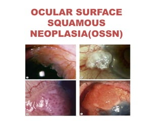
  • 17. • The lesions are described as being slightly elevated, variably shaped, relatively sharply demarcated from the surrounding normal tissues, accompanied by feeder blood vessels and colored from pearly gray to reddish gray depending on the vascularity of the tumor • They most commonly straddle the nasal or temporal limbus between the palpebral fissures
  • 18. • The following appearances may be seen. • a Gelatinous mass with superficial vessels b White leukoplakic plaque that covers the lesion c Papillomatous lesion with corkscrew-like surface blood vessels
  • 19.
  • 20. • In clinical practice, gelatinous type is the commonest. • The gelatinous lesion can again be • circumscribed, which are most common • nodular variety , which has a propensity for rapid growth • diffuse variety, the least common, which can masquerade as chronic conjunctivitis
  • 21. • The corneal side of the lesion may be seen as an opalesence of the epithelium, slightly raised in comparison to adjacent normal epithelium the edges of which are usually sharply defined. • It is best appreciated by retroillumination. • The lesion is usually nonpigmented though pigmented OSSN may also occur. • OSSN may sometimes mimic a pterygium or a pinguecula. It has also been known to occur in a pre-existing pinguecula or pterygium.
  • 22. • It is nearly impossible to categorise OSSN as benign or malignant based on clinical appearance alone. • Larger lesions that are fixated to the underlying tissue are usually malignant.
  • 23.
  • 24.
  • 25.
  • 26. • Staining with fluorescein sodium or Rose Bengal helps in the diagnosis by showing up the papillary or granular nature of the lesion and by delineating its extent • Anterior segment OCT may be done to find out the extent of deep involvement as well as intraocular and angle invasion
  • 27. • Intraocular invasion is rare ,heralded by onset of iritis, glaucoma, scleral thinning or retinal detachment. • Regional and systemic metastases are also uncommon. • Common sites of metastasis include preauricular,submandibular and cervical lymph nodes, parotid gland, lungs, and bone.
  • 28. DIFFERENTIAL DIAGNOSIS • Pterygium • papilloma • pingueculum, • nevus. • Malignant melanoma • pyogenic granuloma • pseudo-epitheliomatous hyperplasia • Corneal pannus from any cause • Mooren's ulcer, • fatty degeneration of the cornea and epithelial dystrophy of the cornea
  • 29.
  • 30. • Papilloma may occur anywhere on the conjunctiva, may be sessile or pedunculated, has a punctate vascular pattern, occurs in younger patients,but ultimately may only be difterentiated by histologic examination
  • 31. • Malignant melanoma has a regular smooth surface, lacks gelatinous or leukoplakic surface disturbances and may become ulcerated
  • 32. • Benign nevi tend to occur in the interpalpebral zone, from the limbus to the caruncle, to occur in younger patients, and to have distinctive cysts on slit-lamp examination
  • 33.
  • 34.
  • 35.
  • 36. HISTOPATHOLOGY • DYSPLASIA: • Mild - less than a third thickness occupied by atypical cells • Moderate - three quarters thickness occupied by atypical cells • Severe -- nearly full thickness occupied by atypical cells • CARCINOMA IN SITU: as above with loss of the normal surface layer • INVASIVE SQUAMOUS CELL CARCINOMA: as above when the basement membrane of the basal epithelial layer has been breached and invasion of the substantia propria has occurred.
  • 37.
  • 38.
  • 39. • Spindle cell, Mucoepidermoid and Adenoid- Squamous are three aggressive variants
  • 41.
  • 42.
  • 43.
  • 44.
  • 45.
  • 47. TOPICAL CHEMOTHERAPY • Topical chemotherapy may be used preoperatively, intraoperatively or postoperatively. Its preoperative use is seen in cases with extensive lesions where it helps to reduce the tumor size and makes the tumor more amenable to surgical excision. Intraoperatively, chemotherapy may have a role in place of adjuvant cryotherapy postoperatively can be beneficial for managing cases with residual disease or positive surgical margins.
  • 48.
  • 49.
  • 50. MITOMYCIN C • Mitomycin C is the most commonly used topical chemotherapy agent • It is a non cell cycle specific ALKYLATING AGENT that acts by alkylating the cross-linked DNA and inhibits DNA, RNA, and protein synthesis. • MMC is used in concentrations of 0.04% qid, 4 days a week for 4 weeks. • Overall, success rates ranging from 87 to 100% have been reported.
  • 51.
  • 52. 5-FU • Is a pyrimidine analogue that acts by integrating with the DNA during S phase. It also interferes with RNA synthesis. • It is used as 1% topical solution. • Side effects are similar to MMC
  • 53. INTERFERONS • Interferons are protein molecules that bind to cell receptors and trigger synthesis of effector proteins that can inhibit viruses, activate immunocompetent cells, and regulate oncogenes. • Reports are emerging on the use of topical IFN- 2b for OSSN. • It is more expensive than MMC and 5 FU, requires prolonged treatment but has a better safety profile
  • 54.
  • 55. RADIOTHERAPY • Plaque brachytherapy using radiation sources like Strontium 90 or Rhuthenium 106 is useful as an adjuvant to support surgical excision. Complications include conjunctival scarring, symblepharon, dry eye, cataract, scleral and corneal ulceration.
  • 56. SUMMARY • [1] Suspected OSSN < 3 clock hours –Excision biopsy + base/ edge cryotherapy + alchoholepitheliectomy is done.
  • 57. • [2] Suspected OSSN 3 – 6 clock hours – As there is a risk of producing limbal stem cell deficiency, excision biopsy + cryotherapy is better avoided. • A diagnostic biopsy is required • In pre-invasive lesions topical chemotherapy is likely to achieve tumour resolution. • If invasive, surgery + cryotherapy is done after chemoreduction with 4 to 6 cycles of topical chemotherapy.
  • 58. • [3] OSSN > 6 clock hours – A diagnostic biopsy is required. • In pre-invasive lesions topical chemotherapy is quite likely to achieve tumour resolution. • If invasive, surgery + cryotherapy is done after chemoreduction with 4 to 6 cycles of topical chemotherapy. • If there is no response to chemotherapy palliative radiotherapy or extensive surgery like enucleation / exenteration may be required.