extracellular matrix
•Als PPTX, PDF herunterladen•
12 gefällt mir•5,300 views

the extra cellular matrix of cell

Empfohlen
Weitere ähnliche Inhalte
Was ist angesagt?
Was ist angesagt? (20)
Ähnlich wie extracellular matrix
Ähnlich wie extracellular matrix (20)
Kürzlich hochgeladen
Kürzlich hochgeladen (20)
extracellular matrix
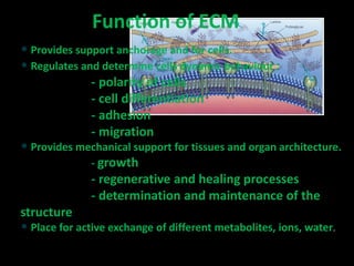
- 1. • Provides support anchorage and for cells. • Regulates and determine cells dynamic behaviour : - polarity of cells - cell differentiation - adhesion - migration • Provides mechanical support for tissues and organ architecture. - growth - regenerative and healing processes - determination and maintenance of the structure • Place for active exchange of different metabolites, ions, water. Function of ECM
- 2. EXTRACELLUJLAR MATRIX The extracellular matrix (ECM) is the noncellular component present within all tissues and organs, and provides not only essential physical scaffolding for the cellular constituents but also initiates crucial biochemical and biomechanical cues that are required for tissue morphogenesis, differentiation and homeostasis Cell adhesion to the ECM is mediated by ECM receptors, such as integrins, discoidin domain receptors and syndecans the ECM directs essential morphological organization and physiological function by binding growth factors (GFs) and interacting with cell-surface receptors to elicit signal transduction and regulate gene transcription.
- 3. The main fibrous ECM proteins are collagens, elastins, fibronectins and laminins ..PGs fill the majority of the extracellular interstitial space within the tissue in the form of a hydrated gel
- 4. • collagen – the main ECM component, forms the main fibres • elastin • proteoglycans - heteropolysacharides • structural glycoproteins - fibronectin, lamininaaaaa Structure of ECM
- 5. • The most abundant protein in the body, making 25%-35% of all the whole-body proteins. • Collagen contributes to the stability of tissues and organs. • It maintains their structural integrity. • It has great tensile strenght. • The main component of fascia, cartilage, ligaments, tendons, bone and skin. • Plays an important role in cell differentiation, polarity, movement • Plays an important role in tissue and organ development Collagen
- 7. Formation of Collagen Networks Fig. 19-52
- 8. Collagen is insoluble glycoprotein (protein + carbohydrate) Collagen polypeptide structure: - G – X – A – G – A – A – G – Y – A – G – A – A – G – X – A – G – A – – A – G – X – A – G – A – A – G – Y – A – G – A – A – G – X – A – G – – A – A – G – X – A – G – A – A – G – Y – A – G – A – A – G – X – A – G - glycine, X - proline or hydroxyproline, Y – lysin or hydroxylysine, A – amino acid • Proline and hydroxyproline constitute about 1/6 of the total sequence, provide the stifness of the polypeptide chain. • Carbohydrates : glucose, galactose Collagen structure
- 9. Collagen Distribution Type Molecule composition Tissue Fibrilar Collagens I [a1(I)2a2(I)] Skin, tendon,bone, ligaments, dentin II [a1(II)]3 Cartilage, vitreus humor III [a1(III)]3 Skin, muscle, blood vessels V [a1(V)]3 Similar to type I, fetal tissue Sheet-forming collagen IV [a1(IV)2a2(IV)] [a1(IV)a2(IV)a3(IV)] All basal laminaes
- 10. • Elastin is a major protein component of tissues that require elasticity such as arteries, lungs, bladder, skin and elastic ligaments and cartilage. • It is composed of soluble tropoelastin protein containing primarily, glycine and valine and modified alanine and proline residues. • Tropoelastin is a ~65kDa protein that is highly cross-linked to form an insoluble complex. • The most common interchain cross-link in elastins is the result of the conversion of the amine groups of lysine to reactive aldehydes by lysyl oxidase. This results in the spontaneous formation of desmosine cross- links. Elastin
- 11. Proteoglycans Proteoglycans represent a special class of glycoproteins that are heavily glycosylated (95%). They consisit of core protein with one or more attached glycosamino glycan chain(s).
- 12. Function of Proteoglycans • organize water molecules - resistant to compression - return to original shape - repel negative molecules • occupy space between cells and collagen • high viscosity - lubricating fluid in the joints • specific binding to other macromolecules • link to collagen fibers - form network - in bone combine with calcium salts (calcium carbonate, hydroxyapatite) • cell migration and adhesion - passageways between cells • anchoring cells to matrix fibers
- 13. • Direct linkage to collagen or proteoglycans - anchoring collagen fibers to cell membrane - covalent attachment to membrane lipid • Major adhesive structural glycoproteins Structural Glycoproteins (Adhesive glycoproteins) • Extracellular – Fibronectins – Laminins • Cell surface – Integrins
- 14. Fibronectin Function • cell adhesion • cell differentiation • anchoring basal laminae to other ECM • blood clothing - clothing process, link to fibrin
- 15. Fibronectin Structure • Dimer connected at C-terminal by S-S linkage • Rigid and flexible domains • Cell binding domain RGDS (arg, gly, asp, ser) - binding receptor in cell membranes • Domain is binding to - collagen type I, II and III - heparin sulfate - hyaluronic acid - fibrin
- 16. Laminin Structure and Function • cross-shaped glycoprotein • 3 polypeptide chains • domain bind - collagen type IV - heparin - heparin sulfate • cell surface receptor • cell adhesion • cell differentiation • anchoring the glycoprotein to basal laminae
- 17. What binds the cells to the ECM?
- 18. Integrins • Groups of transmembran e proteins • Link cytoskeleton to ECM • Fibronectin receptor is best known
Hinweis der Redaktion
- Review of the particular topic , pubmed.com,