Meniscus injury
•Als PPTX, PDF herunterladen•
70 gefällt mir•16,995 views

Orthopedic Knee Injuries Meniscus tear Injury

Empfohlen
Weitere ähnliche Inhalte
Was ist angesagt?
Was ist angesagt? (20)
Ähnlich wie Meniscus injury
Ähnlich wie Meniscus injury (20)
Mehr von Rifhan Kamaruddin
Mehr von Rifhan Kamaruddin (14)
Kürzlich hochgeladen
Kürzlich hochgeladen (20)
Meniscus injury
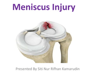
- 1. Meniscus Injury Presented By Siti Nur Rifhan Kamarudin
- 2. ANATOMY • Meniscus is a cushion structure made of cartilage which fits within the knee joint between tibia and femur. • Each Menisci has - Two ends - Two borders - Two surfaces
- 4. MEDIAL MENISCUS • C- Shaped structure and lateral meniscus is more circular. • Anterior horn : Attached to the tibia anterior to the intercondylar eminence to the ACL. • Posterior horn : Anchored immediately in front of the attachment of PCL posterior to the intercondylar eminence.
- 5. Medial Meniscus • Peripheral border attached to the medial capsule through the coronary ligament to the upper border of tibia. • Most of the weight borne on the posterior portion of meniscus
- 6. LATERAL MENISCUS • Circular shaped • The anterior and posterior horns are closer to each other & near insertion of ACL • Anterior Horn : Attached to the tibia in front of the intercondylar eminence. • Posterior Horn : Attached to the posterior aspect of the intercondylar eminence in front of posterior attachment of medial meniscus.
- 7. Lateral Meniscus • The lateral meniscus is mobile and medical meniscus is more fixed -> causing more tears to occurs in medical meniscus • Lateral meniscus is associated with discoid meniscus and meniscal cysts • Lateral meniscus is also assoc. with acute injury to ACL Medial Meniscus • Tears of medical meniscus occurs more with degenerative tears • Associated with a baker’s cyst.
- 8. BLOOD SUPPLY • The blood supply of meniscus decides the healing potential of the meniscus • The outer one-third of meniscus is vascular. It will heal if repaired • The inner one-third is not vascular and is nourished by synovial fluid. • The middle third is red/white and it is avascular. • The blood supply of meniscus originates from medial and lateral genicular arteries
- 9. FUNCTIONS OF MENISCUS • Shock Absorber: Provides load sharing across knee by increasing the contact area and decreasing the contact stress. • Act as joint filler : Compensates for the gross incongruity between tibial and femoral articulating surfaces. • Joint Lubrication: help to distribute Synovial fluid through the joint and aiding the nutrition of articular cartilage.
- 10. OVERVIEW of MENISCAL INJURY • Epidemiology: - Most common indication for knee surgery • Location: Medial Tears - More common - Degenerative tears in older patients usually occur in posterior horn of medial meniscus. Lateral Tears - More common in acute ACL tears
- 11. CLINICAL FEATURES • Pt is usually a young person who sustain twisting injury to the knee • Knee pain (often severe) • Swelling of the knee within 48hours • “Locking” : Sudden inability to extend the knee fully – suggest a ‘bucket-handle tear’. • Popping or clicking within the knee. • Limited motion of knee joint. • Tenderness when pressing on the meniscus (Knee joint line)
- 12. CLASSIFICATION OF MENISCAL TEAR • Based on Location Red Zone: Outer third, vascularized Red-White Zone : Middle Third White Zone : Inner third, Vascularized
- 13. Based On Pattern • Vertical/Longitudinal - Common, esp. with ACL tears • Bucket Handle - Vertical tear which may displace into notch • Horizontal - More common in older population - May be associated with meniscal cysts
- 14. PHYSICAL EXAMINATION • The joint may be held slightly flexed and there is often an effusion. • In late presentations, the quadriceps will be wasted. • Tenderness is localized to the joint line, particularly the medial line. • Flexion is usually full but extension is often limited.
- 15. SPECIAL TESTS 1) Thessaly Test • Standing at 20 degrees of knee flexion on affected limb • Patient twists with knee external and internal rotation. • Positive Test: Clicking, pain or discomfort on joint line.
- 17. 2) McMurrays Test • Principle: To trap the meniscus between the tibia and femur. • Pt needs to be relaxed. • One hand on knee joint line. Other hand holds the foot & ankle. • Flex the knee as far as possible (Hyperflexion) • Externally rotate(Medial Me.) or internally rotate (Lateral Me.) the tibia and then extend the knee. • Positive McMurray’s : Clicking or popping felt associated with pain.
- 19. 2) Apley’s Grinding test • Patient is in prone position • Knee flexed to 90 degrees • The leg is rotated from side to side • Compression force applied • A painful response signifies a torn or degenerate meniscus.
- 21. IMAGING Radiographs • Should be normal in young patient with acute meniscal injury MRI • Most sensitive diagnostic test • Findings - MRI Grade III signal is indicative of a tear - Parameniscal cyst indicates presence of meniscal tear - May see ‘Double PCL” sign that indicates bucket- handle meniscal tear.
- 23. MANAGEMENT NON-OPERATIVE TREATMENT Indication: First line of treatment for degenerative tears : Acute episode without locking but with acute synovitis • Immediate abstinence from weight bearing • Rest • Ice pack application • Compression dressing • NSAIDS • Rehabilitation exercises
- 24. SURGICAL MANAGEMENT 1)Meniscectomy 2)Meniscal Repair 3)Meniscal Transplantation
- 25. OPERATIVE TREATMENT 1) Partial Meniscectomy • Indication: Tears not amenable to repair (complex, degenerative, radial tear patterns) : Repair failure > 2 times • Objective: Remove the torn meniscal fragment and contour the peripheral rim, leaving a balanced, stable rim of meniscal tissue. • Outcomes - >80% satisfactory function • Partial is preferred over total meniscectomy - Shorter operating time, Faster recovery, better post-op function.
- 26. Anthroscopic Meniscal Repair 3 important steps: - Appropriate patient selection : should have documented tear that is able to heal - Tear debridement and local synovial, meniscal and capsular ablation to stimulate a proliferative fibroblastic response - Suture placement to reduce and stabilize the meniscus
- 27. Meniscal Repair Risks: – Saphenous Nerve and Vein damage – Peroneal Nerve – Popliteal Vessels
- 28. 3) Meniscal Transplantation • Attempts at meniscal replacement with - Allograft meniscus - Autograft fascial material - Synthetic meniscus
- 29. REFERENCES • Apley and Solomon’s Concise System of Orthopedic and Trauma, 4th Edition
Hinweis der Redaktion
- if the meniscus is removed or injured, the pt will develop arthritis of knee joint.