Primary open angle glaucoma
•Als PPTX, PDF herunterladen•
219 gefällt mir•50,028 views

POAG ppt

Empfohlen
Weitere ähnliche Inhalte
Was ist angesagt?
Was ist angesagt? (20)
Andere mochten auch
Andere mochten auch (20)
Ähnlich wie Primary open angle glaucoma
Ähnlich wie Primary open angle glaucoma (20)
Kürzlich hochgeladen
Kürzlich hochgeladen (20)
Primary open angle glaucoma
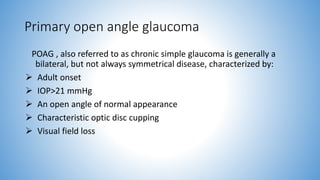
- 1. Primary open angle glaucoma POAG , also referred to as chronic simple glaucoma is generally a bilateral, but not always symmetrical disease, characterized by: Adult onset IOP>21 mmHg An open angle of normal appearance Characteristic optic disc cupping Visual field loss
- 2. Predisposing and risk factors 1. Intraocular pressure( IOP)- most imp risk factor 2. Age - most cases >40 years, unusual <40years 3. Race - blacks >whites 4. Family history and inheritance – sibling > offspring 5. Diabetes mellitus 6. Reduction in perfusion pressure (BP-IOP) 7. Myopia 8. Retinal diseases like central retinal vein occlusion, retinal detachment and retinitis pigmentosa 9. Thyroid disorders 10. Cigarrete smoking 11. Steroid usage – topical (>6 weeks) , systemic
- 3. Pathogenesis of rise in IOP • Rise in IOP occurs due to decrease in the aqueous outflow • Reduced aqueous outflow facility occurs due to failure of aqueous outflow pump mechanism Thickening and sclerosis of trabecular meshwork with faulty collagen tissue Narrowing of intertrabecular spaces Deposition of amorphous material in the juxtacanalicular space Collapse of schlemm’s canal and absence of giant vacuoles in the cells lining it
- 4. Epidemiology of POAG Affects about 1 in 100 of the general population (of either sex) above the age of 40 years Forms about one third cases of all glaucomas PREVALENCE
- 5. Clinical features Symptoms Insidious and asymptomatic disease Gradual painless loss of vision Mild headache, eye ache Visual field defect(SCOTOMA) Frequent change in presbyopic glasses Delayed dark adaptation Significant loss of vision and blindness
- 6. Signs 1. Anterior segment signs - corneal haze - sluggish pupil 2. Intraocular pressure changes Repeated observations of IOP( every 3-4 hour), for 24 hour( diurnal variation test) Different patters of diurnal variation of IOP Morning rise in IOP- 20% of cases Afternoon rise in IOP-25% of cases Biphasic rise in IOP-55% of cases Variation in IOP of over 5 mm Hg (Schiotz) is suspicious and over 8 mm of Hg is diagnostic of glaucoma Fig.Patterns of diurnal variations of IOP: A, normal slight morning rise; B, morning rise seen in 20% cases of POAG; C, afternoon rise seen in 25% cases of POAG; D, biphasic variation seen in 55% cases of POAG
- 7. 3. Optic nerve head evaluation Normal optic nerve head Neuroretinal rim – tissue between the outer edge of the cup and disc margin. A normal rim is orange or pink in colour and follows the ISNT rule Rim-disc ratio Cup-disc ratio- normal vertical C/D ratio= 0.1-0.4, although it varies with the size of the disc Asymmetry between 2 eyes is < 0.2 Blood vessels
- 9. A Normal optic disc (A, Diagrammatic depiction; B, Fundus photograph) B
- 10. Early glaucomatous changes include : Vertically oval, large cup Asymmetry of >0.2 between 2 eyes Large cup i.e., 0.6 or more (normal cup size is 0.3 to 0.4) Pallor of the disc Splinter haemorrhages Atrophy of RNFL C D FIG.optic disc showing early glaucomatous changes (C, Diagrammatic depiction; D, Fundus photograph)
- 11. Advanced glaucomatous changes Marked cupping (cup size 0.7 to 0.9) Thinning of neuroretinal rim Nasal shifting of retinal vessels (Bayonetting sign) Pulsations of the retinal arterioles Lamellar dot sign Glaucomatous optic atrophy All the neural tissue of the disc is destroyed and the optic nerve head appears white and deeply excavated
- 12. Optic disc showing advanced glaucomatous changes (A, diagramatic depiction; B, fundus photograph) and glaucomotous optic atrophy (C, diagramatic depiction; D, fundus photograph A B D D
- 13. 4. Visual field defects Anatomical basis : Distribution of retinal nerve fibres 1. Papillomacular bundle(PMB) 2. Arcuate fibres Superior arcuate fibers(SAF) Inferior arcuate fibers(IAF) 3. Radiating fibres Superior radiating fibers(SRF) Inferior radiating fibers(IRF) Arrangement of nerve fibres within the optic nerve-arcuate fibres occupy temporal portion of the disc and are most susceptible to damage Distribution of retinal nerve fibres Arrangement of nerve fibres within optic nerve head
- 14. Nomenclature of glaucomatous field defects Isopter contraction Baring of blind spot Wing shaped paracentral scotoma-earliest clinically significant field defect Seidel’s scotoma – paracentral scotoma joins the blind spot to form a sickle shaped scotoma Arcuate or Bjerrum’s scotoma Ring or double arcuate scotoma Roenne’s central nasal step Perpheral field defects Advanced glaucomatous field defects Field defects in POAG: A, baring of blind spot; B, superior paracentral scotoma; C, Seidel's scotoma; D, Bjerru-m's scotoma; E, double arcuate scotoma and Roenne's central nasal step
- 15. Criteria to grade glaucomatous field defects • The criteria to label early, moderate and severe glaucomatous field defect from the HFA central 30-2 test, single printout is depicted in Table
- 16. Assessing visual field glaucomatous field defects Camparing individual single field printouts Overview printout Progression analysis software( GPA IN HFA machines and peritrend in octopus perimeter) Visual field index(VFI) • For proper understanding of Table evaluation of the Humphrey single field printout described should be revised
- 17. INVESTIGATIONS 1. Tonometry 2. Central corneal thickness(CCT) 3. Diurnal variation test 4. Gonioscopy 5. Documentation of optic disc changes 6. Slit-lamp examination of anterior segment 7. Perimetry to detect the visual field defects 8. Nerve fibre layer analyzer (NFLA) 9. Provocative tests Water drinking test
- 18. TONOMETRY • The intraocular pressure (IOP) is measured with the help of an instrument called tonometer • Indentation tonometery • Schiotz tonometer • Applanation tonometry • Goldmann tonometer • Perkin’s applanation tonometer • Pneumatic tonometer • Pulse air tonometer • Tono-Pen Schiotz tonometer Perkin’s hand-held applanation tonometer
- 19. Technique of Schiotz tonometry Technique of applanation tonometry End point of applanation tonometry. (A) too small; (B) too large; (C) end point.
- 20. Gonioscopy The technique of biomicroscopic examination of the angle of the anterior chamber using a goniolens. The angle structures seen from behind forward are: 1. Root of the iris 2. Ciliary body band 3. Scleral spur 4. Trabecular meshwork 5. Schwalbe’s line
- 22. PERIMETRY • The visual field is a three-dimensional area of a subject’s surroundings that can be seen at any one time around an object of fixation • Perimetry • It is the procedure for estimating extent of the visual fields • Kinetic versus static perimetry • Peripheral versus central field charting • Peripheral field charting Central field charting • Confrontation method • Perimetery: Lister’s, Goldmann’s and automated • Campimetry or scotometry • Goldmann’s perimetry • Automated field analysis • Manual versus automated perimetry • Manual perimetry • Automated perimetry Extent of normal visual field
- 23. • MANUAL PERIMETRY • Confrontation method • Lister’s perimeter • Campimetry (scotometry) • Goldmann’s perimeter Lister’s perimeter. Bjerrum’s screen Goldmann’s perimeter
- 24. • AUTOMATED PERIMETRY • Automated perimeters are computer assisted and test visual fields by a static method • Commonly used automated perimeters are: Octopus, Field Master and Humphrey field analyser Humphrey field analyser (automated perimeter)
- 25. • Advantages of automated perimetry over manual perimetry • Interpertation of automated perimetry print out field charts • Automated perimeter variables • Testing strategies and programmes • Automated perimeter variables • Background illumination • Stimulus intensity • Stimulus size • Stimulus duration • Testing strategies and programs • Suprathreshold testing • Threshold testing • Full threshold testing • Fast Pac • SITA (Swedish Interactive Threshold Alogarithm) Stimulus intensity scales compared
- 26. • Test programmes- The standard test programmes used with static threshold strategy on the Humphrey’s Field Analyser (HFA) can be grouped as below A. Central field tests • Central 30 - 2 test, • Central 24 - 2 test, • Central 10 - 2 test,and • Macular test B. Peripheral field tests • Peripheral 30/60-1, • Peripheral 30/60-2, • Nasal step, and • Temporal crescent C. Speciality tests • Neurological-20, • Neurological -50, • Central 10-12, and • Macular test D. Custom tests
- 27. • Evaluation of Humphrey single-field print-out I. Patient data and test parameters II. Reliability indices III. Gray scale simulation of the test data is depicted in zone III or part III of the printout IV. Total deviation plots V. Pattern deviation plots VI. Global indices VII. Glaucoma hemifield test (GHT) VIII.Actual threshold values
- 28. DIAGNOSIS 1. Primary open angle glaucoma (POAG) raised IOP(>21 mm of Hg) associated with definite glaucomatous optic disc cupping and visual field changes 2. Ocular hypertension or glaucoma suspect patient has an IOP constantly more than 21 mm of Hg but no optic disc or visual field changes 3. Normal tension glaucoma (NTG) or low tension glaucoma (LTG) diagnosed when typical glaucomatous disc cupping with or without visual field changes is associated with an intraocular pressure constantly below 21 mm of Hg
- 29. Triad of abnormalities in disc, field and intraocular pressure (IOP) for the diagnosis of glaucoma.
- 30. Management The primary aim of treatment is to prevent functional impairment of vision. It is important to perform a good baseline evaluation with which future progress can be compared and documented The initial data should include: visual acuity, slit-lamp examination of anterior segment, tonometry (preferably with applanation tonometer); optic disc evaluation (preferably with fundus photography), gonioscopy and visual field charting GRADING- American Academy of Ophthalmology (AAO) grades severity of glaucoma damage into mild, moderate and severe
- 31. Therapeutic choices 1. Medical therapy 2. Argon or diode laser trabeculoplasty 3. Filteration surgery
- 32. Medical therapy The initial therapy of POAG is still medical, with surgery as the last resort Basic principles of medical therapy of POAG Identification of target pressure Treatment regimes : Single drug therapy One topically instilled anti-glaucoma drug is chosen after considering patient’s medical history and socio-economic background. If the initial drug is ineffective or intolerable, it is to be replaced. Combination therapy If one drug is insufficient to control the IOP, then a combination therapy is advised Monitoring of therapy by disc changes and field changes and tonometry is most essential on regular follow-up
- 33. ANTI-GLAUCOMA DRUGS • Classification • A. Parasympathomimetic drugs (Miotics) • B. Sympathomimetic drugs (Adrenergic agonists) • C. β-blockers • D. Carbonic anhydrase inhibitors • E. Hyperosmotic agents • F. Prostaglandins • G. Calcium channel blockers
- 35. Prostaglandin analogues Mechanism of action: uveoscleral outflow Indication : first line therapy Preparations : 1. Latanoprost (0.005%) - PG F2-α analogue, OD dosage, additive effect with timolol. 2. Bimatoprost (0.03%) - decreases outflow resistance, OD dosage. 3. Travoprost (0.04%) – PG F2-α analogue, OD dosage. 4. Unoprostive isopropyl (0.12%) – dolosanoid related structure similar to PG F2-α, BD dosage.
- 36. Side effects Systemic 1. Upper respiratory tract symptoms (flu like ) 2. Headache and precipitation of migraine in susceptible individuals 3. Muscle and joint pains 4. Skin rash
- 37. Ocular Side Effects 1. Conjunctival hyperaemia and foreign body sensation 2. Eyelash lengthening, thickening, hyperpigmentation, increase in number 3. Iris hyperpigmentation 4. Increase in severity and recurrence of herpetic keratitis 5. Anterior uveitis 6. Cystoid macular edema
- 38. β–adrenergic antagonists Mechanism of action – block β receptors in ciliary processes aqueous production Indication – used as first line therapy for patients who cant PG analogues. Preparations 1. Timolol maleate(0.25%,0.5%) – non selective β blocker, BD dosage “short term escape” and “long term drift” 2. Betaxolol(0.25%,0.5%) – cardioselctive (β1) bocker, BD dosage, useful in asthmatics, less effective than timolol. 3. Levobunolol(0.5%) – non selective β blocker 4. Carteolol(1%, 2%) – lesser incidence of bradycardia 5. Metipranolol(0.1%, 0.3%, 0.6%)
- 39. side effects Systemic 1. Cardiovascular effects – bradycardia, arrhythmia, heart failure, syncope 2. Respiratory reactions – bronchospasm and airway obstruction, especially in asthmatics 3. CNS effects – depression, anxiety, confusion, drowsiness, disorientation 4. Others – nausea, diarrhoea, decreased libido, skin rashes, alopecia
- 40. Ocular 1. Conjuctival hyperaemia 2. Superficial punctate keratopathy 3. Corneal anaesthesia
- 41. Contraindications 1. Bronchial asthma 2. Chronic obstructive pulmonary disease 3. Heart blocks 4. Congestive heart failure 5. Cardiomyopathy
- 42. Parasympathomimetics Classification 1. Agonists (direct acting) – pilocarpine 2. Cholinesterase inhibitors (indirect acting) : Reversible – physostigmine Irreversible – echothiophate iodide, demecarium, diisopropyl- fluoro-phosphate 3. Dual action - e.g. carbachol
- 43. Mechanism of action trabecular outflow Preparations 1. a) Pilocarpine e/d(1%,2%,4%)BD-QID dosage b) ocuserts(pilo-20,pilo-40) c)Pilocarppine gel(4%)HS 2. Carbachol(0.75%,1.5%,3%)BD-TDS dosage, may be useful in pilocarpine sensitivity 3. Echothiophate iodide(0.125%)OD-BD dosage, intense miosis, more GI side effects 4. Demecarium bromide(0.125%,0.25%) 5. Physostigmine(0.5%)
- 44. Side effects Systemic 1. Increased salivation, increased gastric, abdominal cramps, diarrhoea 2. Increased sweating, anxiety 3. Bradycardia
- 45. Ocular 1. Miosis leading to decrease visual acuity in cases of posterior polar cataracts, impairment of night vision 2. Brow ache, head ache 3. Myopia 4. Keratitis 5. Iritis, iris cyst, posterior synechiae 6. Lenticular opacities 7. Retinal detachment
- 46. Carbonic anhydrase inhibitors Mechanism of action aqueous humor production Indications : useful as short term therapy, especially in acute cases
- 47. Preparations Oral 1. Acetazolamide tablet (diamox 250mg, IV diamox5-10mg/kg)BD-QID dosage 2. Dichlorphenamide(50mg) BD-TDS dosage 3. Methazolamide(50 mg) BD-TDS dosage Topical 1. Dorzolamide(2%) BD-TDS dosage, additive effect with timolol 2. Brinzolamide (1%)BD-TDS dosage, less stinging sensation
- 48. Side effects Systemic 1. Paraesthesias, numbmness, lethargy, depression, malaise 2. Metabolic acidosis, hypokalemia, increased serum urate level 3. Urinary frequency 4. Anorexia, cramps, flatulence, weight loss, diarrhoea 5. Sulfonamide related – blood dyscrasias, renal calculi, steven-Johnson syndrome Topical agents are less likely to induce systemic side effects
- 49. Ocular 1. Induced myopia, blurred vision 2. Stinging senstaion 3. Conjunctivitis, keratitis Ocular side effects are seen with topical agents
- 50. Adrenergic agonists Mechanism of action aqueous outflow by both α and β receptor stimulation, aqueous production due to stimulation of α receptors
- 51. Classification and preparations Non selective(α and β receptor stimulation) 1. Epinephrine(0.5%,1%,2%), BD dosage 2. Dipivefrine (0.1%) – prodrug of epinephrine, increased corneal panetration α2 adrenergic agonists Apraclonidine (0.5%,1%)BD-TDS dosage, used prophylactically for prevention of IOP elevation following laser trabeculoplasty, YAG laser iridotomy and posterior capsulotomy Clonidine (0.125%,0.25%) BD dosage, centrally acting anti-hypertensive agent Brimonidine(0.2%)BD-TDS dosage Clinically, nonselective adrenergic agents have been replaced by α2 adrenergic agonists because of their improved efficacy and side effect profile
- 52. Side effects Systemic 1. Hypertension(nonselective agents), hypotension(α2 adrenergic agonists) 2. Headache, fatigue, syncope 3. Anxiety, insomnia, depression
- 53. Ocular 1. Eyelid retraction, lid edema, dermatitis 2. Conjuctival hyperaemia, irritation, allergy, follicular conjuctivitis 3. Mydriasis 4. Cystoid macular edema in aphakics
- 54. Hyperosmotic agents Mechanism of action plasma tonicity osmotic gradient dehydrated the vitreous Indications To control acute episodes of elevated IOP
- 55. Preparations 1. Mannitol IV(20% solution1-2g/kg over 20-30 minutes), to be used cautiously in hypertensives 2. Glycerol oral(50% solution1-1.5g/kg, mixed with equal amoount of water or lime juice), to be used cautiously in diabetics as it is metabolised to glucose 3. Urea IV – not recommended for routine use 4. Isosorbide – metabolically inert
- 56. Side effects Systemic 1. Expansion of blood volume, congestive heart failure 2. Nausea, vomiting, diarrhoea 3. Electrolyte disturbances 4. Renal failure Ocular 1. Rebound increase in IOP 2. Aqueous flare
- 57. Calcium channel blockers Mechanism of action • Might be due to its effects on secretory ciliary epithelium Preparations • Verapamil has been tried as 0.125 percent and 0.25 percent eyedrops twice a day Indications • place in the mangement of patients with POAG, where miotics, beta- blockers and sympathomimetics are all contraindicated NEUROPROTECTIVE AGENTS
- 58. Argon or diode laser trabeculoplasty Indications 1. Avoidance of polypharmacy(>2 preparations) 2. Avoidance of surgery 3. Primary therapy in patients with non-compliance to medical therapy Mechanism of action outflow facility by causing shrinkage of trabecular meshwork
- 59. Technique 40- 50 spots on the anterior half of trabecular meshwork over 180° using a gonioloens Complications 1. Acute rise of IOP 2. Uveitis, haemorrhage, PAS
- 60. Surgical management Indications 1. Uncontrolled glaucoma despite maximal medical therapy(3 drugs) and laser trabeculoplasty 2. Failure of medical therapy and /or laser trabeculaplasty 3. Non-compliance to medical therapy and unavailability of laser trabeculoplasty 4. Advanced disease requiring a very low target pressure may benefit from early surgery 5. Primary line of treatment
- 61. Surgical procedures Trabeculectomy Full – thickness sclerectomy Viscocanalostomy
- 62. Trabeculectomy Creation of a fistula between the angle of anterior chamber and sub-tenon’s space which allows egress of aqueous from AC to a drainage bleb
- 63. Procedure Conjunctival flap Partial thickness scleral flap Excision of trabecular tissue Peripheral iridectomy Closure using 10-0 monofilament sutures Subconj inj dexamethasone and gentamycin
- 66. Use of antimetabolites 5-fluorouracil Mitomycin C Originally advocated for patients with high risk like aphakia, pseudophakia, neovascular glaucoma, H/O failed operations, now also being used as a routinely by many surgeons
- 67. Complications of trabeculectomy Post – op shallow AC Hyphaema Iritis Cataract due to accidental injury to the lens Endophthalmitis
- 68. OCULAR HYPERTENSION Ocular hypertension or glaucoma suspect, either ofthese terms is used when a patient has an IOP constantly more than 21 mm of Hg but no optic disc or visual field changes Glaucoma suspect Defined as an adult having normal open angle on gonioscopy and anyone of the following signs in at least one eye Elevated IOP Suspicious disc changes Visual field defects High risk factors Significant diurnal variation Significantly positive water drinking provocative test Splinter haemorrhages over or near the optic disc IOP constantly more than 28 mm of Hg Retinal nerve fibre large defects Parapapillary changes Central corneal thickness < 555 μm Significant asymmetry in the cup size of the two eyes family history high myopia, diabetes or pigmentary changes in the anterior chamber
- 69. Treatment Patients with high-risk factors should be treated on the lines of POAG Patients with no high risk factors should be annually followed by examination of optic disc, perimetry and record of IOP
- 70. • NORMAL TENSION GLAUCOMA • The term normal tension glaucoma (NTG), also referred to as low tension glaucoma is labelled when typical glaucomatous disc changes with or without visual field defects are associated with an intraocular pressure (IOP) constantly below 21 mm of Hg • It is believed to result from chronic low vascular perfusion, which makes the optic nerve head susceptible to normal IOP • Clinical features • IOP – NORMAL OR LOWER THEN 21 • OPTIC DISC CHANGES • Visual field defects • Differential diagnosis 1. HIGH PRESSURE GLAUCOMAS • POAG • GLAUCOMA with intermitent rise in iop • Previous episodes of glaucoma 2. Non glaucomatous optic neuropathies • Congenital optic disc anomalies • Acquired optic neuropathies
- 71. • Treatment • Medical treatment to lower IOP • Trabeculectomy • Systemic calcium channel blockers • Monitoring of systemic blood pressure