SlideShare ist ein Scribd-Unternehmen logo
1 von 45
Current Management of GBM


      Nishantha Gunasekera
        Neurosurgery, Waikato
                                1
“ better a living problem than a dead certainty ”


                                               2
Epidemiology
 WHO estimates- 100 different types of
brain tumours
 22 500 new cases –malignant primaries
(US ’07)
 Annual incidence of malignant gliomas
5/100000
 GBM: 60-70%

 Anaplastic astros: 10-15%

 Anaplastic oligos/oligoastros: 10%


                                          3
THE   most common primary brain tumour
 Biologically aggressive

 Mean age at presentation 56-64 yr

 Median survival 12-15 months

 Presents unique treatment challenges

We Can do something positive for most


                                          4
 40% commoner in men
 Twice as more in whites than non whites

 The only established risk factor is ionising
radiation
 Head injury, n-nitroso compounds in food,
electromagnetic fields????
 IgE ? protective

 5% have family history

 NF 1,2 ; Li- Fraumeni syn (p53); Turcot’s

 GLIOGENE – an international consortium
       (to study familial gliomas)
                                                 5
Challenges
 The variably disrupted blood-brain barrier
complicating drug delivery
 Tumour capillary leakage – oedema, ICP

 Limited response to therapy

 Neurotoxicity of treatment

 Molecular pathology is complex – but
important recent advances have been made


                                           6
 Localisation of tumours in the brain
 Intrinsic resistance of tumour to
                      conventional therapy
 Limited capacity of the brain to repair itself

 The spread of malignant cells into brain
parenchyma


                                                   7
Predictors of outcome

   Patient age (18 month survival)
           <40y   - 50%
          40-60   - 20%
          >60     - 10%
   Histological features
          median survival 10 months for “classic GBMs ’’
   Performance status (18 month survival)
          KPS>70 34%
          KPS<60 13%

                                                            8
Pathophysiology
  p53 mutation- sets the stage for malignant
transformation
 Allelic loss of ch17p- malignant progression

 Many growth factors/receptors are

     over expressed
         PDGF (platelet)
         FGF (fibroblast)
         VEGF (vascular endothelial)
       EGFR      (epidermal)
   Progression to WHO Gr II is associated with
       Increased   cellularity
       Mitotic activity                     9
   GBM is characterised by
       Dense  cellularity
       High proliferation indices (Ki67-protein marker of prolif.)

       Vascular endothelial proliferation

       Focal necrosis

 Source of mitogenesis is deregulation of
the p16-cdk4-cyclin D1-pRb pathway
 Ch 10p loss is a frequent finding (60-95%)
 1p19q deletion may indicate better
prognosis- (oligo. component)
 Various subsets of GBM exist depending
on the molecular genetics                                             10
11
Histology


            12
13
Spread
   Tracking through white matter
        Corpus  callosum
        Cerebral peduncles – midbrain

        Internal capsule – encroach basal ganglion t. Into
         centrum semiovale
        Uncinate fasciculus – simultaneous frontal and
         temporal
        Interthalamic adhesions – bilateral thalamic gliomas

   CSF pathways
        10-25%   frequency of meningeal and ventricular
        seeding
   V. rarely systemic
                                                           14
Clinical features
 Due to raised ICP
 Progressive focal deficits
       Stroke   in evolution!!
   Headache
       With or without raised ICP
       Worse in the morning – hypoventilation, co2

       relieved by vomiting - ? Hyperventilation

       77% similar to tension h/a; 9% like a migraine

       Only 8% had “classic tumour headache’’

 Siezures (AOE)
 Mental status changes                                  16
Investigations


 CT contrast / non contrast
 MRI / fMRI/ MR spectroscopy

 Stealth imaging (CT/MRI)

 Tractography

 Intra-operative brain mapping

      (for awake surgery)


                                  17
c+ct


       18
mri


      19
fmri


       20
mri spectroscopy

n acytyl aspartate peak = infection
choline peak = tumour


                                      21
tractography


               22
intrapoerative mapping


                         23
Treatment

   Treatment has evolved to include
    1. Maximum safe extent of resection
    2. Fractionated radiotherapy to tumour bed
    3. Alkylating chemotherapy (nitrosoureas:
      BCNU)
       based on trials by BTSG (brain tumour study group)
        and RTOG (radiation therapy oncology group)


                                                        24
25
Treatment issues

 Siezures (levetiracetam does not induce P450)
 Peritumoural oedema (VEGFR inhibitors)
 Venous thromboembolism (20-30%, IVC filters,
anticoagulation)
 Cognitive dysfunction
 Steroid associated probs.
    (Cushings, P. jiroveci etc. etc.)
   Abulia (lack of will or initiative) - ? methylphenidate

                                                              26
 Value of total or near total resection still is
debated but...
 Data from BTSG suggests large volume
resection has a better outcome...
 Value of adjuvant chemotherapy has been
evaluated by meta-analyses (Fine et al,
Stewart et al)
 Modest improvement in 1-2 y survival rates
     (5-6% and 4-5% respectively)
                                                27
   Glioma Outcome Project data ‘05 (560 newly
diagnosed GBM- 58 community and university centres
involved)
 all patients underwent surgery
 87% received radiotherapy

 88% received anticonvulsants

 54 % received chemo

 29% used alternative meds

15% enrolled in clinical trials


                                                     28
 These studies clearly demonstrated a benefit
      for chemotherapy (that is, TMZ) in the initial
treatment of patients with GBM
 Improvement in median (14.6 compared with 12
months) and 2-year survival (27 compared
with 10%) in patients receiving or not   receiving
TMZ.

Consequently, this treatment regimen (TMZ given
concurrently with radiotherapy, followed by six monthly
cycles of TMZ) has become the new standard of care for
patients with newly diagnosed GBM.
                                                          29
Surgery
 Cytoreductive
 Extent of tumour removal and the volume of
      residual tumour on post op imaging have
      a significant effect on time to tumour
progression and median survival
 Mass effect reduction

 CANNOT be cured with surgery.. can it?

 Prolong quality of survival
 In the elderly (>65y), the benefit by surgery is
modest (median survival 17w after biopsy, 30 w after surgery : +
XRT)
                                                              30
 Partial resection of GBMs carries a
significant risk of post op haemorrhage,
oedema with risk of herniation
 Benefit of subtotal resection is deemed
dubious
 Surgical excision should be only considered
      when the goal of gross total resection is
feasible

                                             31
 The following are usually not surgical
candidates
     Extensive dominant lobe GBM
     Lesions with bilateral involvement (butterfly)

     Deep lesions (brain stem, capsule)

     The very elderly

     Patients with poor Karnofsky scores
         In
           general the neurologic condition on steroids is as
         good as its going to get- surgery rarely improves this


                                                             32
Surgical Toys

   Neuronavigation
     Offline

     Realtime   (brain suite- intraopeative MRI)
 Fluorescence guided
 Intraoperative mapping


                                                    33
Brain Suite


              34
Stereotactic Biopsy

 May underestimate by 25%
 Located in deep areas

 Small T. with minimal mass effect

 Patients with poor medical conditions

 Equivocal clinical diagnosis ( lymphoma!)


                                              35
   Stereotactic
     Frame   based


       Frameless- stealth


   Free hand
                             36
Reoperation for recurrence
 Less than 10% recur away from the original
     site
 Reoperation extends survival by an
additional 36 weeks
 Duration of high quality survival was 10
weeks
 Predictors of good quality of survival after
reoperation include
     Age, time from 1st to 2nd surgery, Karnofsky
                                                    37
 Morbidity is higher (5-18%)
 Infection rate is > X3

 Wound dehiscence more

 Neurological deficits more

 Surgical techniques differ


                                38
Radiation

 Usual dose of XRT is 50-60 Gy – whole
brain radiation has not been shown to
increase the median survival compared to
focal XRT but the risks of side effects is
greater
 Brachytherapy has not shown no significant
     benefit as an adjunct to EBRT

                                          39
Chemotherapy
   All current agents have no more than
    30-40% response rate
 Carmustine and cisplatinum have been the
    primary agents
 Temazolamide
     An  oral alkylating agent
     Initial FDA approval for relapse or progression
      of anaplastic astros. while on a nitrosourea
      containing regimen
     Now also used for newly diagnosed GBM and
      AA and progressive low grade gliomas
                                                     40
Chemotherapy
   GLIADEL wafers
     Carmustine   7.7mg in 200mg prolifeprosan 20
      hydrophobic polymer carrier (wafer)
     Following T. removal upto 8 wafers are applied
             to the resection bed (US$12500 for 8)
     Drug is released over 2-3 wks

     Exposes the tumour to 113 times the conc. of
       BCNU achieved by systemic admin.
     Approved by FDA for recurrent GBM


                                                   41
Drug
Resistance


             42
New
Therapies


            43
What next?


     can we prevent?
     can we screen?

     can we immunize?


                         45

Weitere ähnliche Inhalte

Was ist angesagt?

Pediatric medulloblastoma
Pediatric medulloblastomaPediatric medulloblastoma
Pediatric medulloblastoma
nandi_1
 

Was ist angesagt? (20)

Primary CNS Lymphoma
Primary CNS Lymphoma Primary CNS Lymphoma
Primary CNS Lymphoma
 
LOW GRADE GLIOMA controversies in management
LOW GRADE GLIOMA controversies in managementLOW GRADE GLIOMA controversies in management
LOW GRADE GLIOMA controversies in management
 
Recent advances in Glioblastoma Multiforme Management
Recent advances in Glioblastoma Multiforme ManagementRecent advances in Glioblastoma Multiforme Management
Recent advances in Glioblastoma Multiforme Management
 
Glioblastoma
GlioblastomaGlioblastoma
Glioblastoma
 
Low grade glioma evidence based management
Low grade glioma evidence based managementLow grade glioma evidence based management
Low grade glioma evidence based management
 
Glioblastoma
GlioblastomaGlioblastoma
Glioblastoma
 
Approach to the patients with brain metastases
Approach to the patients with brain metastasesApproach to the patients with brain metastases
Approach to the patients with brain metastases
 
Meningeal Hemangiopericytoma
Meningeal HemangiopericytomaMeningeal Hemangiopericytoma
Meningeal Hemangiopericytoma
 
Classification of brain tumors AND MANAGEMENT OG LOW GRADE GLIOMA
Classification of brain tumors AND MANAGEMENT OG LOW GRADE GLIOMAClassification of brain tumors AND MANAGEMENT OG LOW GRADE GLIOMA
Classification of brain tumors AND MANAGEMENT OG LOW GRADE GLIOMA
 
astro guideline on brain mets
 astro guideline on brain mets astro guideline on brain mets
astro guideline on brain mets
 
Tmz ppt
Tmz pptTmz ppt
Tmz ppt
 
Pediatric medulloblastoma
Pediatric medulloblastomaPediatric medulloblastoma
Pediatric medulloblastoma
 
Low Grade Gliomas
Low  Grade  GliomasLow  Grade  Gliomas
Low Grade Gliomas
 
Adjuvant therapy of Glioblastoma in 2020: Marching ahead.
Adjuvant therapy of Glioblastoma in 2020:  Marching ahead.Adjuvant therapy of Glioblastoma in 2020:  Marching ahead.
Adjuvant therapy of Glioblastoma in 2020: Marching ahead.
 
High Grade Glioma
High Grade GliomaHigh Grade Glioma
High Grade Glioma
 
Management of high grade glioma
Management of high grade gliomaManagement of high grade glioma
Management of high grade glioma
 
MEDULLOBLASTOMA
MEDULLOBLASTOMAMEDULLOBLASTOMA
MEDULLOBLASTOMA
 
Management of Low Grade Glioma
Management of Low Grade GliomaManagement of Low Grade Glioma
Management of Low Grade Glioma
 
2021 WHO Classification of brain tumours.pptx
2021 WHO Classification of brain tumours.pptx2021 WHO Classification of brain tumours.pptx
2021 WHO Classification of brain tumours.pptx
 
Glioblastoma Multiforme
Glioblastoma MultiformeGlioblastoma Multiforme
Glioblastoma Multiforme
 

Andere mochten auch

Respiratory chain
Respiratory chainRespiratory chain
Respiratory chain
MUBOSScz
 

Andere mochten auch (15)

Glioblastoma Multiforme
Glioblastoma MultiformeGlioblastoma Multiforme
Glioblastoma Multiforme
 
Glioblastoma multiforme
Glioblastoma multiformeGlioblastoma multiforme
Glioblastoma multiforme
 
Glioblastoma vibhay
Glioblastoma vibhayGlioblastoma vibhay
Glioblastoma vibhay
 
Glioblastoma multiforme
Glioblastoma multiformeGlioblastoma multiforme
Glioblastoma multiforme
 
Glioma
GliomaGlioma
Glioma
 
Radiation for Glioblastoma
Radiation for GlioblastomaRadiation for Glioblastoma
Radiation for Glioblastoma
 
HIGH GRADE GLIOMA MANAGEMENT
HIGH GRADE GLIOMA MANAGEMENTHIGH GRADE GLIOMA MANAGEMENT
HIGH GRADE GLIOMA MANAGEMENT
 
Brain tumors
Brain tumorsBrain tumors
Brain tumors
 
Brain tumor
Brain tumorBrain tumor
Brain tumor
 
Pathology of CNS Tumors
Pathology of CNS TumorsPathology of CNS Tumors
Pathology of CNS Tumors
 
Pathology of CNS Tumors
Pathology of CNS TumorsPathology of CNS Tumors
Pathology of CNS Tumors
 
Brain Tumor And Its Types
Brain Tumor And Its TypesBrain Tumor And Its Types
Brain Tumor And Its Types
 
Future direction in the management of high risk LOW GRADE GLIOMA
Future direction in the management of high risk LOW GRADE GLIOMAFuture direction in the management of high risk LOW GRADE GLIOMA
Future direction in the management of high risk LOW GRADE GLIOMA
 
Respiratory chain
Respiratory chainRespiratory chain
Respiratory chain
 
Brain tumour patient forum Paul de Souza Tests related to management
Brain tumour patient forum Paul de Souza Tests related to managementBrain tumour patient forum Paul de Souza Tests related to management
Brain tumour patient forum Paul de Souza Tests related to management
 

Ähnlich wie Glioblastoma Multiforme.Dr NG NeuroEdu

Alberto fara Skullbase
Alberto fara SkullbaseAlberto fara Skullbase
Alberto fara Skullbase
Sabrina Mameli
 

Ähnlich wie Glioblastoma Multiforme.Dr NG NeuroEdu (20)

Evidence based management in High grade gliomas
Evidence based management in High grade gliomasEvidence based management in High grade gliomas
Evidence based management in High grade gliomas
 
High grade glioma kiran
High grade glioma  kiranHigh grade glioma  kiran
High grade glioma kiran
 
Presentation
PresentationPresentation
Presentation
 
Alberto fara Skullbase
Alberto fara SkullbaseAlberto fara Skullbase
Alberto fara Skullbase
 
Brain_mets_drvikash.pdf
Brain_mets_drvikash.pdfBrain_mets_drvikash.pdf
Brain_mets_drvikash.pdf
 
CNS tumors_MG
CNS tumors_MGCNS tumors_MG
CNS tumors_MG
 
Radiosurgery for brain metastases
Radiosurgery for brain metastasesRadiosurgery for brain metastases
Radiosurgery for brain metastases
 
Controversies in management of low grade glioma
Controversies in management of low grade gliomaControversies in management of low grade glioma
Controversies in management of low grade glioma
 
Soft tissue sarcomas, treatment (surgical, radiation, chemotherapy)
Soft tissue sarcomas, treatment (surgical, radiation, chemotherapy)Soft tissue sarcomas, treatment (surgical, radiation, chemotherapy)
Soft tissue sarcomas, treatment (surgical, radiation, chemotherapy)
 
Primary cns lymphoma main
Primary cns lymphoma mainPrimary cns lymphoma main
Primary cns lymphoma main
 
Role of radiation in benign conditions
Role of radiation in benign conditionsRole of radiation in benign conditions
Role of radiation in benign conditions
 
Medulloblastoma
Medulloblastoma Medulloblastoma
Medulloblastoma
 
Pituitary
PituitaryPituitary
Pituitary
 
Presentation...Non-surgical management of brain tumors
Presentation...Non-surgical management of brain tumorsPresentation...Non-surgical management of brain tumors
Presentation...Non-surgical management of brain tumors
 
Dr Vikash_re irradiation_brain tumors.pdf
Dr Vikash_re irradiation_brain tumors.pdfDr Vikash_re irradiation_brain tumors.pdf
Dr Vikash_re irradiation_brain tumors.pdf
 
High grade gliomas 8 august 2016
High grade gliomas 8 august 2016High grade gliomas 8 august 2016
High grade gliomas 8 august 2016
 
MANAGEMENT OF PITUITARY TUMORS.pptx
MANAGEMENT OF PITUITARY  TUMORS.pptxMANAGEMENT OF PITUITARY  TUMORS.pptx
MANAGEMENT OF PITUITARY TUMORS.pptx
 
Metastatic diseases of nervous system
Metastatic diseases of nervous systemMetastatic diseases of nervous system
Metastatic diseases of nervous system
 
Brain tumors Imaging
Brain tumors Imaging Brain tumors Imaging
Brain tumors Imaging
 
primary CNS lymphoma
primary CNS lymphomaprimary CNS lymphoma
primary CNS lymphoma
 

Glioblastoma Multiforme.Dr NG NeuroEdu

  • 1. Current Management of GBM Nishantha Gunasekera Neurosurgery, Waikato 1
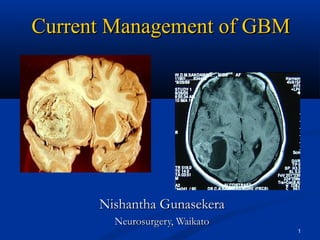
  • 2. “ better a living problem than a dead certainty ” 2
  • 3. Epidemiology  WHO estimates- 100 different types of brain tumours  22 500 new cases –malignant primaries (US ’07)  Annual incidence of malignant gliomas 5/100000  GBM: 60-70%  Anaplastic astros: 10-15%  Anaplastic oligos/oligoastros: 10% 3
  • 4. THE most common primary brain tumour  Biologically aggressive  Mean age at presentation 56-64 yr  Median survival 12-15 months  Presents unique treatment challenges We Can do something positive for most 4
  • 5.  40% commoner in men  Twice as more in whites than non whites  The only established risk factor is ionising radiation  Head injury, n-nitroso compounds in food, electromagnetic fields????  IgE ? protective  5% have family history  NF 1,2 ; Li- Fraumeni syn (p53); Turcot’s  GLIOGENE – an international consortium  (to study familial gliomas) 5
  • 6. Challenges  The variably disrupted blood-brain barrier complicating drug delivery  Tumour capillary leakage – oedema, ICP  Limited response to therapy  Neurotoxicity of treatment  Molecular pathology is complex – but important recent advances have been made 6
  • 7.  Localisation of tumours in the brain  Intrinsic resistance of tumour to conventional therapy  Limited capacity of the brain to repair itself  The spread of malignant cells into brain parenchyma 7
  • 8. Predictors of outcome  Patient age (18 month survival)  <40y - 50%  40-60 - 20%  >60 - 10%  Histological features  median survival 10 months for “classic GBMs ’’  Performance status (18 month survival)  KPS>70 34%  KPS<60 13% 8
  • 9. Pathophysiology  p53 mutation- sets the stage for malignant transformation  Allelic loss of ch17p- malignant progression  Many growth factors/receptors are over expressed  PDGF (platelet)  FGF (fibroblast)  VEGF (vascular endothelial)  EGFR (epidermal)  Progression to WHO Gr II is associated with  Increased cellularity  Mitotic activity 9
  • 10. GBM is characterised by  Dense cellularity  High proliferation indices (Ki67-protein marker of prolif.)  Vascular endothelial proliferation  Focal necrosis  Source of mitogenesis is deregulation of the p16-cdk4-cyclin D1-pRb pathway  Ch 10p loss is a frequent finding (60-95%)  1p19q deletion may indicate better prognosis- (oligo. component)  Various subsets of GBM exist depending on the molecular genetics 10
  • 11. 11
  • 12. Histology 12
  • 13. 13
  • 14. Spread  Tracking through white matter  Corpus callosum  Cerebral peduncles – midbrain  Internal capsule – encroach basal ganglion t. Into centrum semiovale  Uncinate fasciculus – simultaneous frontal and temporal  Interthalamic adhesions – bilateral thalamic gliomas  CSF pathways  10-25% frequency of meningeal and ventricular seeding  V. rarely systemic 14
  • 15.
  • 16. Clinical features  Due to raised ICP  Progressive focal deficits  Stroke in evolution!!  Headache  With or without raised ICP  Worse in the morning – hypoventilation, co2  relieved by vomiting - ? Hyperventilation  77% similar to tension h/a; 9% like a migraine  Only 8% had “classic tumour headache’’  Siezures (AOE)  Mental status changes 16
  • 17. Investigations  CT contrast / non contrast  MRI / fMRI/ MR spectroscopy  Stealth imaging (CT/MRI)  Tractography  Intra-operative brain mapping (for awake surgery) 17
  • 18. c+ct 18
  • 19. mri 19
  • 20. fmri 20
  • 21. mri spectroscopy n acytyl aspartate peak = infection choline peak = tumour 21
  • 24. Treatment  Treatment has evolved to include 1. Maximum safe extent of resection 2. Fractionated radiotherapy to tumour bed 3. Alkylating chemotherapy (nitrosoureas: BCNU)  based on trials by BTSG (brain tumour study group) and RTOG (radiation therapy oncology group) 24
  • 25. 25
  • 26. Treatment issues  Siezures (levetiracetam does not induce P450)  Peritumoural oedema (VEGFR inhibitors)  Venous thromboembolism (20-30%, IVC filters, anticoagulation)  Cognitive dysfunction  Steroid associated probs. (Cushings, P. jiroveci etc. etc.)  Abulia (lack of will or initiative) - ? methylphenidate 26
  • 27.  Value of total or near total resection still is debated but...  Data from BTSG suggests large volume resection has a better outcome...  Value of adjuvant chemotherapy has been evaluated by meta-analyses (Fine et al, Stewart et al)  Modest improvement in 1-2 y survival rates (5-6% and 4-5% respectively) 27
  • 28. Glioma Outcome Project data ‘05 (560 newly diagnosed GBM- 58 community and university centres involved)  all patients underwent surgery  87% received radiotherapy  88% received anticonvulsants  54 % received chemo  29% used alternative meds 15% enrolled in clinical trials 28
  • 29.  These studies clearly demonstrated a benefit for chemotherapy (that is, TMZ) in the initial treatment of patients with GBM  Improvement in median (14.6 compared with 12 months) and 2-year survival (27 compared with 10%) in patients receiving or not receiving TMZ. Consequently, this treatment regimen (TMZ given concurrently with radiotherapy, followed by six monthly cycles of TMZ) has become the new standard of care for patients with newly diagnosed GBM. 29
  • 30. Surgery  Cytoreductive  Extent of tumour removal and the volume of residual tumour on post op imaging have a significant effect on time to tumour progression and median survival  Mass effect reduction  CANNOT be cured with surgery.. can it?  Prolong quality of survival  In the elderly (>65y), the benefit by surgery is modest (median survival 17w after biopsy, 30 w after surgery : + XRT) 30
  • 31.  Partial resection of GBMs carries a significant risk of post op haemorrhage, oedema with risk of herniation  Benefit of subtotal resection is deemed dubious  Surgical excision should be only considered when the goal of gross total resection is feasible 31
  • 32.  The following are usually not surgical candidates  Extensive dominant lobe GBM  Lesions with bilateral involvement (butterfly)  Deep lesions (brain stem, capsule)  The very elderly  Patients with poor Karnofsky scores  In general the neurologic condition on steroids is as good as its going to get- surgery rarely improves this 32
  • 33. Surgical Toys  Neuronavigation  Offline  Realtime (brain suite- intraopeative MRI)  Fluorescence guided  Intraoperative mapping 33
  • 35. Stereotactic Biopsy  May underestimate by 25%  Located in deep areas  Small T. with minimal mass effect  Patients with poor medical conditions  Equivocal clinical diagnosis ( lymphoma!) 35
  • 36. Stereotactic  Frame based  Frameless- stealth  Free hand 36
  • 37. Reoperation for recurrence  Less than 10% recur away from the original site  Reoperation extends survival by an additional 36 weeks  Duration of high quality survival was 10 weeks  Predictors of good quality of survival after reoperation include Age, time from 1st to 2nd surgery, Karnofsky 37
  • 38.  Morbidity is higher (5-18%)  Infection rate is > X3  Wound dehiscence more  Neurological deficits more  Surgical techniques differ 38
  • 39. Radiation  Usual dose of XRT is 50-60 Gy – whole brain radiation has not been shown to increase the median survival compared to focal XRT but the risks of side effects is greater  Brachytherapy has not shown no significant benefit as an adjunct to EBRT 39
  • 40. Chemotherapy  All current agents have no more than 30-40% response rate  Carmustine and cisplatinum have been the primary agents  Temazolamide  An oral alkylating agent  Initial FDA approval for relapse or progression of anaplastic astros. while on a nitrosourea containing regimen  Now also used for newly diagnosed GBM and AA and progressive low grade gliomas 40
  • 41. Chemotherapy  GLIADEL wafers  Carmustine 7.7mg in 200mg prolifeprosan 20 hydrophobic polymer carrier (wafer)  Following T. removal upto 8 wafers are applied to the resection bed (US$12500 for 8)  Drug is released over 2-3 wks  Exposes the tumour to 113 times the conc. of BCNU achieved by systemic admin.  Approved by FDA for recurrent GBM 41
  • 44.
  • 45. What next?  can we prevent?  can we screen?  can we immunize? 45