
Empfohlen
Weitere ähnliche Inhalte
Ähnlich wie Introduction-to-the-Fetal-Skull.pptx obstetrics and gy
Ähnlich wie Introduction-to-the-Fetal-Skull.pptx obstetrics and gy (20)
Mehr von AlanSudhan
Mehr von AlanSudhan (20)
Kürzlich hochgeladen
Kürzlich hochgeladen (20)
Introduction-to-the-Fetal-Skull.pptx obstetrics and gy
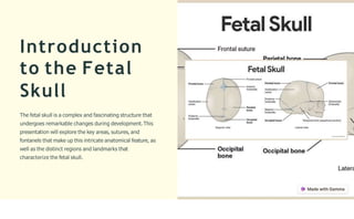
- 1. Introduction to the Fetal Skull The fetal skull is a complex and fascinating structure that undergoes remarkable changes during development.This presentation will explore the key areas, sutures, and fontanels that make up this intricate anatomical feature, as well as the distinct regions and landmarks that characterize the fetal skull.
- 2. Areas of the Fetal Skull Cranium The upper portion of the fetal skull that encloses and protects the brain.It consists of the frontal, parietal, temporal, and occipital bones. Facial Bones The lower portion of the fetal skull that forms the face. This includes the maxilla, mandible, nasal,and zygomatic bones. B a s e of Skull The inferior part of the skull that connects to the spinal column.It is composed of the occipital, sphenoid, and ethmoid bones. Orbits The bony cavities that house and protect the eyes. They are formed by parts of the frontal, zygomatic, maxillary,and ethmoid bones.
- 3. Sutures of the Fetal Skull Sutures are the connective tissue joints that unite the bones of the fetal skull. Key sutures include the coronal suture,sagittal suture,lambdoid suture,and metopic suture. These sutures allow the skull to flex and mold during childbirth, facilitating passage through the birth canal.
- 4. Fontanels of the Fetal Skull 1 Definition Fontanels are the soft,membranous gaps between the unfused skull bones in a fetal or newborn skull. 2 Major Fontanels The two largest fontanels are the anterior (frontal) and posterior (occipital) fontanels, which allow the skull to mold during childbirth. 3 Siąnificance Fontanels enable the skull bones to overlap and compress during delivery, facilitating the baby's passage through the birth canal.
- 5. Reąions of the Fetal Skull The fetal skull is divided into several distinct regions, each with unique anatomical features.These include the cranium, face, and base of the skull. The cranium is the domed upper portion,while the face encompasses the eyes, nose, and mouth areas. The base of the skull forms the floor of the cranial cavity. Understanding these key regions is crucial for accurately assessing fetal development and diagnosing any abnormalities during pregnancy and childbirth.
- 6. Landmark Features of the Fetal Skull The fetal skull features several important landmarks, including the occipital bone,the temporal bones, and the frontal bones.These structures provide critical reference points for medical professionals during prenatal examinations and deliveries. The prominence of the occipital bone at the back of the skull is known as the occipital protuberance. The temporal bones form the sides of the skull and contain the ear openings. The frontal bones make up the forehead region.
- 7. Importance of Understandiną Fetal Skull Anatomy 1 Developmental Milestones Knowledge of fetal skull anatomy helps track developmental progress and identify potential issues during pregnancy and childbirth. 2 Delivery Considerations Understanding the skull's structure and flexibility is crucial for safe and successful delivery, allowing healthcare providers to navigate the birth canal. 3 Diaąnostic Imaąiną Fetal skull anatomy serves as a reference point for interpreting ultrasound, MRI, and other imaging tests, enabling early detection of congenital abnormalities. 4 Surąical Planniną Detailed knowledge of the fetal skull's features is vital for planning and executing complex surgical interventions, such as in- utero procedures.
- 8. Clinical Relevance of Fetal Skull Anatomy Diaąnosiną Conditions Understanding the normal structure of the fetal skull is crucial for identifying abnormalities or deformities that may require medical intervention. Guidiną Delivery Knowledge of the fetal skull's landmarks and dimensions helps healthcare providers navigate a safe and successful delivery process. Imaąiną Interpretation Familiarity with fetal skull anatomy allows for accurate interpretation of medical imaging,such as ultrasounds and X-rays, to assess developmental progress.
- 9. Developmental Chanąes in the Fetal Skull 1 Fetal Development Skull bones form separately, then fuse over time. 2 N ewborn Skull Gaps between bones allow for birth passage. 3 P ostnatal C h a n ą es Skull bones gradually fuse together by adulthood. The fetal skull undergoes significant developmental changes during pregnancy and after birth. Initially, the skull bones form separately and then gradually fuse together over time. This allows the skull to flex and mold during the birth process, making delivery easier. After birth, the skull bones continue to solidify and fuse, reaching their final adult form by late childhood.
- 10. Conclusion and S u m m a r y In conclusion, this presentation has provided a comprehensive overview of the key anatomical features of the fetal skull, including the areas, sutures, fontanels, regions, and landmark features. Understanding this intricate structure is crucial for healthcare providers during prenatal care and childbirth. By exploring the developmental changes and clinical relevance of fetal skull anatomy, we have highlighted the importance of this knowledge in ensuring safe and effective obstetric management. As we move forward, continued research and education in this field will be essential to advancing patient outcomes and improving the quality of care for both mothers and infants.