SlideShare ist ein Scribd-Unternehmen logo
Complete Supply of Abdomen & Pelvis.
ABDOMEN
& PELVIS Nerve Supply Blood Supply Venous Drainage Lymphatic Drainage
Anterolater
al
abdominal
wall
Skin & muscles of anterolateral
abdominal wall are supplied by:
 Thoracoabdominal (T7-T11):
- Continuation of lower (7th
–
11th
) intercostal nerves.
- Supply: Muscles of
anterolateral abdominal wall
& overlying skin.
 Lateral cutaneous (thoracic)
cutaneous branches ( 7th
– 9th
):
- Supply : Skin of right & left
hypochondriac regions.
 Subcostal (anterior ramus of T12):
- Supply: Muscles of
anterolateral abdominal wall
& overlying skin, superior to
iliac crest & inferior to
umbilicus.
 Iliohypogastric & Ilioinguinal
nerves:
( terminal branches of anterior ramus
of spinal nerve L1)
- Ilihypogastric: Supply: Skin
overlying iliac crest , upper
inguinal & hypogastric regions,
internal oblique & transverses
abdominis muscles.
- Ilioinguinal : Supply: Skin of
lower inguinal region , mons
pubis, anterior scrotum or
Cutaneous Arteries ( Deep &
superficial ):
1.Musculophrenic Artery:
Origin:
Internal thoracic artery
Distribution:
(a) Superficial & deep abdominal
wall of hypochondriac region.
(b) Anterolateral diaphragm.
2.Superior epigastric artery:
Origin:
Internal thoracic artery
Distribution:
(a) Rectus abdominis muscle
(b) Superficial & deep abdominal
wall of epigastric & upper
umbilical regions.
3.10th & 11th posterior
intercostals arteries:
Origin:
Aorta
Distribution:
(a).Superficial & Deep abdominal wall
of lateral region.
4.Subcostal Artery:
Origin:
Aorta
Distribution:
(a).Superficial & deep abdominal wall
1. Venous drainage
passes above mainly
into axillary vein via
lateral thoracic vein.
2. Venous drainage
passes below mainly
into femoral vein via
superficial epigastric
& great saphenous
vein.
Lymphatic drainage of
anterolateral abdominal
wall follows the following
pattern.
 Superficial Lymphatic
vessels:
- Superior to
transumbilical
plane: Drains into
axillary lymph
nodes, however a
few drain into
parasternal lymph
nodes
- Inferior to
transumbilical
plane: Drain into
superficial inguinal
lymph nodes.
 Deep Lymphatic vessels:
- Drain into –
external iliac
Common iliac
Right & left
lumbar (caval & aortic)
lymph nodes.
labium majus & adjacent
medial thigh, inferior most
internal oblique & transverses
abdominis.
of lateral region.
5.Inferior epigastric artery:
Origin:
External Iliac artery
Distribution:
(a).Rectus Abdominis muscle
(b).Deep abdominal wall of pubic &
inferior umbilical regions.
Muscles of
Anterolater
al
abdominal
wall
1. External oblique:
Thoracoabdominal & subcostal
nerve.
2. Internal oblique:
Thoracoabdominal & first
lumbar nerve
3. Tranversus abdominis:
Thoracoabdominal & first
lumbar nerve.
4. Rectus abdominis:
Thoracoabdominal nerves.
6.Deep circumflex iliac artery:
Origin:
External Iliac artery
Distribution:
(a).Iliacus muscle
(b).Deep abdominal wall of inguinal
region
(c).Iliac fossa
7.Superficial circumflex iliac
artery:
Origin:
Femoral artery
Distribution:
(a).Superficial abdominal wall of
inguinal region & adjacent anterior
thigh
8.Superficial epigastric artery:
Origin:
Femoral artery
Distribution:
(a).Superficial abdominal wall of
pubic & inferior umbilical regions.
Penis Sensory nerve supply : sensory nerve
supply is derived from
1. Dorsal nerve of penis
(1).Internal pudendal artery gives
off 3 branches which supply the
penis:
1. Superficial dorsal
vein:
1. Deep Inguinal
lymph nodes –
2. Ilioinguinal nerve
Autonomic : Autonomic nerves are
derived from Pelvic plexus via
prostatic plexus.
- Sympathetic nerves are
vasodilator
- Parasympathetic nerves
(S2,S3,S4) are vasodilator.
 Autonomic fibers are distributed
through pudendal nerve.
Muscles of Penis: supplied by perineal
branch of pudendal nerve.
(i) Deep artery of penis
(ii) Dorsal artery of Penis
(iii) Artery of the bulb of penis.
(2).Femoral artery gives off the
superficial external pudendal
artery which supplies the skin &
fasciae of penis.
- Drain prepuce &
penile skin
- Open into
external
pudendal vein
2. Deep dorsal vein:
- Drains glans penis
& corpora
cavernosa penis
Divide into left & right
branches which connect
with internal pudendal vein
& enters the prostatic
plexus.
glans penis
2. Superficial inguinal
lymph nodes – rest
of penis
Scrotum The nerves of scrotum include
 Branches of lumbar plexus to
anterolateral surface.
 Branches of sacral plexus to
posterior & inferior surfaces.
1. Anterolateral surface: Genital
branch of genitofemoral nerve
(L1 & L2).
2. Anterior surface: Anterior
scrotal nerves (branches of
ilioinguinal nerve L1)
3. Posterior surface: Posterior
scrotal nerves (branches of
perineal branch of pudendal
nerve S2,S3,S4)
4. Posteroinferior surface:
Perineal branches of posterior
cutaneous nerve of thigh
S2,S3.
Dartos muscle: Genital branch of
1. Posterior scrotal branches
of the perineal artery, a
branch of internal
pudendal artery
2. Anterior scrotal branches
of deep external pudendal
artery, a branch of femoral
artery
3. Cremestaric artery ,a
branch of inferior
epigastric artery
Scrotal veins accompany the
arteries
Superficial inguinal lymph
nodes
genitofemoral nerve.
Testis The autonomic nerves of testis arise
as testicular plexus of nerves &
contain:
1. Vagal parasympathetic &
visceral afferent fibers.
2. Sympathetic fibers from T7
segment of spinal cord.
Testicular artery ( branch of
abdominal aorta)
Veins emerging from the
testis & Epididymis form
Pampiniform venous plexus,
a network of 8-12 veins lying
anterior to the ductus
deferens & surrounding the
testicular artery in the
spermatic cord.
The veins of each
pampiniform plexus
converge superiorly forming
right testicular vein which
enters inferior vena cava &
a left testicular vein which
enters the left renal vein.
1. Right & left lumbar
(caval/aortic)
lymph nodes
Preaortic lymph nodes
Epididymis Testicular plexus of nerves Testicular artery Same as testis. 1. Right & left lumbar
(caval/aortic)
lymph nodes
2. Preaortic lymph
nodes
Esophagus Esophagus is innervated by the
esophageal plexus formed by:
1. Vagal trunks
2. Thoracic sympathetic trunks
via greater splanchnic nerves
& peri-arterial plexus around
left gastric & inferior phrenic
arteries.
1. Left gastric artery (branch
of celiac trunk)
2. Left inferior phrenic artery
The venous drainage from
the submucosal veins of this
part of esophagus is both to
- Portal venous
system through
left gastric vein
Systemic venous system
through esophageal veins
entering the azygous vein
Left gastric lymph nodes (
Efferent lymphatic vessels
from these nodes drain
mainly to celiac lymph
nodes)
Stomach Sympathetic nerve supply : From T6-
T9 segments of spinal cord passes to
celiac plexus through greater
Along lesser curvature by :
1. Left gastric artery(br. of
celiac trunk)
Veins of stomach parallel the
arteries in position & course
-Right & left gastric veins
Stomach can be divided
into 4 lymphatic
territories:
splanchnic nerve & is distributed
through the plexus around gastric &
gastro-omental arteries.
Parasympathetic nerve supply: From
- Anterior vagal trunk (derived
mainly from left vagus nerve):
Gives off: Hepatic, duodenal &
anterior gastric branches.
- Posterior vagal trunk ( derived
mainly from right vagus
nerve):
Gives off: a celiac branch ,
posterior gastric branches &
branches to anterior &
posterior surfaces of stomach.
2. Right gastric artery ( br. of
proper hepatic artery)
Along greater curvature by:
1. Right gastro-omental
artery(right gastroepiploic
artery), branch of
gastroduodenal artery.
2. Left gastro-omental
artery(left gastroepiploic
artery), branch of splenic
artery.
Fundus:
- 5-7 short gastric arteries
(branches of splenic
artery)
drain into hepatic portal vein
-Short gastric veins & left
gastroepiploic (gastro-
omental) veins drain into
splenic vein which joins
superior mesenteric vein
(SMV) to form hepatic portal
vein
-Right gastroepiploic (gastro-
omental) vein empties into
SMV.
Prepyloric vein ascends over
the pylorus to the right
gastric vein.
(a) Upper part of left
1/3rd
–
pancreaticosplenic
nodes
(b) Right 2/3rd
– left
gastric nodes
(c) Lower part of left
1/3rd
– Right
gastroepiploic
nodes
Pyloric part – pyloric
lymph nodes
Duodenum Sympathetic nerve supply: T9 & T10
segments of spinal cord.
Parasympathetic nerve supply:
Derived from vagus & greater and
lesser splanchnic nerves by the way of
celiac & superior mesenteric plexuses
& reach the duodenum along its
arteries.
Upto the opening of bile duct:
- Superior
pancreaticoduodenal
artery ( br.of
gastroduodenal artery)
Below the opening of bile duct:
Inferior pancreaticoduodenal
artery (br.of superior mesenteric
artery).
1. Superior
pancreaticoduodenal
vein – drains into
portal vein
2. Inferior
pancreaticoduodenal
vein – joins the SMV
1. Anterior lymphatic
vessels : Drain
upward into
pancreaticoduode
nal lymph nodes &
into pyloric lymph
nodes
2. Posterior
lymphatic vessels:
Drain downward
via
pancreaticoduode
nal nodes to the
superior
mesenteric lymph
nodes
 Efferent lymphatic
vessels of all above lymph
nodes drain into celiac
lymph nodes.
Jejunum &
Ileum
(Reference from
BD Chaurasia)
Nerve supply is derived from
sympathetic & parasympathetic
nerves
- Sympathetic: T9-T11
- Parasympathetic: from vagus
nerve
(Reference from
Keith.L.Moore)
- The sympathetic fibers
originate in the T8-T10
segments of spinal cord &
reach the superior mesenteric
nerve plexus through
sympathetic trunks & thoracic
abdominopelvic
(greater,lesser & least)
splanchnic nerves.
- The parasympathetic fibers
derived from posterior vagal
trunks.
Superior mesenteric artery
(Lowest part of ileum is also
supplied by Ileocolic artery)
Superior mesenteric vein
(SMV)
Lymph passes through 3
groups of lymph nodes:
1. Juxta-intestinal
lymph nodes
2. Mesenteric lymph
nodes
3. Superior central
lymph nodes
 Efferent lymphatic
vessels from the
mesenteric lymph nodes
drain into superior
mesenteric lymph nodes.
Cecum Sympathetic: originate in lower
thoracic part of spinal cord.
Parasympathetic: derive from vagus
nerve.
 Branches from sympathetic &
parasympathetic nerves form the
superior mesenteric plexus.
Anterior & posterior rectal
arteries from the ileocolic artery (
Branch of superior mesenteric
artery)
Venous drainage from
cecum flow through a
tributary of SMV, the
ileocolic vein.
Lymph passes from
several mesenteric lymph
nodes to ileocolic lymph
nodes , then to superior
mesenteric lymph nodes
Appendix Same as cecum Appendicular artery , branch of
ileocolic artery
SMV (same as cecum) Same as cecum in
addition to appendicular
nodes
Ascending
colon
Sympathetic & parasympathetic
(vagus) nerves from superior
mesenteric plexus.
1. Ileocolic artery
2. Right colic branches of
superior mesenteric
arteries
SMV Lymph passes from :
Epicolic &
paracolic➔Ileocolic &
intermediate right colic
nodes➔superior
mesenteric lymph nodes.
Transverse
colon
Superior mesenteric nerve plexus via
peri-arterial plexuses of right & left
colic arteries.
These nerves transmit sympathetic,
parasympathetic (vagal) & visceral
afferent nerve fibers.
Middle colic artery (branch of
superior mesenteric artery)
SMV Middle colic lymph
nodes➔Superior
mesenteric lymph nodes
Descending
& sigmoid
colon
Sympathetic: From lumbar part of
sympathetic trunk via :
- Lumbar splanchnic nerves
- Superior mesenteric plexus
- Peri-arterial plexus
Parasympathetic: From pelvic
splanchnic nerves via:
- Inferior hypogastric plexus &
nerves.
1. Left colic artery ( br.of
inferior mesenteric artery)
2. Sigmoid arteries (br.of
Inferior mesenteric artery)
From inferior mesenteric
vein ➔ Splenic vein ➔
hepatic portal vein.
1. Intermediate colic
lymph nodes
2. Inferior
mesenteric lymph
nodes
 Lymph from left colic
flexure may also drain to
superior mesenteric lymph
nodes.
Pancreas Sympathetic: Splanchnic nerves
through the plexus.
Parasympathetic: Vagus
1. Pancreatic branches of
splenic artery
2. Superior
pancreaticoduodenal
artery
3. Inferior
pancreaticoduodenal
artery
1. Splenic vein
2. Superior mesenteric
vein (SMV)
3. Portal vein
1. Pancreaticosplenic
lymph nodes
2. Celiac lymph
nodes
3. Superior
mesenteric lymph
nodes
Liver Nerves of liver derived from Hepatic Liver receives blood Hepatic sinusoids drain into 1. Superficial
plexus ( derivative of celiac plexus)
Which consists of
- Sympathetic fibers from celiac
plexus
- Parasympathetic fibers from
anterior & posterior vagal
trunks.
1. 20-25% through hepatic
artery
2. 75-80% through portal
vein
interlobular veins ➔ which
join to form sublobular veins
➔ which join to form
hepatic veins ➔ which drain
into inferior vena cava (IVC)
lymphatics – caval
, hepatic,
paracardial &
celiac lymph
nodes.
2. Deep lymphatics –
inferior vena cava
& hepatic lymph
nodes.
Gall
bladder &
Cystic duct
Nerves to gall bladder & custic duct
from:
1. Celiac plexus – sympathetic &
visceral afferent fibers.
2. Vagus nerve –
parasympathetic fibers
3. Right phrenic nerve – Somatic
afferent fibers.
Cystic artery ( arise from right
phrenic artery)
From cystic veins to hepatic
portal pancreaticoduodenal
vein to hepatic portal
vein.vein
1. Hepatic lymph
nodes ➔ celiac
lymph nodes
2. Cystic lymph
nodes ➔ celiac
lymph nodes
Bile Duct
1. Cystic artery
2. Right hepatic artery
3. Posterior superior
pancreaticoduodenal
artery
4. Gastroduodenal artery
The posterior superior
pancreaticoduodenal vein
drains the distal part of the
bile duct and empties into
the hepatic portal vein or
one of its tributaries.
Cystic veins
1. Cystic nodes ➔
celiac lymph nodes
2. Lymph node of
omental foramen
➔ celiac lymph
nodes
3. Hepatic lymph
nodes ➔ celiac
lymph nodes
Kidneys Renal nerve plexus which consists of
- Sympathetic T10-L1 &
parasympathetic fibers.
Renal artery divides to form
various segmental arteries:
1. Superior segmental artery
to Superior segment
2. Anterosuperior segmental
artery to Anterosuperior
segment
3. Anteroinferior segmental
artery to antero inferior
Right & Left renal veins
 Interlobular veins ➔
arcuate veins ➔ interlobar
veins ➔ renal vein ➔ IVC
Right & left lumbar ( caval
& aortic ) lymph nodes
segment
4. Inferior segmental artery
to inferior segment
5. Posterior segmental artery
to posterior segment of
the kidney
Ureters 1. Renal plexus
2. Abdominal aortic plexus
3. Superior hypogastric plexus
1. Upper part – Renal artery
2. Middle part- (i)
Testicular/ovarian arteries
(ii)
Abdominal aorta
(iii) Common
iliac arteries
3. Lower part-
(i) Vesical artery
(ii) Middle rectal artery
(iii) Uterine vessels
1. Renal veins
2. Gonadal
(testicular/ovarian)
veins.
1. Superior part –
Lumbar lymph
nodes
2. Middle part –
common iliac
lymph nodes
3. Inferior part –
common, external
or internal iliac
lymph nodes.
Suprarenal
glands
1. Celiac plexus
2. Abdominopelvic (greater,
lesser & least splanchnic
nerves)
1. Superior suprarenal
arteries (6-8) from inferior
phrenic arteries
2. Middle suprarenal arteries
(≤1) from abdominal aorta
3. Inferior suprarenal arteries
(≤1 from Renal artery)
 Suprarenal veins
1. Right suprarenal vein
➔ IVC
2. Left suprarenal vein
➔ joins inferior
phrenic vein ➔
drains into Left renal
vein.
Lateral aortic ( lumbar)
lymph nodes
Diaphragm Motor nerve supply: Phrenic nerves
(ventral rami C3,C4,C5)
Sensory nerve supply:
- Central part: Phrenic nerve
- Peripheral part: lower 6
thoracic nerves
Muscles of
posterior
Psoas major : Anterior rami of lumbar
nerves L1, L2, L3.
abdominal
wall
Iliacus: Femoral nerve (L2-L4)
Quadratus lumborum: Anterior
branches of T12 & L1-L4 nerves.
Urinary
bladder
Urinary bladder is supplied by vesical
plexus of nerves which is made up of
nerves derived from inferior
hypogastric plexus. Vesical plexus
contains both sympathetic &
parasympathetic components, each of
which contains motor(efferent) &
sensory(afferent) fibers.
- Parasympathetic efferent
fibers S3,S3,S4
- Sympathetic efferent fibers
T11-L2.
- Somatic: pudendal nerve
(S2,S3,S4)
- Sensory nerves [For
detail see page#378 BD
Chaurasia]
1. Superior & Inferior vesical
arteries (branches of
anterior trunk of internal
iliac artery)
 In females from uterine
& vaginal arteries instead
of inferior vesical.
2. Obturator & Inferior
Gluteal arteries (
additional supply to
bladder)
Vesical venous plexus ➔
which drains into Internal
iliac veins
1. External iliac
nodes
2. Internal
iliac/lateral aortic
nodes
Male
urethra
Nerves are derived from prostatic
plexus ( one of the pelvic plexus)
Parasympathetic nerves are from 2nd
to 4th
sacral segments.
1. Urethral artery arises from
internal pudendal artery
2. Dorsal penile artery via
circumflex branches on
each side
Anterior urethra:
Dorsal vein of penis➔
internal pudendal vein ➔
prostatic venous plexus.
Posterior urethra:
Prostatic & vesical venous
plexus ➔ internal iliac veins.
1. Prostatic urethra –
internal iliac
lymph nodes
2. Membranous
urethra – internal
iliac lymph nodes
3. Anterior urethra –
deep inguinal
lymph nodes.
Female
urethra
Vesical plexus & pudendal nerve.
Explanation:
- Parasympathetic preganglionic
fibers from 2nd
to 4th
sacral
segments of spinal cord.
Synapse in vesical venous
plexus. Post-ganglionic fibers
1. Superior vesical arteries
2. Vaginal arteries
Venous plexus around
urethra ➔ vesical venous
plexus ➔ internal pudendal
vein
1. Internal iliac
lymph nodes
2. External iliac
lymph nodes
reach smooth muscles.
- Somatic fibers from S2-S4. Do
not synapse in vesical venous
plexus.
- Sensory fibers in pelvic
splanchnic nerves reach to 2nd
to 4th
sacral segments of spinal
cord.
- Postganglionic sympathetic
fibers arise from plexus
around vagina arteries.
Rectum Nerve supply to rectum is derived
from sympathetic & parasympathetic
systems.
- Sympathetic: from lumbar
spinal cord (L1-L2) conveyed
via:
Lumbar splanchnic nerve
Hypogastric/pelvic plexus
Peri-arterial plexus (of
inferior mesenteric & superior
rectal arteries)
- Parasympathetic: from S2-S4
passing via
Pelvic splanchnic nerves
Left & right inferior
hypogastric plexus to the
rectal (pelvic) plexus.
1. Superior rectal artery –
continuation of inferior
mesenteric artery
2. Middle rectal arteries –
arise from anterior division
of internal iliac artery
3. Median sacral artery –
small branch arising from
back of aorta
1. Superior rectal vein –
tributeries of this
vein arise from
internal rectal
venous plexus.
2. Middle rectal vein –
open into internal
iliac veins.
3. Median sacral vein –
joins left common
iliac vein
1. Upper half of
rectum : Inferior
mesenteric nodes
after passing
through pararectal
& sigmoid nodes
2. Lower half of
rectum: Internal
iliac nodes
Anal canal Above pectinate line:
- Sympathetic: Inferior
hypogastric plexus
- Parasympathetic: Pelvic
splanchnic nerve S2,S3.
Above pectinate line – superior
rectal artery
Below pectinate line – Inferior
rectal artery
1. Internal rectal
venous plexus
2. External rectal
venous plexus
3. Anal veins
1. Above pectinate
line: Internal iliac
lymph nodes ➔
common iliac &
lumbar lymph
Below pectinate line:
- Somatic: inferior rectal nerve
(S2,S3,S4) branch of pudendal
nerve
External anal sphincter:
- Inferior rectal nerve
- Perineal branch of 4th
sacral
nerve
nodes
2. Below pectinate
line : Superficial
inguinal lymph
nodes.
Ovaries &
uterine
tubes
1. Ovarian plexus : Derived from
renal, aortic & hypogastric
plexus. Contains both
sympathetic &
parasympathetic nerve fibers.
- Sympathetic: T10-T11 are
afferent for pain as well as
efferent or vasomotor.
- Parasympathetic: S2,S3,S4 are
vasodilator.
2. Uterine plexus
1. Ovarian arteries (arise
from abdominal aorta)
2. Tubal branches of uterine
arteries ( Branch of
internal iliac arteries)
Veins drainig the ovary form
a vine like pampiniform
plexus of veins. The plexus
condenses into a single
ovarian vein near pelvic
inlet.
 Right ovarian vein drains
into inferior vena cava
 Left ovarian vein drains
into left renal vein.
Uterine Tube:
Tubal veins : drain into
pampiniform venous plexus
& uterine venous plexus.
Lateral aortic & para
aortic lymph nodes.
Uterus Sympathetic & parasympathetic
nerves through Inferior hypogastric &
ovarian plexus.
- Sympathetic nerves are from
T12 & L1.
- Parasympathetic nerves are
S2,S3,S4.
1. Uterine arteries
2. Ovarian arteries
The veins form a plexus.
Plexus drains through
Uterine , ovarian & vaginal
veins into internal iliac veins.
3 lymphatic territories:
1. Upper lymphatics:
Drain into aortic &
superficial inguinal
lymph nodes.
2. Middle
lymphatics: Drain
into external iliac
lymph nodes.
3. Lower lymphatics:
External iliac,
internal iliac &
sacral nodes.
Vagina Lower 1/3rd
: Supplied by pudendal
nerve through inferior rectal &
posterior labial branches of perineal
nerve.
Upper 2/3rd
: Supplied by
- Sympathetic – L1,L2
- Parasympathetic – S2,S3,S4.
Nerves are derived from inferior
hypogastric & uterovaginal plexus.
1. Vagina branch of internal
iliac artery
2. In addition
- The upper part is supplied
by cevicovaginal branch of
uterine artery.
- Lower part by middle
rectal & internal pudendal
arteries.
 Branches of these arteries
anastomose to form anterior &
posterior midline vessels called
the vaginal azygos arteries.
Vaginal venous plexus
drains into internal iliac
veins.
Upper 1/3rd
: External iliac
nodes
Middle 1/3rd
: Internal
iliac nodes
Lower 1/3rd
: Medial
group of superficial
inguinal lymph nodes.
Prostate
Gland
Prostate is supplied by Prostatic
plexus of nerves which is derives from
lower part of inferior hypogastric
plexus.
Supplied by branches from
1. Inferior vesical
2. Middle rectal
3. Internal pudendal arteries
- These arteries form a large
outer or subcapsular
plexus, and a small inner
or periurethral plexus.
Greater part of gland is
supplied by subcapsular
plexus.
Veins form a rich plexus
which communicates with
vesical plexus & with
internal pudendal vein &
drains into vesical & internal
iliac veins.
1. Internal iliac
lymph nodes.
2. External iliac
lymph nodes.
3. Sacral lymph
nodes.
Reference: K.L.M & B.D chaurasia

Weitere ähnliche Inhalte

Ähnlich wie ##ABDOMEN PELVIS COMPLETE Supply (1).pdf

269034 mediastinum
269034 mediastinum269034 mediastinum
269034 mediastinum
YoAmoNYC
 
269034 mediastinum
269034 mediastinum269034 mediastinum
269034 mediastinum
abctutor
 
Aorta&its branches . pptx.pptx
Aorta&its branches . pptx.pptxAorta&its branches . pptx.pptx
Aorta&its branches . pptx.pptx
KannurLion
 
anatomy (mediastinum)(0).pptx
anatomy (mediastinum)(0).pptxanatomy (mediastinum)(0).pptx
anatomy (mediastinum)(0).pptx
Funandstudy1
 
anatomy (mediastinum).pptx
anatomy (mediastinum).pptxanatomy (mediastinum).pptx
anatomy (mediastinum).pptx
SalsabeelArif
 

Ähnlich wie ##ABDOMEN PELVIS COMPLETE Supply (1).pdf (20)

Back to Basic: Artery, Vein of the male reproductive system.pdf
Back to Basic: Artery, Vein of the male reproductive system.pdfBack to Basic: Artery, Vein of the male reproductive system.pdf
Back to Basic: Artery, Vein of the male reproductive system.pdf
 
Pelvic vessels,nerves and lymph nodes.
Pelvic vessels,nerves and lymph nodes.Pelvic vessels,nerves and lymph nodes.
Pelvic vessels,nerves and lymph nodes.
 
Aorta by Dr. Karle Geetanjali
Aorta by Dr. Karle GeetanjaliAorta by Dr. Karle Geetanjali
Aorta by Dr. Karle Geetanjali
 
Mediastinum
MediastinumMediastinum
Mediastinum
 
269034 mediastinum
269034 mediastinum269034 mediastinum
269034 mediastinum
 
269034 mediastinum
269034 mediastinum269034 mediastinum
269034 mediastinum
 
Blood supply & innervation of upper limb
Blood supply & innervation of upper limbBlood supply & innervation of upper limb
Blood supply & innervation of upper limb
 
Anatomy of the thorax
Anatomy of the thoraxAnatomy of the thorax
Anatomy of the thorax
 
The Sympathetic Nervous System
The Sympathetic Nervous SystemThe Sympathetic Nervous System
The Sympathetic Nervous System
 
Anterior abdominal wall
Anterior abdominal wallAnterior abdominal wall
Anterior abdominal wall
 
Comparative study of Blood vessels of vertebrates
Comparative study of Blood vessels of vertebratesComparative study of Blood vessels of vertebrates
Comparative study of Blood vessels of vertebrates
 
SPINAL CORD.pptx
SPINAL CORD.pptxSPINAL CORD.pptx
SPINAL CORD.pptx
 
Aorta&its branches . pptx.pptx
Aorta&its branches . pptx.pptxAorta&its branches . pptx.pptx
Aorta&its branches . pptx.pptx
 
2 b. ABDOMINAL AORTA1.ppt
2 b. ABDOMINAL AORTA1.ppt2 b. ABDOMINAL AORTA1.ppt
2 b. ABDOMINAL AORTA1.ppt
 
blood and nerve supply of the pelvis 2.pptx
blood and nerve supply of the pelvis 2.pptxblood and nerve supply of the pelvis 2.pptx
blood and nerve supply of the pelvis 2.pptx
 
The Anatomy of the Systemic Circulation.ppt
The Anatomy of the Systemic Circulation.pptThe Anatomy of the Systemic Circulation.ppt
The Anatomy of the Systemic Circulation.ppt
 
Radiological anatomy of chest including lungs,mediastinum and thoracic cage
Radiological anatomy of chest including lungs,mediastinum and thoracic cageRadiological anatomy of chest including lungs,mediastinum and thoracic cage
Radiological anatomy of chest including lungs,mediastinum and thoracic cage
 
anatomy (mediastinum)(0).pptx
anatomy (mediastinum)(0).pptxanatomy (mediastinum)(0).pptx
anatomy (mediastinum)(0).pptx
 
anatomy (mediastinum).pptx
anatomy (mediastinum).pptxanatomy (mediastinum).pptx
anatomy (mediastinum).pptx
 
Subclavian vessels.pptx
Subclavian vessels.pptxSubclavian vessels.pptx
Subclavian vessels.pptx
 

Mehr von YasirRehman33 (6)

DOC-20240128-WA0009..pptx constitution 1973
DOC-20240128-WA0009..pptx constitution 1973DOC-20240128-WA0009..pptx constitution 1973
DOC-20240128-WA0009..pptx constitution 1973
 
Presentation1.pdf. pharmacology. Katzung
Presentation1.pdf. pharmacology. KatzungPresentation1.pdf. pharmacology. Katzung
Presentation1.pdf. pharmacology. Katzung
 
Other Beta Lactam Drugs.pptx pharmacology
Other Beta Lactam Drugs.pptx pharmacologyOther Beta Lactam Drugs.pptx pharmacology
Other Beta Lactam Drugs.pptx pharmacology
 
Beta_Lactam_Antibiotics1.ppt
Beta_Lactam_Antibiotics1.pptBeta_Lactam_Antibiotics1.ppt
Beta_Lactam_Antibiotics1.ppt
 
BIO BLOGGING.pptx
BIO BLOGGING.pptxBIO BLOGGING.pptx
BIO BLOGGING.pptx
 
LIMBIC SYSTEM PPT.pptx
LIMBIC SYSTEM PPT.pptxLIMBIC SYSTEM PPT.pptx
LIMBIC SYSTEM PPT.pptx
 

Kürzlich hochgeladen

Kürzlich hochgeladen (20)

Features of Video Calls in the Discuss Module in Odoo 17
Features of Video Calls in the Discuss Module in Odoo 17Features of Video Calls in the Discuss Module in Odoo 17
Features of Video Calls in the Discuss Module in Odoo 17
 
Application of Matrices in real life. Presentation on application of matrices
Application of Matrices in real life. Presentation on application of matricesApplication of Matrices in real life. Presentation on application of matrices
Application of Matrices in real life. Presentation on application of matrices
 
How to the fix Attribute Error in odoo 17
How to the fix Attribute Error in odoo 17How to the fix Attribute Error in odoo 17
How to the fix Attribute Error in odoo 17
 
How to Manage Notification Preferences in the Odoo 17
How to Manage Notification Preferences in the Odoo 17How to Manage Notification Preferences in the Odoo 17
How to Manage Notification Preferences in the Odoo 17
 
Pragya Champions Chalice 2024 Prelims & Finals Q/A set, General Quiz
Pragya Champions Chalice 2024 Prelims & Finals Q/A set, General QuizPragya Champions Chalice 2024 Prelims & Finals Q/A set, General Quiz
Pragya Champions Chalice 2024 Prelims & Finals Q/A set, General Quiz
 
Basic Civil Engg Notes_Chapter-6_Environment Pollution & Engineering
Basic Civil Engg Notes_Chapter-6_Environment Pollution & EngineeringBasic Civil Engg Notes_Chapter-6_Environment Pollution & Engineering
Basic Civil Engg Notes_Chapter-6_Environment Pollution & Engineering
 
“O BEIJO” EM ARTE .
“O BEIJO” EM ARTE                       .“O BEIJO” EM ARTE                       .
“O BEIJO” EM ARTE .
 
Championnat de France de Tennis de table/
Championnat de France de Tennis de table/Championnat de France de Tennis de table/
Championnat de France de Tennis de table/
 
The Art Pastor's Guide to Sabbath | Steve Thomason
The Art Pastor's Guide to Sabbath | Steve ThomasonThe Art Pastor's Guide to Sabbath | Steve Thomason
The Art Pastor's Guide to Sabbath | Steve Thomason
 
[GDSC YCCE] Build with AI Online Presentation
[GDSC YCCE] Build with AI Online Presentation[GDSC YCCE] Build with AI Online Presentation
[GDSC YCCE] Build with AI Online Presentation
 
Gyanartha SciBizTech Quiz slideshare.pptx
Gyanartha SciBizTech Quiz slideshare.pptxGyanartha SciBizTech Quiz slideshare.pptx
Gyanartha SciBizTech Quiz slideshare.pptx
 
INU_CAPSTONEDESIGN_비밀번호486_업로드용 발표자료.pdf
INU_CAPSTONEDESIGN_비밀번호486_업로드용 발표자료.pdfINU_CAPSTONEDESIGN_비밀번호486_업로드용 발표자료.pdf
INU_CAPSTONEDESIGN_비밀번호486_업로드용 발표자료.pdf
 
The Benefits and Challenges of Open Educational Resources
The Benefits and Challenges of Open Educational ResourcesThe Benefits and Challenges of Open Educational Resources
The Benefits and Challenges of Open Educational Resources
 
slides CapTechTalks Webinar May 2024 Alexander Perry.pptx
slides CapTechTalks Webinar May 2024 Alexander Perry.pptxslides CapTechTalks Webinar May 2024 Alexander Perry.pptx
slides CapTechTalks Webinar May 2024 Alexander Perry.pptx
 
size separation d pharm 1st year pharmaceutics
size separation d pharm 1st year pharmaceuticssize separation d pharm 1st year pharmaceutics
size separation d pharm 1st year pharmaceutics
 
Removal Strategy _ FEFO _ Working with Perishable Products in Odoo 17
Removal Strategy _ FEFO _ Working with Perishable Products in Odoo 17Removal Strategy _ FEFO _ Working with Perishable Products in Odoo 17
Removal Strategy _ FEFO _ Working with Perishable Products in Odoo 17
 
Advances in production technology of Grapes.pdf
Advances in production technology of Grapes.pdfAdvances in production technology of Grapes.pdf
Advances in production technology of Grapes.pdf
 
How to Break the cycle of negative Thoughts
How to Break the cycle of negative ThoughtsHow to Break the cycle of negative Thoughts
How to Break the cycle of negative Thoughts
 
50 ĐỀ LUYỆN THI IOE LỚP 9 - NĂM HỌC 2022-2023 (CÓ LINK HÌNH, FILE AUDIO VÀ ĐÁ...
50 ĐỀ LUYỆN THI IOE LỚP 9 - NĂM HỌC 2022-2023 (CÓ LINK HÌNH, FILE AUDIO VÀ ĐÁ...50 ĐỀ LUYỆN THI IOE LỚP 9 - NĂM HỌC 2022-2023 (CÓ LINK HÌNH, FILE AUDIO VÀ ĐÁ...
50 ĐỀ LUYỆN THI IOE LỚP 9 - NĂM HỌC 2022-2023 (CÓ LINK HÌNH, FILE AUDIO VÀ ĐÁ...
 
An Overview of the Odoo 17 Discuss App.pptx
An Overview of the Odoo 17 Discuss App.pptxAn Overview of the Odoo 17 Discuss App.pptx
An Overview of the Odoo 17 Discuss App.pptx
 

##ABDOMEN PELVIS COMPLETE Supply (1).pdf

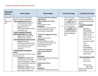
  • 1. Complete Supply of Abdomen & Pelvis. ABDOMEN & PELVIS Nerve Supply Blood Supply Venous Drainage Lymphatic Drainage Anterolater al abdominal wall Skin & muscles of anterolateral abdominal wall are supplied by:  Thoracoabdominal (T7-T11): - Continuation of lower (7th – 11th ) intercostal nerves. - Supply: Muscles of anterolateral abdominal wall & overlying skin.  Lateral cutaneous (thoracic) cutaneous branches ( 7th – 9th ): - Supply : Skin of right & left hypochondriac regions.  Subcostal (anterior ramus of T12): - Supply: Muscles of anterolateral abdominal wall & overlying skin, superior to iliac crest & inferior to umbilicus.  Iliohypogastric & Ilioinguinal nerves: ( terminal branches of anterior ramus of spinal nerve L1) - Ilihypogastric: Supply: Skin overlying iliac crest , upper inguinal & hypogastric regions, internal oblique & transverses abdominis muscles. - Ilioinguinal : Supply: Skin of lower inguinal region , mons pubis, anterior scrotum or Cutaneous Arteries ( Deep & superficial ): 1.Musculophrenic Artery: Origin: Internal thoracic artery Distribution: (a) Superficial & deep abdominal wall of hypochondriac region. (b) Anterolateral diaphragm. 2.Superior epigastric artery: Origin: Internal thoracic artery Distribution: (a) Rectus abdominis muscle (b) Superficial & deep abdominal wall of epigastric & upper umbilical regions. 3.10th & 11th posterior intercostals arteries: Origin: Aorta Distribution: (a).Superficial & Deep abdominal wall of lateral region. 4.Subcostal Artery: Origin: Aorta Distribution: (a).Superficial & deep abdominal wall 1. Venous drainage passes above mainly into axillary vein via lateral thoracic vein. 2. Venous drainage passes below mainly into femoral vein via superficial epigastric & great saphenous vein. Lymphatic drainage of anterolateral abdominal wall follows the following pattern.  Superficial Lymphatic vessels: - Superior to transumbilical plane: Drains into axillary lymph nodes, however a few drain into parasternal lymph nodes - Inferior to transumbilical plane: Drain into superficial inguinal lymph nodes.  Deep Lymphatic vessels: - Drain into – external iliac Common iliac Right & left lumbar (caval & aortic) lymph nodes.
  • 2. labium majus & adjacent medial thigh, inferior most internal oblique & transverses abdominis. of lateral region. 5.Inferior epigastric artery: Origin: External Iliac artery Distribution: (a).Rectus Abdominis muscle (b).Deep abdominal wall of pubic & inferior umbilical regions. Muscles of Anterolater al abdominal wall 1. External oblique: Thoracoabdominal & subcostal nerve. 2. Internal oblique: Thoracoabdominal & first lumbar nerve 3. Tranversus abdominis: Thoracoabdominal & first lumbar nerve. 4. Rectus abdominis: Thoracoabdominal nerves. 6.Deep circumflex iliac artery: Origin: External Iliac artery Distribution: (a).Iliacus muscle (b).Deep abdominal wall of inguinal region (c).Iliac fossa 7.Superficial circumflex iliac artery: Origin: Femoral artery Distribution: (a).Superficial abdominal wall of inguinal region & adjacent anterior thigh 8.Superficial epigastric artery: Origin: Femoral artery Distribution: (a).Superficial abdominal wall of pubic & inferior umbilical regions. Penis Sensory nerve supply : sensory nerve supply is derived from 1. Dorsal nerve of penis (1).Internal pudendal artery gives off 3 branches which supply the penis: 1. Superficial dorsal vein: 1. Deep Inguinal lymph nodes –
  • 3. 2. Ilioinguinal nerve Autonomic : Autonomic nerves are derived from Pelvic plexus via prostatic plexus. - Sympathetic nerves are vasodilator - Parasympathetic nerves (S2,S3,S4) are vasodilator.  Autonomic fibers are distributed through pudendal nerve. Muscles of Penis: supplied by perineal branch of pudendal nerve. (i) Deep artery of penis (ii) Dorsal artery of Penis (iii) Artery of the bulb of penis. (2).Femoral artery gives off the superficial external pudendal artery which supplies the skin & fasciae of penis. - Drain prepuce & penile skin - Open into external pudendal vein 2. Deep dorsal vein: - Drains glans penis & corpora cavernosa penis Divide into left & right branches which connect with internal pudendal vein & enters the prostatic plexus. glans penis 2. Superficial inguinal lymph nodes – rest of penis Scrotum The nerves of scrotum include  Branches of lumbar plexus to anterolateral surface.  Branches of sacral plexus to posterior & inferior surfaces. 1. Anterolateral surface: Genital branch of genitofemoral nerve (L1 & L2). 2. Anterior surface: Anterior scrotal nerves (branches of ilioinguinal nerve L1) 3. Posterior surface: Posterior scrotal nerves (branches of perineal branch of pudendal nerve S2,S3,S4) 4. Posteroinferior surface: Perineal branches of posterior cutaneous nerve of thigh S2,S3. Dartos muscle: Genital branch of 1. Posterior scrotal branches of the perineal artery, a branch of internal pudendal artery 2. Anterior scrotal branches of deep external pudendal artery, a branch of femoral artery 3. Cremestaric artery ,a branch of inferior epigastric artery Scrotal veins accompany the arteries Superficial inguinal lymph nodes
  • 4. genitofemoral nerve. Testis The autonomic nerves of testis arise as testicular plexus of nerves & contain: 1. Vagal parasympathetic & visceral afferent fibers. 2. Sympathetic fibers from T7 segment of spinal cord. Testicular artery ( branch of abdominal aorta) Veins emerging from the testis & Epididymis form Pampiniform venous plexus, a network of 8-12 veins lying anterior to the ductus deferens & surrounding the testicular artery in the spermatic cord. The veins of each pampiniform plexus converge superiorly forming right testicular vein which enters inferior vena cava & a left testicular vein which enters the left renal vein. 1. Right & left lumbar (caval/aortic) lymph nodes Preaortic lymph nodes Epididymis Testicular plexus of nerves Testicular artery Same as testis. 1. Right & left lumbar (caval/aortic) lymph nodes 2. Preaortic lymph nodes Esophagus Esophagus is innervated by the esophageal plexus formed by: 1. Vagal trunks 2. Thoracic sympathetic trunks via greater splanchnic nerves & peri-arterial plexus around left gastric & inferior phrenic arteries. 1. Left gastric artery (branch of celiac trunk) 2. Left inferior phrenic artery The venous drainage from the submucosal veins of this part of esophagus is both to - Portal venous system through left gastric vein Systemic venous system through esophageal veins entering the azygous vein Left gastric lymph nodes ( Efferent lymphatic vessels from these nodes drain mainly to celiac lymph nodes) Stomach Sympathetic nerve supply : From T6- T9 segments of spinal cord passes to celiac plexus through greater Along lesser curvature by : 1. Left gastric artery(br. of celiac trunk) Veins of stomach parallel the arteries in position & course -Right & left gastric veins Stomach can be divided into 4 lymphatic territories:
  • 5. splanchnic nerve & is distributed through the plexus around gastric & gastro-omental arteries. Parasympathetic nerve supply: From - Anterior vagal trunk (derived mainly from left vagus nerve): Gives off: Hepatic, duodenal & anterior gastric branches. - Posterior vagal trunk ( derived mainly from right vagus nerve): Gives off: a celiac branch , posterior gastric branches & branches to anterior & posterior surfaces of stomach. 2. Right gastric artery ( br. of proper hepatic artery) Along greater curvature by: 1. Right gastro-omental artery(right gastroepiploic artery), branch of gastroduodenal artery. 2. Left gastro-omental artery(left gastroepiploic artery), branch of splenic artery. Fundus: - 5-7 short gastric arteries (branches of splenic artery) drain into hepatic portal vein -Short gastric veins & left gastroepiploic (gastro- omental) veins drain into splenic vein which joins superior mesenteric vein (SMV) to form hepatic portal vein -Right gastroepiploic (gastro- omental) vein empties into SMV. Prepyloric vein ascends over the pylorus to the right gastric vein. (a) Upper part of left 1/3rd – pancreaticosplenic nodes (b) Right 2/3rd – left gastric nodes (c) Lower part of left 1/3rd – Right gastroepiploic nodes Pyloric part – pyloric lymph nodes Duodenum Sympathetic nerve supply: T9 & T10 segments of spinal cord. Parasympathetic nerve supply: Derived from vagus & greater and lesser splanchnic nerves by the way of celiac & superior mesenteric plexuses & reach the duodenum along its arteries. Upto the opening of bile duct: - Superior pancreaticoduodenal artery ( br.of gastroduodenal artery) Below the opening of bile duct: Inferior pancreaticoduodenal artery (br.of superior mesenteric artery). 1. Superior pancreaticoduodenal vein – drains into portal vein 2. Inferior pancreaticoduodenal vein – joins the SMV 1. Anterior lymphatic vessels : Drain upward into pancreaticoduode nal lymph nodes & into pyloric lymph nodes 2. Posterior lymphatic vessels: Drain downward via pancreaticoduode nal nodes to the superior
  • 6. mesenteric lymph nodes  Efferent lymphatic vessels of all above lymph nodes drain into celiac lymph nodes. Jejunum & Ileum (Reference from BD Chaurasia) Nerve supply is derived from sympathetic & parasympathetic nerves - Sympathetic: T9-T11 - Parasympathetic: from vagus nerve (Reference from Keith.L.Moore) - The sympathetic fibers originate in the T8-T10 segments of spinal cord & reach the superior mesenteric nerve plexus through sympathetic trunks & thoracic abdominopelvic (greater,lesser & least) splanchnic nerves. - The parasympathetic fibers derived from posterior vagal trunks. Superior mesenteric artery (Lowest part of ileum is also supplied by Ileocolic artery) Superior mesenteric vein (SMV) Lymph passes through 3 groups of lymph nodes: 1. Juxta-intestinal lymph nodes 2. Mesenteric lymph nodes 3. Superior central lymph nodes  Efferent lymphatic vessels from the mesenteric lymph nodes drain into superior mesenteric lymph nodes. Cecum Sympathetic: originate in lower thoracic part of spinal cord. Parasympathetic: derive from vagus nerve.  Branches from sympathetic & parasympathetic nerves form the superior mesenteric plexus. Anterior & posterior rectal arteries from the ileocolic artery ( Branch of superior mesenteric artery) Venous drainage from cecum flow through a tributary of SMV, the ileocolic vein. Lymph passes from several mesenteric lymph nodes to ileocolic lymph nodes , then to superior mesenteric lymph nodes
  • 7. Appendix Same as cecum Appendicular artery , branch of ileocolic artery SMV (same as cecum) Same as cecum in addition to appendicular nodes Ascending colon Sympathetic & parasympathetic (vagus) nerves from superior mesenteric plexus. 1. Ileocolic artery 2. Right colic branches of superior mesenteric arteries SMV Lymph passes from : Epicolic & paracolic➔Ileocolic & intermediate right colic nodes➔superior mesenteric lymph nodes. Transverse colon Superior mesenteric nerve plexus via peri-arterial plexuses of right & left colic arteries. These nerves transmit sympathetic, parasympathetic (vagal) & visceral afferent nerve fibers. Middle colic artery (branch of superior mesenteric artery) SMV Middle colic lymph nodes➔Superior mesenteric lymph nodes Descending & sigmoid colon Sympathetic: From lumbar part of sympathetic trunk via : - Lumbar splanchnic nerves - Superior mesenteric plexus - Peri-arterial plexus Parasympathetic: From pelvic splanchnic nerves via: - Inferior hypogastric plexus & nerves. 1. Left colic artery ( br.of inferior mesenteric artery) 2. Sigmoid arteries (br.of Inferior mesenteric artery) From inferior mesenteric vein ➔ Splenic vein ➔ hepatic portal vein. 1. Intermediate colic lymph nodes 2. Inferior mesenteric lymph nodes  Lymph from left colic flexure may also drain to superior mesenteric lymph nodes. Pancreas Sympathetic: Splanchnic nerves through the plexus. Parasympathetic: Vagus 1. Pancreatic branches of splenic artery 2. Superior pancreaticoduodenal artery 3. Inferior pancreaticoduodenal artery 1. Splenic vein 2. Superior mesenteric vein (SMV) 3. Portal vein 1. Pancreaticosplenic lymph nodes 2. Celiac lymph nodes 3. Superior mesenteric lymph nodes Liver Nerves of liver derived from Hepatic Liver receives blood Hepatic sinusoids drain into 1. Superficial
  • 8. plexus ( derivative of celiac plexus) Which consists of - Sympathetic fibers from celiac plexus - Parasympathetic fibers from anterior & posterior vagal trunks. 1. 20-25% through hepatic artery 2. 75-80% through portal vein interlobular veins ➔ which join to form sublobular veins ➔ which join to form hepatic veins ➔ which drain into inferior vena cava (IVC) lymphatics – caval , hepatic, paracardial & celiac lymph nodes. 2. Deep lymphatics – inferior vena cava & hepatic lymph nodes. Gall bladder & Cystic duct Nerves to gall bladder & custic duct from: 1. Celiac plexus – sympathetic & visceral afferent fibers. 2. Vagus nerve – parasympathetic fibers 3. Right phrenic nerve – Somatic afferent fibers. Cystic artery ( arise from right phrenic artery) From cystic veins to hepatic portal pancreaticoduodenal vein to hepatic portal vein.vein 1. Hepatic lymph nodes ➔ celiac lymph nodes 2. Cystic lymph nodes ➔ celiac lymph nodes Bile Duct 1. Cystic artery 2. Right hepatic artery 3. Posterior superior pancreaticoduodenal artery 4. Gastroduodenal artery The posterior superior pancreaticoduodenal vein drains the distal part of the bile duct and empties into the hepatic portal vein or one of its tributaries. Cystic veins 1. Cystic nodes ➔ celiac lymph nodes 2. Lymph node of omental foramen ➔ celiac lymph nodes 3. Hepatic lymph nodes ➔ celiac lymph nodes Kidneys Renal nerve plexus which consists of - Sympathetic T10-L1 & parasympathetic fibers. Renal artery divides to form various segmental arteries: 1. Superior segmental artery to Superior segment 2. Anterosuperior segmental artery to Anterosuperior segment 3. Anteroinferior segmental artery to antero inferior Right & Left renal veins  Interlobular veins ➔ arcuate veins ➔ interlobar veins ➔ renal vein ➔ IVC Right & left lumbar ( caval & aortic ) lymph nodes
  • 9. segment 4. Inferior segmental artery to inferior segment 5. Posterior segmental artery to posterior segment of the kidney Ureters 1. Renal plexus 2. Abdominal aortic plexus 3. Superior hypogastric plexus 1. Upper part – Renal artery 2. Middle part- (i) Testicular/ovarian arteries (ii) Abdominal aorta (iii) Common iliac arteries 3. Lower part- (i) Vesical artery (ii) Middle rectal artery (iii) Uterine vessels 1. Renal veins 2. Gonadal (testicular/ovarian) veins. 1. Superior part – Lumbar lymph nodes 2. Middle part – common iliac lymph nodes 3. Inferior part – common, external or internal iliac lymph nodes. Suprarenal glands 1. Celiac plexus 2. Abdominopelvic (greater, lesser & least splanchnic nerves) 1. Superior suprarenal arteries (6-8) from inferior phrenic arteries 2. Middle suprarenal arteries (≤1) from abdominal aorta 3. Inferior suprarenal arteries (≤1 from Renal artery)  Suprarenal veins 1. Right suprarenal vein ➔ IVC 2. Left suprarenal vein ➔ joins inferior phrenic vein ➔ drains into Left renal vein. Lateral aortic ( lumbar) lymph nodes Diaphragm Motor nerve supply: Phrenic nerves (ventral rami C3,C4,C5) Sensory nerve supply: - Central part: Phrenic nerve - Peripheral part: lower 6 thoracic nerves Muscles of posterior Psoas major : Anterior rami of lumbar nerves L1, L2, L3.
  • 10. abdominal wall Iliacus: Femoral nerve (L2-L4) Quadratus lumborum: Anterior branches of T12 & L1-L4 nerves. Urinary bladder Urinary bladder is supplied by vesical plexus of nerves which is made up of nerves derived from inferior hypogastric plexus. Vesical plexus contains both sympathetic & parasympathetic components, each of which contains motor(efferent) & sensory(afferent) fibers. - Parasympathetic efferent fibers S3,S3,S4 - Sympathetic efferent fibers T11-L2. - Somatic: pudendal nerve (S2,S3,S4) - Sensory nerves [For detail see page#378 BD Chaurasia] 1. Superior & Inferior vesical arteries (branches of anterior trunk of internal iliac artery)  In females from uterine & vaginal arteries instead of inferior vesical. 2. Obturator & Inferior Gluteal arteries ( additional supply to bladder) Vesical venous plexus ➔ which drains into Internal iliac veins 1. External iliac nodes 2. Internal iliac/lateral aortic nodes Male urethra Nerves are derived from prostatic plexus ( one of the pelvic plexus) Parasympathetic nerves are from 2nd to 4th sacral segments. 1. Urethral artery arises from internal pudendal artery 2. Dorsal penile artery via circumflex branches on each side Anterior urethra: Dorsal vein of penis➔ internal pudendal vein ➔ prostatic venous plexus. Posterior urethra: Prostatic & vesical venous plexus ➔ internal iliac veins. 1. Prostatic urethra – internal iliac lymph nodes 2. Membranous urethra – internal iliac lymph nodes 3. Anterior urethra – deep inguinal lymph nodes. Female urethra Vesical plexus & pudendal nerve. Explanation: - Parasympathetic preganglionic fibers from 2nd to 4th sacral segments of spinal cord. Synapse in vesical venous plexus. Post-ganglionic fibers 1. Superior vesical arteries 2. Vaginal arteries Venous plexus around urethra ➔ vesical venous plexus ➔ internal pudendal vein 1. Internal iliac lymph nodes 2. External iliac lymph nodes
  • 11. reach smooth muscles. - Somatic fibers from S2-S4. Do not synapse in vesical venous plexus. - Sensory fibers in pelvic splanchnic nerves reach to 2nd to 4th sacral segments of spinal cord. - Postganglionic sympathetic fibers arise from plexus around vagina arteries. Rectum Nerve supply to rectum is derived from sympathetic & parasympathetic systems. - Sympathetic: from lumbar spinal cord (L1-L2) conveyed via: Lumbar splanchnic nerve Hypogastric/pelvic plexus Peri-arterial plexus (of inferior mesenteric & superior rectal arteries) - Parasympathetic: from S2-S4 passing via Pelvic splanchnic nerves Left & right inferior hypogastric plexus to the rectal (pelvic) plexus. 1. Superior rectal artery – continuation of inferior mesenteric artery 2. Middle rectal arteries – arise from anterior division of internal iliac artery 3. Median sacral artery – small branch arising from back of aorta 1. Superior rectal vein – tributeries of this vein arise from internal rectal venous plexus. 2. Middle rectal vein – open into internal iliac veins. 3. Median sacral vein – joins left common iliac vein 1. Upper half of rectum : Inferior mesenteric nodes after passing through pararectal & sigmoid nodes 2. Lower half of rectum: Internal iliac nodes Anal canal Above pectinate line: - Sympathetic: Inferior hypogastric plexus - Parasympathetic: Pelvic splanchnic nerve S2,S3. Above pectinate line – superior rectal artery Below pectinate line – Inferior rectal artery 1. Internal rectal venous plexus 2. External rectal venous plexus 3. Anal veins 1. Above pectinate line: Internal iliac lymph nodes ➔ common iliac & lumbar lymph
  • 12. Below pectinate line: - Somatic: inferior rectal nerve (S2,S3,S4) branch of pudendal nerve External anal sphincter: - Inferior rectal nerve - Perineal branch of 4th sacral nerve nodes 2. Below pectinate line : Superficial inguinal lymph nodes. Ovaries & uterine tubes 1. Ovarian plexus : Derived from renal, aortic & hypogastric plexus. Contains both sympathetic & parasympathetic nerve fibers. - Sympathetic: T10-T11 are afferent for pain as well as efferent or vasomotor. - Parasympathetic: S2,S3,S4 are vasodilator. 2. Uterine plexus 1. Ovarian arteries (arise from abdominal aorta) 2. Tubal branches of uterine arteries ( Branch of internal iliac arteries) Veins drainig the ovary form a vine like pampiniform plexus of veins. The plexus condenses into a single ovarian vein near pelvic inlet.  Right ovarian vein drains into inferior vena cava  Left ovarian vein drains into left renal vein. Uterine Tube: Tubal veins : drain into pampiniform venous plexus & uterine venous plexus. Lateral aortic & para aortic lymph nodes. Uterus Sympathetic & parasympathetic nerves through Inferior hypogastric & ovarian plexus. - Sympathetic nerves are from T12 & L1. - Parasympathetic nerves are S2,S3,S4. 1. Uterine arteries 2. Ovarian arteries The veins form a plexus. Plexus drains through Uterine , ovarian & vaginal veins into internal iliac veins. 3 lymphatic territories: 1. Upper lymphatics: Drain into aortic & superficial inguinal lymph nodes. 2. Middle lymphatics: Drain into external iliac lymph nodes. 3. Lower lymphatics: External iliac, internal iliac & sacral nodes.
  • 13. Vagina Lower 1/3rd : Supplied by pudendal nerve through inferior rectal & posterior labial branches of perineal nerve. Upper 2/3rd : Supplied by - Sympathetic – L1,L2 - Parasympathetic – S2,S3,S4. Nerves are derived from inferior hypogastric & uterovaginal plexus. 1. Vagina branch of internal iliac artery 2. In addition - The upper part is supplied by cevicovaginal branch of uterine artery. - Lower part by middle rectal & internal pudendal arteries.  Branches of these arteries anastomose to form anterior & posterior midline vessels called the vaginal azygos arteries. Vaginal venous plexus drains into internal iliac veins. Upper 1/3rd : External iliac nodes Middle 1/3rd : Internal iliac nodes Lower 1/3rd : Medial group of superficial inguinal lymph nodes. Prostate Gland Prostate is supplied by Prostatic plexus of nerves which is derives from lower part of inferior hypogastric plexus. Supplied by branches from 1. Inferior vesical 2. Middle rectal 3. Internal pudendal arteries - These arteries form a large outer or subcapsular plexus, and a small inner or periurethral plexus. Greater part of gland is supplied by subcapsular plexus. Veins form a rich plexus which communicates with vesical plexus & with internal pudendal vein & drains into vesical & internal iliac veins. 1. Internal iliac lymph nodes. 2. External iliac lymph nodes. 3. Sacral lymph nodes. Reference: K.L.M & B.D chaurasia