
Empfohlen
Weitere ähnliche Inhalte
Was ist angesagt?
Was ist angesagt? (20)
Andere mochten auch
Andere mochten auch (20)
Ähnlich wie Homer.necklumps
Ähnlich wie Homer.necklumps (20)
Kürzlich hochgeladen
Kürzlich hochgeladen (20)
Homer.necklumps
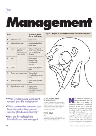
- 1. ENT Management Area Structures giving Figure 1 Regions of neck and tissues from which neck lumps arise rise to neck lumps A Submental area Lymph nodes B Submandibular area Submandibular gland, lymph nodes C Upper Jugular chain Tail of parotid gland, (deep to lymph nodes, branchial sternocleidoma- cyst, carotid sheath stoid muscle) structures D Mid Lymph nodes, branchial cyst, carotid sheath structures E Lower Lymph nodes, thyroid gland F Central neck compartment Thyroid gland, thyroglossal cyst, lymph nodes G Posterior triangle Lymph nodes, parotid gland H Supraclavicular fossa Lymph nodes (infraclavicular drainage), mediastinal structures •What symptoms and signs point N JARROD J HOMER eck lumps are common in MD, FRCS, FRCS(ORL-HNS) patients of all ages, and can towards possible malignancy? Consultant Otolaryngologist/Head-and-Neck be due to a variety of Surgeon, Manchester Royal Infirmary & Christie pathologies in a number of different Hospital, Manchester. Mr Homer is Honorary •What conservative measures can Clinical Lecturer, University of Manchester. His main clinical interest is in head and neck structures in the neck. Most are benign, but it is essential to promptly be addressed to help prevent oncology/tumour surgery investigate and treat patients with salivary gland calculi forming? PRIYA SILVA potentially serious disease. In the past, patients with neck MRCS lumps have been referred to a vari- •How are thyroglossal and Senior House Officer in Otolaryngology, ety of clinics for investigation, Department of Otolaryngology-Head and Neck bronchial cysts best managed? Surgery, Manchester Royal Infirmary sometimes with either sub-optimal or even dangerous management. 726 THE PRACTITIONER, SEPTEMBER 2003, VOL 247
- 2. Neck lumps of neck lumps Clinical focus •Salivary gland neoplasias are anterior to the sternomastoid more common with increasing muscle at the junction of the age but not rare in young upper third and lower two- adults. They are, however, thirds rare in children. Often, the lump itself is asymptomatic •In thyroglossal cysts the history and discovered incidentally is of a painless midline neck swelling. On examination a •In sialolithiasis, calculi form midline (usually) cystic neck within the duct system of the lump is most often situated salivary gland. The calculus can near the hyoid bone. It moves often be merely a sludge. The on both swallowing and history is very characteristic. tongue protrusion There is swelling and tenderness of salivary gland •Solitary thyroid nodules may when eating or just before occur at any age from young eating. The treatment is usually adulthood onwards. The to remove the calculus, if history is of a painless lump in present and distal enough. This the neck with or without procedure is peroral, and compression symptoms. On usually carried out under local examination there is a solitary anaesthetic thyroid lump. Cervical lymphadenopathy or vocal •In branchial cysts the history is cord palsy on laryngoscopy of a painless lump in the neck, suggest malignancy. The lump with or without recurrent should be investigated by infection. They arise usually FNAC; an ultrasound scan is during the second to forth required to delineate the lump decade of life. A cystic lump is further. Thyroid function found most commonly just should also be tested Examples include failure to exam- Figure 2 investigation and further manage- symptoms will, in many cases, give ine the upper aerodigestive tract, Parotid lump – ment by the neck lump clinic. the underlying pathology away. delayed diagnosis and inappropri- pleomorphic GDifferentiation of neck lumps ate open biopsy. Today, however, adenoma In the majority of cases it is possible COMMON, CLASSIC AND most otolaryngology/head-and- to make a diagnosis based upon the IMPORTANT CAUSES OF neck-surgery departments have history, site and characteristics of NECK LUMPS dedicated neck-lump clinics, and neck lumps, and the age of the These are: the situation has improved. patient. GMajor salivary gland lumps, This article aims to outline the In general terms the clinician including parotid and sub- causes of neck lumps. We have needs to consider where the neck mandibular glands It is usually excluded acute disease, but empha- lump is and what structures lie in straightforward to identify the sise the common, classic or serious that area (see figure 1). Then con- anatomical origin of parotid (see M pathologies. We also briefly describe sideration of the age of patient and figure 2) or submandibular gland THE PRACTITIONER, SEPTEMBER 2003, VOL 247 727
- 3. Neck lumps lumps. The main alternative in both enlargement, pain, paraesthesia Figure 3 benign tumours will continue to sites is cervical lymphadenopathy. and facial nerve dysfunction Branchial cyst, grow and become more difficult to • Salivary gland neoplasias These (parotid). cystic lump just excise, and there is a small risk of most commonly occur in the Examination reveals a firm lump anterior to malignant transformation with parotid gland. Some 80 per cent are within the gland of origin. Facial upper part of time. The facial nerve should benign; the common histologies nerve paralysis, skin tethering or sternocleido- always be preserved. are pleomorphic adenoma and ade- skin involvement would suggest mastoid. If Malignant cases should be nolymphoma (Warthin’s tumour). malignancy. solid it would referred to the head and neck The commonest malignant types Fine needle aspiration cytology be massive oncology multidisciplinary clinic are adenoid cystic carcinoma and (FNAC) may be used but the results lymphaden- for radical excision of the tumour adenocarcinoma. are not as accurate as for other opathy and (with facial nerve preservation in Salivary gland neoplasias are neck lumps. Diagnosis may not be possibly due to most cases), neck dissection (of more common with increasing age made until after surgery. lymphoma or lymph nodes) and possible post- but not rare in young adults. They MR scan is the optimal way of upper operative radiotherapy. are, however, rare in children. imaging, but ultrasound will suffice aerodigestive •Sialolithiasis (salivary gland Often, the lump itself is asympto- in some cases. carcinoma calculi) and sialectasis (duct sys- matic and discovered incidentally. Benign neoplasias are treated by tem distortion) In sialolithiasis cal- Features that raise the possibility complete excision on the basis that: culi form within the duct system of of malignancy include noticeable a definitive diagnosis can be made, the salivary gland. The calculus can 728 THE PRACTITIONER, SEPTEMBER 2003, VOL 247
- 4. Neck lumps often be merely a sludge. The Figure 4 (centre) minutes or hours before settling often therapeutic. obstruction causes the gland to Thyroglossal down, or it may last for a few days, If, at the time of assessment, swell, especially when saliva produc- cyst in which case a secondary infection there has been no recent episode tion is increased, usually when eat- is suggested. and there is no calculus to palpate, ing, and can lead to secondary Sialolithiasis may occur at any no investigation may be necessary. infection. Figure 5 (top age from the teens onwards. The treatment is usually to These most commonly occur (80 right) When examined, the gland itself remove the calculus, if present and per cent of cases) in the sub- Solitary thyroid may be normal between episodes, distal enough. This procedure is mandibular gland. Predisposing nodule or generally enlarged and with peroral, and usually carried out factors include reduced flow rates, some tenderness. Palpation biman- under local anaesthetic. duct obstruction, especially after ually of the relevant salivary duct Conservative measures are used chronic inflammation (sialectasis), may reveal the responsible stone. if there is no detectable calculus changes in salivary pH and dehy- Massage of the gland occasionally and the symptoms are recurrent; dration. produces a purulent secretion. these include regular gland and The history is very characteristic. On plain X-ray most sub- duct massage, maintenance of good There is swelling and tenderness of mandibular gland calculi are radio- hydration and regular saliva stimu- the salivary gland when eating or opaque. Sialography will reveal a lation with sialogues (a lemon or a just before eating. This can be filling defect due to intraductal lemon sweet may be used) to ‘flush’ M recurrent, that is, it may last for stones, and flushing of the duct is the duct system out. THE PRACTITIONER, SEPTEMBER 2003, VOL 247 729
- 5. Neck lumps The terminal duct may be mar- tous disorders, such as TB or sar- Figure 6 Goitre during the second to forth decade supialised if the symptoms are coid, and autoimmune disease, of life. recurrent and there is a stenosis in such as Sjögren’s syndrome. On examination, a cystic lump is this duct. Investigation is geared towards Figure 7 (centre) found most commonly just anterior The gland is removed if the excluding either these specific dis- Lymphoma. to the sternomastoid muscle at the symptoms are recurrent and sialog- orders or a tumour. Management is Multiple junction of the upper third and raphy shows proximal calculi generally conservative. posterior lower two-thirds (see figure 3). and/or intraglandular duct disrup- GBranchial cysts The classic triangle FNAC will show the lump is cystic. tion, or if other treatments fail. explanation of branchial cysts is lymphadenopathy Some ‘branchial cysts’ will be •Sialadenitis is a chronic inflam- that they are a congenital persis- necrotic metastatic lymph nodes mation of the salivary glands. In tence of cervical sinus during from primary undiagnosed head- sialectasis there is resultant disrup- branchial cleft development, but a and-neck cancer. It is hence manda- tion to the duct system and symp- more modern explanation is that tory that the patient undergoes toms of sialolithiasis. Clinically they result from degeneration of thorough head and neck examina- there may generalised gland cervical lymph nodes that have con- tion including fibreoptic laryn- enlargement with or without ten- genital epithelial inclusions. gopharyngoscopy, and that the derness. The history is of a painless lump aspirate is submitted for cytology. Most cases are non-specific, but in the neck, with or without recur- Treatment is by surgical exci- specific causes include granuloma- rent infection. They arise usually sion. Every patient over 30 years of 730 THE PRACTITIONER, SEPTEMBER 2003, VOL 247
- 6. Neck lumps age should undergo panendoscopy Figure 8 The history is of a painless mid- These patients should also be inves- of the upper aerodigestive tract at Squamous cell line neck swelling with or without a tigated by FNAC. the same time to look for a possible carcinoma history of recurrent enlargement Treatment is to excise the cyst primary in case the branchial cyst is metastasis – and tenderness caused by infection. and the tract that extends from the a lymph node metastasis. firm nodal Thyroglossal cysts are usually cyst superiorly around the hyoid At-risk patients (those over the mass upper seen in the first two decades of life, bone to the tongue base (the latter age of 40 with tobacco or excessive jugular chain but presentations later in life are includes resection of the middle alcohol consumption) should be not uncommon. portion of the hyoid bone). This considered for panendoscopy as On examination a midline (usu- minimises the chance of recur- well as on-table frozen section ally) cystic neck lump is most often rences that can be difficult to man- pathology with the option to pro- situated near the hyoid bone. It age. ceed to definitive neck dissection if moves on both swallowing and GThyroid lumps When a patient malignancy is found. tongue protrusion (see figure 4). has a thyroid mass, it is necessary to GThyroglossal cyst This is a con- Ultrasound investigation identi- distinguish a solitary or dominant genital condition. The thyroid fies and and delineates the cyst and thyroid nodule (see figure 5) from gland descends in utero from the ensures the thyroid tissue is normal. a goitre (a generalised enlargement tongue as the thyroglossal duct. A This is important because there is a of the entire gland – see figure 6). thyroglossal cyst arises from persis- small chance that the ‘cyst’ will rep- •Goitre The pathology may be M tent epithelial tissue along the duct. resent an ectopic thyroid gland. physiological (in which case it THE PRACTITIONER, SEPTEMBER 2003, VOL 247 731
- 7. Neck lumps typically arises during puberty or smooth or nodular goitre. Thyroid Figure 9 anaplastic carcinomas. Other pregnancy), or due to autoimmune function tests reveal thyroid autoan- Primary causes include a benign neoplasm disease (often with hypo- or hyper- tibodies. If there appears to be squamous cell (follicular adenoma), colloid nod- thyroidism), or due to hyperplasia, some possibility of a dominant nod- carcinoma of ule, cyst or an autonomous hyper- (which is usually euthyroid and can ule, ultrasound examination may tongue – easily functional adenoma. be smooth or multinodular with be required(see below). Computed detected if Solitary thyroid nodules may colloid nodules). tomography is needed if there are looked for in occur at any age from young adult- Hyperplasia is usually idiopathic thoracic inlet compression symp- any setting hood onwards. The history is of a but can be drug-induced, for exam- toms. painless lump in the neck with or ple, from use of carbimazole, or Most cases need no treatment; without compression symptoms. due to iodine deficiency. however, any underlying thyroid Figure 10 On examination there is a soli- The history is of a painless mid- dysfunction should be treated in (centre) tary thyroid lump. Cervical lym- line neck lump. There may be the usual way. Surgery (sub- or Primary phadenopathy or vocal cord palsy symptoms of hypo- or hyper- near-total thyroidectomy) may be squamous cell on laryngoscopy suggest malig- thyroidism; there may also be mass necessary for compression symp- carcinoma of nancy. effect from the goitre, most com- toms, cosmetic concerns or failed larynx – only The lump should be investigated monly in the form of a perception endocrine treatment of Graves’ dis- detectable by by FNAC; an ultrasound scan is of a lump in the throat. ease. laryngoscopy required to delineate the lump fur- Rarely, there may be dysphagia •Solitary or dominant thyroid in neck lump ther or to enhance the accuracy of or stridor. The condition is much nodule Some five per cent of these clinic FNAC. Thyroid function should more common in females. are malignant neoplasms and may also be tested. On examination there is a be papillary, follicular, medullary or There is no longer any indica- 732 THE PRACTITIONER, SEPTEMBER 2003, VOL 247
- 8. Neck lumps tion for the routine use of radio- Figure 11 lumps, with or without constitu- •Cervical lymph node metastasis isotope scans for diagnosis. Apparatus for tional symptoms, such as fever, from head and neck cancer These Where the FNAC reveals malig- fine needle night sweats, fatigue and weight are mostly a squamous cell carci- nancy the patient is referred to a aspiration loss. They may occur at any age, noma (SCC) arising in the upper head-and-neck oncology team for cytology including childhood. aerodigestive tract (oral cavity, definitive surgical resection. This is (FNAC) On examination there is a rub- oropharynx, larynx, hypopharynx, usually total thyroidectomy with or bery lymphadenopathy often cervical oesophagus or nasophar- without neck dissection. involving the posterior triangle (see ynx) that has metastasised to the Where the FNAC result is suspi- figure 7). There may be infraclavic- cervical lymph nodes. More cious or non-diagnostic it should be ular lymph nodes or hepatospleno- uncommonly, metastases may be repeated. If the result is the same, a megaly. from a cutaneous SCC. diagnostic thyroid lobectomy is FNAC may produce suggestive The history is of a lump in the required. results, but biopsy is necessary for neck with or without symptoms Where the FNAC result is benign definitive diagnosis and accurate from the primary tumour, such as it should be repeated in 6–12 histiotyping. A chest X-ray is useful hoarseness, dysphagia, odonopha- months, because of the small possi- at an early stage when this diagnosis gia (painful swallowing), sore bility of a false-negative result. is suspected. throat or mouth ulcer. GCervical lymph node The treatment depends on his- The patient is usually middle- • Lymphoma These are either tiotype and stage (after CT scans, aged and older, and may have a Hodgkin’s disease or non- bone marrow trephine), and will high-risk history with regard to Hodgkin’s lymphoma. the history is comprise chemotherapy or radio- tobacco and alcohol consumption. M of one or more painless neck therapy, or both. On examination there is a firm THE PRACTITIONER, SEPTEMBER 2003, VOL 247 733
- 9. Neck lumps ‘When cervical lymphadenitis is chronic, it is usually reactive to ongoing local non-specific viral or bacterial infections’ neck lump, especially in the jugular Specific underlying disease include (C,D,E), anterior compartment of chain (see figure 8). There may be Epstein-Barr virus, HIV, granuloma- the neck (F), back up the jugular signs of a primary tumour in the tous disorders such as TB or cat chain but now posterior to the ster- oral cavity (see figure 9), orophar- scratch fever, and protozoal agents nocleidomastoid (C,D,E again) and ynx or in the larynx, hypopharynx, such as toxoplasmosis. finally the posterior triangle of the tongue base, nasopharynx (on flex- For persistent lymphadenopathy neck (G) ending up in the ible endoscopy, see figure 10), or in in adults, a full work-up will include supraclavicular fossa (H). the skin or scalp. thorough head and neck examina- It is essential that the examina- Investigations should include tion, FNAC plus or minus serologi- tion includes the oral cavity and FNAC, biopsies of suspected or pos- cal blood testing. oropharynx, and skin and scalp. In sible primary sites, a CT or MR scan If there is doubt raised in the primary care, further examination for more accurate staging, and a FNAC or if there is clinical suspi- of the rest of the upper aerodiges- chest X-ray or CT of the thorax to cion of a specific underlying disor- tive tract will not be possible. exclude synchronous primary lung der, biopsy will help with the cancer or lung metastasis. diagnosis. THE NECK LUMP CLINIC Treatment starts with referral to Secondary care referral should be to a multidisciplinary head and neck CHILDREN a dedicated neck lump clinic. Such a oncology team. When there is a cer- The above comments regarding service should be based upon: vical lymph node metastasis, treat- cervical lymphadenopathy apply to •Clinical examination including a ment usually involves surgical neck children. In most cases FNAC is not thorough examination of the dissection, whatever the modality of possible in children. The norm is, upper aerodigestive tract, and treatment of the primary tumour therefore, repeated examination flexible laryngopharyngoscopy. (surgery or radiotherapy). with excision biopsy reserved for •FNAC (see figure 11), preferably •Cervical lymph node metastasis large nodes, especially in the poste- with a ‘hotlab’ facility offering from cancer below the clavicles rior triangles or supraclavicular fos- results available in about one Neck lumps that are supraclavicular sae, rapid increase in size or other hour. The experience and audit lymph nodes may be metastases worrying history. within a dedicated neck lump from infraclavicular sites, such as clinic will optimise accuracy of intra-abdominal malignancy, or SUBCUTANEOUS LESIONS FNAC. from the breast or lungs. Subcutaneous lesions arise from •Ultrasound examination, prefer- FNAC will often suggest malig- many areas including the neck, the ably available within the clinic. nancy but not give histological most common being lipoma and •Access to pan-endoscopy and information beyond ‘carcinoma’. sebaceous cysts. The former can biopsy of upper aerodigestive In this situation primary head and often be left alone if they do not tract under general anaesthetic. neck cancers, including thyroid, bother the patient and the latter •Links to a multidisciplinary head must be excluded. The search for a are usually excised. and neck oncology team. infraclavicular primary will include basic investigations, such as chest EXAMINATION WARNING X-ray and abdominal ultrasound, It is advised that a systematic The practice of early excision and specific investigations accord- approach is adopted towards the biopsy of all neck lumps without ing to symptoms, sex, age of patient examination of neck lumps. A sug- thorough work-up is to be con- and so on. Incisional biopsy may be gested approach is the move from demned, but still occurs and is usu- required. one area to another, considering ally performed by surgeons who are •Inflammatory cervical lym- which tissue pathology may arise not head-and-neck specialists. At phadenitis Cervical lymphadenitis from each area. best, this may be unnecessary, but at occurs universally at some point in In figure 1 one would move from worst, in the case of metastasis from life, especially during the first areas A to B and C (including head-and-neck cancer, excision decade. When chronic, it is usually parotid), then down the jugular results in the need to perform reactive to ongoing local non-spe- chain feeling initially anterior to more radical salvage surgery and cific viral or bacterial infections. the sternocleidomastoid muscle confers a worse prognosis. I 734 THE PRACTITIONER, SEPTEMBER 2003, VOL 247