1. http://www.roche-applied-science.com/sis/apoptosis/docs/manual_apoptosis.pdf - ACESSO em 13/09/2010.
Roche Applied Science
Apoptosis, Cell Death, and
Cell Proliferation
3rd edition
Overview of this Guide
01How this guide can help you study cell death and cell proliferation?
02When and why do cells die? Does the concentration of environmental
03pollutants exert cytotoxic or cytostatic effects on cells? What factors
04influence the rate and timing of cell proliferation? Researchers in basic,
05industrial, and medical research are asking these questions and looking for
06answers. Understanding the normal regulation of cell death and cell
07proliferation will be critical e.g., for the development of new and more
08successful therapies for preventing and treating cancer and for the
09screening of new anti-cancer compounds.
10Many assays exist to measure cell death and cell proliferation. However, if
11you have only recently become interested in cell death or cel
12proliferation, you may find the diversity of such assays bewildering. You
13may not be able to determine what each assay measures nor decide which
14assays are best for your purposes. This guide is designed to help you make
15such decisions. It presents a brief overview of cell death and cell
16proliferation, along with the major assays currently available to measure
17each. In addition, it clearly lists the advantages and the disadvantages of
18these assays.
19For those who want to eliminate radioactivity from their laboratories, this
20review also describes a number of non-radioactive assays that can serve as
21alternatives to radioactive assays. Wherever possible, the review will
22compare the sensitivity of the radioactive and non-radioactive assays.
Cell Death VI
by Andrew H. Wyllie
01Over the past five or six years there has been a near-exponential increase
02in publications on apoptosis. Around 30 new molecules have been discovered
03whose known functions are exclusively to do with the initiation or
04regulation of apoptosis. A further 20 molecules at least, although already
05associated with important roles in signalling or DNA replication,
06transcription or repair, have been recognised as affecting the regulation
07of apoptosis.
08This article is dedicated to young scientists thinking of entering this
09exploding area of biology, and to those more mature ones who happened to be
10looking elsewhere when the blast reached them, and consequently are in need
11of a rapid introduction to the present state of affairs.
12The term apoptosis first appeared in the biomedical literature in 1972, to
13delineate a structurally-distinctive mode of cell death responsible for
14cell loss within living tissues1.
2. 15The cardinal morphological features are cell shrinkage, accompanied by
16transient but violent bubbling and blebbing from the surface, and
17culminating in separation of the cell into a cluster of membrane-bounded
18bodies. Organellar structure is usually preserved intact, but the nucleus
19undergoes a characteristic condensation of chromatin, initiated at
20sublamellar foci and often extending to generate toroidal or cap-like,
21densely heterochromatic regions. Changes in several cell surface molecules
22also ensure that, in tissues, apoptotic cells are immediately recognised
23and phagocytosed by their neighbours. The result is that many cells can be
24deleted from tissues in a relatively short time with little to show for it
25in conventional microscopic sections.
26This remarkable process is responsible for cell death in development,
27normal tissue turnover, atrophy induced by endocrine and other stimuli,
28negative selection in the immune system, and a substantial proportion of T-
29cell killing. It also accounts for many cell deaths following exposure to
30cytotoxic compounds, hypoxia or viral infection. It is a major factor in
31the cell kinetics of tumors, both growing and regressing. Many cancer
32therapeutic agents exert their effects through initiation of apoptosis, and
33even the process of carcinogenesis itself seems sometimes to depend upon a
34selective, critical failure of apoptosis that permits the survival of cells
35after mutagenic DNA damage. Apoptosis probably contributes to many chronic
36degenerative processes, including Alzheimer’s disease, Parkinson’s disease
37and heart failure. So how does it work?
38Molecular genetic studies on the hard-wired developmental cell death
39programme of the nematode Caenorhabditis elegans led to discovery of a set
40of proteins, widely represented by homologues in other species, and
41responsible for turning on or off the final commitment to death2. In the
42nematode these proteins include the products of the ced3 and ced4 genes
43(which initiate cell suicide), ced9 (which prevents it) and a series of
44some seven genes involved in recognition and phagocytosis of the doomed
45cell.
46CED3 is the prototype of a family of around a dozen mammalian proteases,
47called caspases because of the obligatory cysteine in their active site and
48their predilection for cutting adjacent to aspartate residues. Mammalian
49caspases appear to constitute an autocatalytic cascade, some members
50(notably caspase 8 or FLICE) being “apical” and more susceptible to
51modification by endogenous regulatory proteins, whilst others (notably
52caspase 3 – also called CPP32, Yama and apopain) enact the final,
53irreversible commitment to death. Study of caspase substrates is providing
54interesting insights into the ways in which cells dismantle their structure
55and function. Such substrates include – not surprisingly – cytoskeletal
56proteins such as actin and fodrin and the nuclear lamins, but also an array
57of regulatory and chaperone-like proteins whose function is altered by
58cleavage in subtle and suggestive ways3. A recent example is the nuclease
59chaperone ICAD, whose cleavage permits nuclear entry by a distinctive
60apoptosis nuclease responsible for chromatin cleavage to oligonucleosome
61fragments4.
VII
61Caspases appear to be present in most if not all cells in inactive pro-
62enzyme form, awaiting activation by cleavage. One of the killing mechanisms
63of cytotoxic T cells is a protease, granzyme B, that is delivered to the
64target cell by the T cell granules and triggers these latent pro-enzymes.
65There are endogenous triggers also, and the first to be discovered – the C.
66elegans CED4 protein and its mammalian homologue – is particularly
3. 67intriguing because of its mitochondrial origin5. Thus CED4 could be the
68signal that initiates apoptosis under conditions of shut-down of cellular
69energy metabolism, or when there is a critical level of cell injury
70affecting mitochondrial respiration. In this way CED4 may act as the link
71between agents long known to be associated with mitochondrial injury, such
72as calcium and reactive oxygen species, and the initiation of apoptosis.
73A second mitochondrial protein of enormous significance in apoptosis is
74BCL2, a mammalian homologue of the nematode CED9 protein. BCL2 has the
75tertiary structure of a bacterial pore-forming protein, and inserts into
76the outer membrane of mitochondria. It abrogates apoptosis, probably
77through binding CED4 and another protein BAX, with which it forms
78heterodimers and which, like CED4, is also a “killer” protein6. Both BCL2
79and BAX have several structurally and functionally similar homologues and
80some of this family at least also tap into other cell membranes such as the
81outer nuclear membrane and the endoplasmic reticulum.
82So are there other sources of death transducers, activating the caspase
83cascade because of injury to or signals arising in other parts of the cell
84than mitochondria? There are already examples that show that the answer is
85yes. Thus, the oncosuppressor protein p53 is activated following some types
86of DNA damage and can trigger apoptosis. One way – but only one of several
87– whereby this happens is through transcriptional activation of BAX7.
88The second messenger ceramide, a product of membrane-linked acid
89sphingomyelinase activation, may act as a signal for plasma membrane
90damage8. And a powerful caspase activating system is mediated by cytokine
91receptors of the tumor necrosis factor family, notably fas/apo-1/CD95, TNF
92receptor I, and others. These receptors, on receiving a death stimulus from
93binding their ligand, initiate a series of protein-protein interactions,
94building a complex (the death initiating signalling complex or DISC) which
95eventually recruits and activates caspase 89.
96These mechanisms for coupling cell injury to apoptosis have mostly depended
97on activation of pre-formed proteins. Apoptosis can also be initiated (and
98forestalled) by transcriptional mechanisms, although rather little is known
99about most of them. An outstanding example is the Drosophila gene reaper,
100transcriptionally activated around two hours prior to developmental and
101injury-induced deaths in this organism. Drosophila apoptosis can occur
102without reaper transactivation, but requires very substantially enhanced
103stimuli, suggesting that reaper adjusts a threshold for apoptosis
104initiation10.
105Another gene whose transcription can initiate death is the familiar
106immediate early gene c-myc11. Transcriptional activation of c-myc
107initiates entry into DNA synthesis and is required for sustained re-entry
108in repeated cell cycles, but c-myc activation in the absence of concurrent
109cytokine support triggers apoptosis. This can also be interpreted as a
110“threshold regulatory” effect: – c-myc expression increases the cellular
111requirement for survival factors such as IGF-1.
112Impressive confirmation of the significance of these pathways to apoptosis
113is available from study of transforming viruses. These are hardened
114survivors in the labyrinth of cell regulation, and have found keys to
115allow escape from cell death in a variety of ways. Thus the transforming
116papovavirus SV40, adenovirus type 12, Human Papilloma Virus type 16 and
117Epstein-Barr Virus all have proteins that inactivate apoptosis through
118inactivation of p53 or binding of BAX12. Even lytic viruses possess
4. 119mechanisms to postpone death, such as the cowpox crmA serpin protein and
120the baculovirus p35 protein, which are caspase inhibitors.
121So far so good: there are transcriptional and non-transcriptional pathways
122for activation of apoptosis, and they play through common effector events
123mediated by caspases and regulated by members of the BCL2 family.
124Underlying this simple scheme, however, is an extraordinary complexity.
125Thus, inactivation of fas signalling appears to neuter the ability of both
126c-myc and p53 to initiate apoptosis13,14. Maybe fas signaling is yet
127another example of “threshold regulation”. New proteins have been
128discovered that are recruited to the DISC but appear to inhibit rather
129than activate death15, some of them of viral origin. 130 Many of the
proteins mentioned above have alternative splice variants that 131have
opposite effects. And we still have little idea of the relevance of
132intracellular location or of cell lineage to the activity of most of the
133apoptosis proteins. Susceptibility to apoptosis can be influenced by many
134other gene products, including oncoproteins such as RAS and ABL16, but in
135some cases a single oncoprotein may either increase or decrease
136susceptibility depending on the context. Perhaps it is not surprising that
137a cellular function as important and irreversible as death should be
138subject to a huge range of coarse and fine controls. The reagents and
139protocols in this book should help unravel these.
Andrew H. Wyllie FRS,
Professor of Experimental Pathology,
Sir Alastair Currie CRC Laboratories, University Medical School,
Edinburgh, Scotland
5. 1 Introduction
1.1 Terminology of cell death
Cell death can occur by either of two distinct1,2 mechanisms, necrosis or
apoptosis. In addition, certain chemical compounds and cells are said to be
cytotoxic to the cell, that is, to cause its death.
Someone new to the field might ask, what’s the difference between these
terms? To clear up any possible confusion, we start with some basic
definitions.
Necrosis and apoptosis
The two mechanisms of cell death may briefly be defined:
Necrosis (“accidental” cell death) is the pathological process which occurs
when cells are exposed to a serious physical or chemical insult.
Apoptosis (“normal” or “programmed” cell death) is the physiological process
by which unwanted or useless cells are eliminated during development and
other normal biological processes.
Cytotoxicity
Cytotoxicity is the cell-killing property of a chemical compound (such as a
food, cosmetic, or pharmaceutical) or a mediator cell (cytotoxic T cell). In
contrast to necrosis and apoptosis, the term cytotoxicity does not indicate a
specific cellular death mechanism.
For example, cell-mediated cytotoxicity (that is, cell death mediated by
either cytotoxic T lymphocytes [CTL] or natural killer [NK] cells) combines
some aspects of both necrosis and apoptosis3,4.
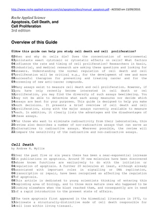
Figure 1: Illustration of the morphological features of necrosis and apoptosis.
1.2 Differences between necrosis and apoptosis
6. There are many observable morphological (Figure 1, Table 1) and biochemical
differences (Table 1) between necrosis and apoptosis2.
Necrosis occurs when cells are exposed to extreme variance from physiological
conditions (e.g., hypothermia, hypoxia) which may result in damage to the
plasma membrane. Under physiological conditions direct damage to the plasma
membrane is evoked by agents like complement and lytic viruses.
Necrosis begins with an impairment of the cell’s ability to maintain
homeostasis, leading to an influx of water and extracellular ions.
Intracellular organelles, most notably the mitochondria, and the entire cell
swell and rupture (cell lysis). Due to the ultimate breakdown of the plasma
membrane, the cytoplasmic contents including lysosomal enzymes are released
into the extracellular fluid. Therefore, in vivo, necrotic cell death is
often associated with extensive tissue damage resulting in an intense
inflammatory response5.
Apoptosis, in contrast, is a mode of cell death that occurs under normal
physiological conditions and the cell is an active participant in its own
demise (“cellular suicide”). It is most often found during normal cell
turnover and tissue homeostasis, embryogenesis, induction and maintenance of
immune tolerance, development of the nervous system and endocrine-dependent
tissue atrophy.
Cells undergoing apoptosis show characteristic morphological and biochemical
features6. These features include chromatin aggregation, nuclear and
cytoplasmic condensation, partition of cytoplasm and nucleus into membrane
bound-vesicles (apoptotic bodies) which contain ribosomes, morphologically
intact mitochondria and nuclear material. In vivo, these apoptotic bodies are
rapidly recognized and phagocytized by either macrophages or adjacent
epithelial cells7. Due to this efficient mechanism for the removal of
apoptotic cells in vivo no inflammatory response is elicited. In vitro, the
apoptotic bodies as well as the remaining cell fragments ultimately swell and
finally lyse. This terminal phase of in vitro cell death has been termed
“secondary necrosis” (Figure 1).
Differences between necrosis and apoptosis
7. Table 1: Differential features and significance of necrosis and apoptosis.
1.3 Apoptotic Pathways
Scientists now recognize that most, if not all, physiological cell death
occurs by apoptosis, and that alteration of apoptosis may result in a variety
of malignant disorders. Consequently, in the last few years, interest in
apoptosis has increased greatly. Great progress has been made in the
understanding of the basic mechanisms of apoptosis and the gene products
involved (Figure 2 below, Table 20, see Appendix, page 123).
Figure 2: Apoptotic pathways. This apoptotic pathways chart
represents a compendium of information on different cell lines, from
various sources. As the dynamic field of apoptosis changes, the
information shown here will likely change. Table 20 in the Appendix,
page 123 contains a list of sources that can be consulted for more
information about the items on this chart. Have a look on our
website www.roche-applied-science/apoptosis to find this chart under
Scientific Information-Apoptotic Pathways and click on the name of a
molecule to get information about its function.
8. Key elements of the apoptotic pathway include:
Death receptors
Apoptosis has been found to be induced via the stimulation of several
different cell surface receptors in association with caspase activation. For
example, the CD95 (APO-1, Fas) receptor ligand system is a critical mediator
of several physiological and pathophysiological processes, including
homeostasis of the peripheral lymphoid compartment and CTL-mediated target
cell killing. Upon cross-linking by ligand or agonist antibody, the Fas
receptor initiates a signal transduction cascade which leads to caspase-
dependent programmed cell death.
Membrane alterations
In the early stages of apoptosis, changes occur at the cell surface and
plasma membrane. One of these plasma membrane alterations is the
translocation of phosphatidylserine (PS) from the inner side of the plasma
membrane to the outer layer, by which PS becomes exposed at the external
surface of the cell.
Protease cascade
Signals leading to the activation of a family of intracellular cysteine
proteases, the caspases, (Cysteinyl-aspartate-specific proteinases) play a
pivotal role in the initiation and execution of apoptosis induced by various
stimuli. Different members of caspases in mammalian cells have been
identified. Among the best-characterized caspases is caspase-1 or ICE
(Interleukin-1 -Converting Enzyme), which was originally identified as a
cysteine protease responsible for the processing of interleukin 1 .
Mitochondrial changes
Mitochondrial physiology is disrupted in cells undergoing either apoptosis or
necrosis. During apoptosis mitochondrial permeability is altered and
apoptosis specific protease activators are released from mitochondria.
Specifically, the discontinuity of the outer mitochondrial membrane results
in the redistribution of cytochrome C to the cytosol followed by subsequent
depolarization of the inner mitochondrial membrane. Cytochrome C (Apaf-2)
release further promotes caspase activation by binding to Apaf-1 and
there-fore activating Apaf-3 (caspase 9). AIF (apoptosis inducing factor),
released in the cytoplasm, has proteolytic activity and is by itself
sufficient to induce apoptosis.
DNA fragmentation
The biochemical hallmark of apoptosis is the fragmentation of the genomic
DNA, an irreversible event that commits the cell to die and occurs before
changes in plasma membrane permeability (prelytic DNA fragmentation). In many
systems, this DNA fragmentation has been shown to result from activation of
an endogenous Ca2+ and Mg2+- dependent nuclear endonuclease. This enzyme
selectively cleaves DNA at sites located between nucleosomal units (linker
DNA) generating mono- and oligonucleosomal DNA fragments.
Note: For more information about the elements of the pathways as well as
synonyms and abbreviations, please see Table 20 in the Appendix, page
123.