Alimentary canal
•Als PPTX, PDF herunterladen•
7 gefällt mir•12,924 views

gnm

Empfohlen
Weitere ähnliche Inhalte
Was ist angesagt?
Was ist angesagt? (20)
Andere mochten auch
Andere mochten auch (11)
Ähnlich wie Alimentary canal
Ähnlich wie Alimentary canal (20)
Mehr von Kalinga Institute of Medical Sciences
Mehr von Kalinga Institute of Medical Sciences (20)
Kürzlich hochgeladen
Kürzlich hochgeladen (20)
Alimentary canal
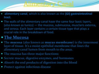
- 1. Alimentary Canal alimentary canal, which is also known as the (GI) gastrointestinal tract. The walls of the alimentary canal have the same four basic layers, (also known as tunics) — the mucosa, submucosa, musclaris externa, and serosa. Each layer contains a certain tissue type that plays a crucial role in the breakdown of food. The Mucosa The mucosa (also known as mucus membrane) is the innermost layer of tissue. It’s a moist epithilial membrane that lines the alimentary canal lumen from mouth to the anus. The mucosa has three major functions: Secrete mucus, digestive enzymes, and hormones Absorb the end products of digestion into the blood Protect against infectious disease
- 2. In certain regions of the alimentary canal, the mucosa may perform one or all three of these functions. Digestive mucosa is made up of three sublayers: (1) a lining epithelium, (2) a lamina propria, and (3) a musclularis mucosae. Except in the mouth, esophagus, and anus where it’s stratified squamous, the epithelium of mucosa is a simple columnar epithelium rich in mucus-secreting cells. The mucus it produces protects certain digestive organs from being digested by enzymes working within the same cavity, it also eases food passage along the GI tract. The lamina propria, which underlies the epithelium, is loose areolar connective tissue. Its capillaries nourish the epithelium and absorb
- 3. digested nutrients. mucosa associated lymphoid tissue) help defend against bacteria and other pathogens, which have free access to our digestive tract. Large collections of lymphoid follicles occur in the pharynx (tonsils) and appendix. External to the lamina propria is the musularis mucosae, a layer of smooth muscle cells that produces local movements of mucosa. In the small intestine, this muscle layer’s tone throws the mucosa into a series of small folds that immensely increases its surface area.
- 4. The Submucosa The submucosa, just external to the mucosa, is areolar connective tissue containing a rich supply of blood and lymphatic vessels, lymphoid follicles, and nerve fibers which supply the surrounding tissues of the GI tract wall. Its elastic fibers enable the stomach to regain its normal shape after temporarily storing a large meal. The Muscularis Externa Th muscularis externa, (also called the muscularis) surrounds the submucosa. The muscularis is responsible for segmentation and peristalsis. It typically has an inner circular layer and an outer longitudal layer of smooth muscle cells. In several places along the tract, the circular layer thickens and forms sphincters that act as valves that control food passage from one organ to the next, they also prevent backflow.
- 5. The Serosa The serosa is the outermost layer of the intraperitoneal organs (it’s also considered the visceral peritoneum). In most alimentary canal organs, its made up of areolar connective tissue covered with mesothelium, a single layer of squamous epithelial cells. In the esophagus, which is located in the thoracic instead of the abdominopelvic cavity, the serosa is replaced by an adventitia, ordinary fibrous connective tissue that binds the esophagus to surrounding structures. Retroperitoneal organs have both a serosa (facing the peritoneal cavity) and an adventia (on the side abutting the dorsal body wall).