
Empfohlen
Weitere ähnliche Inhalte
Was ist angesagt?
Was ist angesagt? (20)
Ähnlich wie Embryology seminar
Ähnlich wie Embryology seminar (20)
Kürzlich hochgeladen
Kürzlich hochgeladen (20)
Embryology seminar
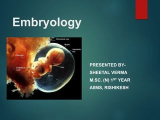
- 1. Embryology PRESENTED BY- SHEETAL VERMA M.SC. (N) 1ST YEAR AIIMS, RISHIKESH
- 2. Embryology is the study of events occurring during prenatal period to gain an understanding of how the anatomical structures formed.
- 3. Embryonic period vs. Fetal period Embryonic – first 8 weeks • Development of the three primary germ layers give rise to all structures • Basic body plan takes shape Fetal period – remaining 30 weeks Structures and organs continue to grow and develop, increasing in complexity
- 6. Major Events of First Week Conception – in lateral third of uterine tube Fusion of female and male pronuclei = amphimixis Zygote (46 chromosomes) moves toward the uterus Blastomeres – daughter cells formed from zygote
- 7. Morula (means mulberry) – cluster of 12–16 Blastomeres Blastophere– about 60 cells Inner cavity is called the blastocoele Trophoblast – layer separating blastocoele from external environment
- 8. Oogenesis
- 11. Fertilization The process of fertilization implies the union of mature germ cells, the ovum and spermatozoa.
- 12. Fertilization life span of oocyte ranges from 12 hours to 24 hours Similarly, fertilizable life span of spermatozoa ranges between 48 and 72 hours. Majority of pregnancies occur when coitus takes place within 3 days prior to ovulation
- 13. Events in sperm-egg interaction There are three types of glycoprotein in zona pellucida. These are known as ZP1, ZP2 and ZP3 of which ZP3 is the most abundant. ZP3 is the primary ligand for the sperm and ZP2 is responsible for Zona Reaction following sperm penetration to prevent polyspermy. Penetration through the zona is rapid and mediated by Acrosin, a trypsin like proteinase.
- 15. Spermatozoa enters perivitelline space at an angle. Then there is binding between inner acrosomal membrane of the sperm head and oolemma (outer membrane of ooplasm). This induces cortical and zona reactions which prevent entry of another spermatozoon into the oocyte, thereby blocking polyspermy.
- 17. Pronucleus Formation- Syngamy- Embryonic Cleavage Approximately, 3 hours after entry of sperm head in the oocyte, the second meiotic division is completed and the second polar body is released with a haploid complement of oocyte chromosomes
- 18. The remaining haploid number of chromosomes in the oocyte will form the female pronucleus. The nucleus of sperm head undergoes decondensation and the male nuclei migrates towards each other. When they come in close proximity, the limiting membrane breakdown. There is an exchange of chromosome material between the male and female pronucleus. The process is known as Syngamy.
- 19. A spindle is formed on which chromosomes become aligned. The stages for first cell division has now been organised and with first cell division a zygote is formed. Embryonic genomic cell activity starts between 4 and 8 cell stage of cleavage, 2-3 days after fertilization. Normal embryonic genomic activity will now control further cell division into Morula and blastocyst.
- 20. Preimplantation Preparatory Changes Prior to implantation both developing endometrium and incoming embryo undergo some preparatory changes.
- 21. Endometrial preparation for implantation (endometrial receptivity) Endometrial preparation for implantation (endometrial receptivity) is a complex procedure which includes interaction of several molecules generated by following changes within the endometrium. Endocrine regulation (E2 and progesterone) Physiological changes (pinopod formation) Biochemical changes (integrin, selectin) Immunomodulatory alteration (formation of protective cytokines interleukin – 3,5,6,10,13 and suppression of natural killer. Endometrial genetic expression (troponin, transformation growth factor alpha, PDG-a etc.)
- 22. Morula After the zygote formation, typical mitotic division of the nucleus occurs producing to Blastomeres. The two cell stage is reached approximately after 30 hours of fertilisation. Each contains equal cytoplasmic volume and chromosome numbers.
- 23. The Blastomeres continue to divide by binary division through 4,8,16 cell stage until a cluster of cells is formed and is called Morula, resembling a mulberry. As the total volume of the cell mass is not increased and the zona pellucida remains intact, the Morula after spending about 3 days in the uterine tube enters the uterine cavity through the narrow uterine ostium (1mm) on the 4th day in the 16-64 cell stage.
- 25. The transport is slow process and is controlled by muscular contraction and movement of the cilia. The central cell of the Morula is known as inner cell mass which forms the embryo proper and the peripheral cells are called outer cell mass which will form protective and nutritive membranes of the embryo.
- 27. Blastocyst While the Morula remains free in the uterine cavity on the 4th and 5th day it is covered by a film of mucus. The fluid passes through the canaliculi of the zona pellucida which separates the cells of the Morula and is now termed blastocyst.
- 29. Zona hatching is the next step so that trophectoderm cells interact with endometrial cells and implantation occurs. Due to Blastocyst enlargement the zona pellucida becomes stretched, thin and gradually disappears. Lysis of zona and escape of embryo is called zona hatching. The cells on the outer side of the Morula become trophectoderm and the inner cell become inner cell mass by the mediation of epithelial cadherin(protein).
- 30. Trophectoderm differentiates into chorion and the inner cell mass into the embryo. Completely undifferentiated cells are called pluripotent embryonic stem cells (ES cells). ES cells are able to produce mature somatic cells of any germ layers (ectoderm, mesoderm and endoderm).
- 33. Implantation Implantation occurs in the endometrium of the anterior or posterior wall of the body near the fundus on the 6th day which corresponds, to the 20th day of regular menstrual cycle.
- 34. IMPLANTATION occurs through four stages: Apposition Adhesion Penetration Invasion
- 35. Changes in the Blastocyst Adhesion of trophoblast cells to the endometrial cells The factors responsible for blastocyst attachment are P.selectin, heparin sulphate, proteoglycan, integrin trophinin, tasin and others.
- 36. Apposition Occurs through pinopod formation. These pinopod absorbs the endometrial fluid which is secreted by the endometrial gland cells. This fluid rich in glycogen and mucin provides nutrition to the blastocyst initially. Unless this fluid is absorbed, adhesion phase cannot occur. Adhesion of blastocyst to the endometrium occurs through the adhesion molecules like integrin, selectin and cadherin (glycoproteins).
- 37. Penetration Actual penetration occurs through the stromal cells in between the glands and is facilitated by the histolytic action of the blastocyst. With increasing lysis of the stromal cells, the blastocyst is burrowed more and more inside the stratum compactum of the decidua. Concurrently, the syncytical cells penetrate deeper into stroma and erode the endothelium of the maternal capillaries.
- 38. Further penetration is stopped probably by the maternal immunological factor and the original point of entry is sealed by fibrin clot and later by epithelium. The process is completed by 10th or 11th day which corresponds to D 24-25 from LMP.
- 39. The process of implantation is controlled by the Immunomodulatory role of various cytokines, many local peptides like epidermal growth factor and prostaglandins. Both the decidua and embryo synthesize these molecules.
- 40. TROPHOBLAST differentiation into an outer trophectoderm and inner cell mass. The trophectoderm is further differentiated into an inner mononuclear cellular layer cell called cytotrophoblast or langhan’s layer and an outer layer of multinucleated syncyticum called syncytiotrophoblast. Placenta and fetal membranes are developed from the trophoblast. It serves as- Invasion Nutrition Hormones production
- 41. Trophoblasts (from Greek trephein: to feed, and blastos: germinator) are cells forming the outer layer of a blastocyst, which provide nutrients to the embryo and develop into a large part of the placenta.
- 42. Decidua The decidua is the endometrium of the pregnant uterus. It is so named because of it is shed following delivery. Decidual reaction – the increased structural and secretory activity of the endometrium that is brought about in response to progesterone following implantation is known as decidual reaction.
- 43. The well-developed decidua differentiates into three layers- Superficial compact layer Intermediate spongy layer Thin basal layer
- 44. After the interstitial implantation of the blastocyst into the compact layer of the decidua, the different portions of the decidua are renamed as- Decidua basalis (contact with the basal layer of the blastocyst) Decidua capsularis (thin superficial layer covering the blastocyst) Decidua Vera (lining the uterine cavity outside the site of implantation )
- 46. Functions of decidua good nidus for the implantation of the blastocyst It supplies nutrition the early stage of the growing ovum by its rich source of glycogen and fat Deeper penetration of the trophoblast is controlled by local peptides, cytokines and integrins. Decidua basalis takes part in the formation of the placenta and for the nourishment of the developing embryo.
- 47. Chorion and chorionic villi It consists of two embryonic layer- outer trophoblast and inner primitive mesenchyme which appears on the 9th day.
- 48. Formation of chorionic villi- Development of primary stem villi by syncytotrophoblast Lacunar space by Mesodermal cell is known as intervillous space Formation of secondary villi formation on 16th day Development of villous capillary system from the Mesodermal cells on 21st day also known as 21st day
- 49. Development of inner cell mass Along with the changes in the trophoblast, on the 8th day, the embryoblast differentiates into bilaminar germ disc which consist of dorsal ectoderm layer and ventral endodermal layer. Two cavities appear one on each side of the germ disc- Amniotic cavity (formed by extension of epiblast) Yolk sac (extension of hypoblast) Formation of trilaminar germ layer Proliferation of Ectodermal cells in midline leads to formation of primitive streak
- 54. During the embryonic stage which extends from the 4th to 8th week individual differentiation of the germ layers and the formation of the folds of the embryo occur. Most of the tissues and organs are developed during this period. The major structures which are developed from the three germinal layers from which embryo can be differentiated as human at 8th week
- 56. Gastrulation is the process of formation of three germ layers during 3rd week. Ectodermal layer central and peripheral nervous system epidermis of the skin with its appendages pituitary gland, salivary glands, mucous lining of the nasal cavity, paranasal sinus, roof of the mouth etc.
- 57. Mesodermal layer bones, cartilages, muscles, kidney, gonads, spleen, peritoneal cavity and cardiovascular system.
- 58. Endodermal layer Epithelial lining of the GI tract, liver, gall bladder, pancreas and mucous membrane of urinary bladder and urethra etc. The embryonic endoderm is the first of three germ layers from which all tissues (bone, muscle, connective tissue), organs and structures derive. Structures such as fetal membranes, umbilical cord and part of the placenta also develops from these layers.
- 60. At the time of implantation (6th day) the embryo known as bilaminar embryo because the embryo disc arising from the inner cell mass has two layers of cells- a) epiblast b) hypoblast
- 62. From the beginning of the third week, the primitive stalk, arising from the embryonic disk, is the growth centre for embryo for about two weeks after which it disintegrates. During the third week, the neural tube, coelomic spaces primitive blood cells develop.
- 63. During 4th week The heart starts to beat at the beginning of 4th week. During the 4th week longitudinal and transverse folding of the embryonic disk takes place which converts the embryo from straight to a curve form. By the end of 4th week, the embryo has assumed its often called salamander look and has the rudiments of ear, arm, legs, facial and neck structures.
- 64. During the 5th week rapid development of brain results in extensive growth of the head and makes it much larger in relation to the rest of the body. Development takes place from cephalic to caudal, with the development of legs almost a week behind development of the arms. The eyes begin to develop with the lens, vesicles, optic cups and retinal pigments.
- 65. During 6th week Nose, mouth and palate begin to take form and the eyelids visible. Arms and legs undergo extensive development and by the end of 7th week arms and legs are formed with clearly defined fingers and toes.
- 66. During 7th week the neck region is established, the abdomen is less protuberant and urogenital development begins. The external ears are evident The embryo by the end of 7th week has distinctive human characteristics. The end of the 7th week also marks the end of the embryonic period. All essential internal and external structures are formed. They undergo further elaboration and growth, including the replacement of cartilage with the bone cells
- 67. The embryonic period is a critical period during which any teratogen (e.g. drugs, X-rays, viruses) may either be lethal or cause any major congenital malformations
- 68. By the end of the first trimester ,the intestines are fully into the abdomen, the external genitalia have male or female characteristics but neither are fully formed, the anus has formed and the facial characteristics of the fetus now look undeniably human. The fetus at this stage can swallow and make respiratory movements, urinate, move specific parts of limbs and open and shut his or her mouth.
- 71. Q-1 The part of the sperm containing proteolytic enzymes to digest the zona pellucida is the: A. capacitor B. head C. corona D. acrosome
- 72. Q-2: The testes descend to the scrotum during the fetal period at: a) The 3rd month b) The 6th month c) The 4th month d) Just before the labor
- 73. Q-3 The first week of human development is characterized by formation of the: A. inner cell mass B. hypoblast C. Trophoblast D. blastocyst E. all of the above
- 74. Q-4 With the light microscope, the zona pellucida appears as a translucent membrane surrounding the: A. primary oocyte B. zygote C. Morula D. very early blastocyst E. all of the above
- 75. Q-5 The first two intraembryonic germ layers to differentiate are the: A. ectoderm and hypoblast B. epiblast and hypoblast C. ectoderm and endoderm D. ectoderm and mesoderm
- 76. Q-6 The most important region of the decidua for the nourishment of the conceptus is the decidua ___________. A. frondosum B. capsularis C. parietalis D. basalis
- 77. Q-7 Failure of the brain to grow may result in: A. Plagiocephaly B. craniostenosis C. acrocephaly D. microcephaly
- 78. Q-8 Embryonic period is up to: a) 8 days b) 4 weeks c) 8 weeks d) 12 weeks
- 79. Q-9 Implantation Occurs a) On 24th day b) 4 weeks c) 3rd day d) 6th day
- 80. Q-10 Identify the picture