SlideShare ist ein Scribd-Unternehmen logo
1 von 6
Downloaden Sie, um offline zu lesen
Embryology overview C. Riedinger                                                     1


                   Overview of what becomes what.

               Epiblast      Ectoderm
                                    Mesoderm
Embryoblast

               Hypoblast  Endoderm


Ectoderm
   -   Overall : epidermis, nervous system
   -   neural crest cells      glial and schwann cells
                               melanocytes
                               parts of meninges
                               parts of teeth
                               dorsal root, cranial, enteric, autonomic ganglia
                               connective tissue of face
                               bones of skull
                               adrenal medulla
   -   neural tube             brain + spinal cord

Mesoderm
   -   overall: skeletal, muscle, connective
   -   axial                   notochord  nucleus pulposus of vertebrae
   -   paraxial                somites       myotome, scleratome, dermatome
   -   intermediate            urogenital
   -   lateral plate (continuous with extra-embryonic mesoderm of
       amniotic cavity         parietal/somatic layer
       and yolk sac            visceral/splanchnic layer)
                               blood, peritoneum
                               intraembryonic coelum  pericardial, pleural and
                                                           peritoneal cavities

Endoderm
   -   overall:  gut tube, lining of GI tract and respiratory system (epithelium)
   -   pharyngeal arches
   -   thyroid gland (from foramen caecum)
   -   lung           (from respiratory diverticulum in foregut)
   -   liver          (hepatic diverticulum in duodenum)
   -   pancreas
   -   foregut:       bucopharyngeal membrane to midgut
   -   midgut:        foregut to hindgut, initially connected to yolk sac
   -   hindgut:       midgut to cloacal membrane, connecting stalk
                      (later to urogenital openings, allantois= “überhindgut”)
   -   parenchymal cells of liver, pancreas, parathyroid

Yolk sack  vitelline duct  meckel’s diverticulum (if not obliterated)
Embryology overview C. Riedinger                                                       2


Pharyngeal arches
       Pharyngeal          Pharyngeal          Pharyngeal             Pharyngeal
       arches              cartilages          pouches (inside)       clefts (outside)
  1    Vc:                 Aliphenoid          Tympanic cavity        External
       Mastication         malleolus incus     Eustachian tube        auditory
       MATT                Meckel’s                                   meatus
                           cartilage
  2    VII:                Stapes              Palatine tonsils
       facial expression   styloid process
       POSS                stylohyoid
                           ligament                                   Cervical sinus
                           lesser cornu of                            (obliterated)
                           hyoid
  3    XI:                 Greater cornu of    Inferior parathyroid
       stylopharyngeus     hyoid               thymus
                           body of hyoid
  4    Sup. laryngeal:     Thyroid cartilage   Superior
       cricothyroid        cricoid cartilage   parathyroid
       Middle + inferior   (could also be 6)   ultimo-branchial
       constrictor                             body (parafollicular
                                               calcitonin-
                                               producing cells)
  6    Recurrent
       laryngeal:
       larynx


Cross-talk between epithelium (endoderm) and mesenchyme
(mesoderm)
   -   mesenchyme = undifferentiated loose connective tissue derived mainly from
       mesoderm (some from neural crest cells though which are ectodermal)
   -   parenchyma = functional parts of an organ in the body
   -   stroma = structural tissue (connective, supportive)
   -   differentiation of endodermal epithelium dictated by signals from
       mesoderm (mesenchyme)
   -   stomach:       gastric glands
   -   intestines:    villi
   -   liver:         hepatic cords
   -   pancreas:
   -   lung:          branching morphogenesis
       branching (in bronchi) or inhibition of branching (trachea)
   -   kidney:        dichotomous branching
       ureteric bud induces mesoderm to become metanephric blastema
       /mesenchyme, which in turn induces further buds
Embryology overview C. Riedinger                                                 3


Endodermal derivatives
Lung
  - lung diverticulum (from gut endoderm) grows into splanchnopleuric/visceral
     mesoderm
  - branching morphogenesis: guided by FGF10, antagonist: sonic hedgehog
  - stages of lung growth: embryonic, pseudoglandular, canalicular, sacuular,
     alveolar

Stomach
   - thickening of foregut tube (differential growth)
            more on left  greater curvature
            less on right  lesser curvature
   - 90* clockwise rotation so that:
            left vagus       ant
            right            post
            ventral mesentery  right               lesser omentum
            dorsal mesentery         left          greater omentum
   - pylorus rises, this makes duodenum C-shaped
   - duodenum is half foregut half midgut

Liver
   - diverticulum from duodenal endoderm
   - pushes into septum transversum  ventral mesentery
   - gall bladder = ventral outpouching
   -
Pancreas
   - outgrowth of hepatic diverticulum
   - dorsal bud  accessory pancreatic duct / minor papilla
   - ventral bud  uncinate process, manjor papilla along with bile

Small intestine
  - rapid enlongation of midgut causes physiological umbilical hernia
  - 1* rotation, then another 90*, another 180*, all anticlockwise

Bladder
   - at cloacal membrane (no mesoderm) urogenital septum grows in to divide
      hindgut from allantois
   - urogenital septum  perineum (?)
   - widening of gut on allantoic side = urogenital sinus  bladder, urethra
      male: only prostatic and membranous urethra
      female: entire urethra
   - allantois  urachus  median umbilical ligament
Embryology overview C. Riedinger                                                  4


Mesodermal derivatives

Development of heart
   ♥ from angiogenic cell clusters in extra-embryonic mesoderm
   ♥ Two heart tubes form single tube during folding

   ♥   Truncus arteriosus           spiral septum  aorta + pulmonary trunk
   ♥   Bulboventricular groove
   ♥   Bulbis cordis                   conus cordis  RV / infundibulum
   ♥   Ventricle                       LV (trabeculated part)
   ♥   Atrioventricular groove         atrioventricular valves
                                       endocard. cushions sept. intermedium
       (septum intermedium between right and left AV canal)
       spiral septum eventually fuses with septum intermedium and muscular
       ventricular septum
   ♥   Atrium                                 auricles
   ♥   Sinus venosus (right sinus horn)  RA
   ♥   Left sinus horn                        coronary sinus
   ♥   Septum primum has osteum primum (which closes) and then osteum
       secundum, septum
   ♥   Right/left directionality determined by nodal gene

Fetal circulation:
   ♥ 3 shunts:
            o ductus venosus: closure within 5 days
            o foramen ovale
            o ductus arteriosus: closure within 10 days
   ♥ changes at birth:
            o lungs inflate, blood enters them and returns to the LA
            o p in LA > p in RA
            o foramen ovale shuts
            o prostaglandin levels decrease as no more flow from umbilical vein
   ♥ umbilical vein            ligamentum teres
   ♥ ductus venosus            ligamentum venosum
   ♥ foramen ovale             fossa ovalis
   ♥ ductus arteriosus         ligamentum arteriosum (left recurrent laryngeal
        winds around it)

Blood vessels:
   - vasculogenesis:        differentiation from within a cell mass
   - angiogenesis:          invasion of tissue from existing blood vessels
Embryology overview C. Riedinger                                              5


Septum transversum
   - thickened sheet of mesoderm between cardiogenic area and cranial margin of
      disc, later caudal and anterior to gut tube
   - septum transversum  central tendon of diaphragm
   - septum transversum also makes VENTRAL MESENTERY for caudal portion
      of foregut: liver, stomach, spleen
   - complete diaphragm develops from:
          o septum transversum
          o somatic mesoderm from body wall
          o mesentery of oesophagus
          o pleuroperitoneal membrane
          o myoblasts from cervical somites

Kidney
   - from intra-embryonic intermediate mesoderm
   - nephric part or urogenital ridge
   - pronephros             regresses early, non-functional
   - mesonephros            functional, regresses
   - metanephros            definite kidney
   - duct from pronephros through mesonephros to urogenital sinus = mesonephric
      duct (Wolffian duct)
   - mesonephric duct outpouching/metanephric diverticulum
       ureteric bud  metanephros
Embryology overview C. Riedinger                                                    6


Urogenital system
   - same origin as kidney, from from intra-embryonic intermediate mesoderm
   - gonadal part of urogenital ridge
   - migrating primordial germ cells enter and induce sex-specific differentiation =
      end of indifferent stage (germ cells originate from epiblast?)
      germ cells  spermatogonia / oocytes
   - SRY (XY gene product), SOX9 crucial for development of testes

male:
   - mesonephric duct                 vas deferens
                                      epididymis
                                      seminal vesicle
   -   paramesonephric duct           regresses to prostratic utricle, appendix of
                                     the testes, ejaculatory duct
   -   mesonephric mesenchyme  Leydig cells (make androgens!)
   -   making testosterone requires 5-alpha reductase
   -   sex cords                      sertoli cells (Muellerian inhibitory substance
                                     to suppress formation of femal genitalia!) +
                                     seminiferous tubules (spermatogenesis)
   -   gubernaculum guides descent of testes          gubernaculum
                                                      scrotal ligament
   -   genital tubercle / urogenital folds            penis
                                                      corpora cavernosa
                                                      corpus spongiosum
   -   labioscrotal swellings /folds                  scrotum


female:
   - mesonephric duct                 regresses to Gartner’s cyst in wall of vagina
   - paramesonephric duct             fallopian tubes
                                      uterus
                                      top of vagina
       (inf end of vagina develops from urogenital sinus (sinovaginal bulb))
   -   mesonephric mesenchyme  thecal cells (make corpuls luteum to make
                                     progesterone but also androgens)
   -   sex cords break up and condense around germ cells  primary follicles
   -   gubernaculum                   round ligament of ovary and uterus
   -   genital tubercle               clitoris
                                      corpus cavernosa
                                      bulbospongiosum
   -   urethral folds                 labia minora
   -   labioscrotal swellings         labia majora

Weitere ähnliche Inhalte

Was ist angesagt?

Development of respiratory system
Development of respiratory systemDevelopment of respiratory system
Development of respiratory system
Mohamed Autifi
 

Was ist angesagt? (20)

Histology of trachea and lung
Histology of trachea and lungHistology of trachea and lung
Histology of trachea and lung
 
Pharyngeal "Branchial" Arches (Special Embryology)
Pharyngeal "Branchial" Arches (Special Embryology)Pharyngeal "Branchial" Arches (Special Embryology)
Pharyngeal "Branchial" Arches (Special Embryology)
 
Anatomy of trachea & lungs
Anatomy of  trachea & lungsAnatomy of  trachea & lungs
Anatomy of trachea & lungs
 
Previous year question on pharyngeal arches embryology based on neet pg, usml...
Previous year question on pharyngeal arches embryology based on neet pg, usml...Previous year question on pharyngeal arches embryology based on neet pg, usml...
Previous year question on pharyngeal arches embryology based on neet pg, usml...
 
Pharyngeal arches
Pharyngeal archesPharyngeal arches
Pharyngeal arches
 
11.10.08(d): Histology of the Respiratory Tract
11.10.08(d): Histology of the Respiratory Tract11.10.08(d): Histology of the Respiratory Tract
11.10.08(d): Histology of the Respiratory Tract
 
Nasal Cavity Anatomy
Nasal Cavity AnatomyNasal Cavity Anatomy
Nasal Cavity Anatomy
 
Overview Cranial Foramina
Overview Cranial ForaminaOverview Cranial Foramina
Overview Cranial Foramina
 
Pharyngeal apparatus
Pharyngeal apparatusPharyngeal apparatus
Pharyngeal apparatus
 
Development of respiratory system
Development of respiratory systemDevelopment of respiratory system
Development of respiratory system
 
Thorax bones
Thorax bonesThorax bones
Thorax bones
 
2. gastrulation
2. gastrulation2. gastrulation
2. gastrulation
 
1. second week of development
1. second week of development1. second week of development
1. second week of development
 
Development of Respiratory System (Special Embryology)
Development of Respiratory System (Special Embryology)Development of Respiratory System (Special Embryology)
Development of Respiratory System (Special Embryology)
 
Formation of the body cavities
Formation of the body cavitiesFormation of the body cavities
Formation of the body cavities
 
Thoracic part of Sympathetic Chain (Anatomy of the Thorax)
Thoracic part of Sympathetic Chain (Anatomy of the Thorax)Thoracic part of Sympathetic Chain (Anatomy of the Thorax)
Thoracic part of Sympathetic Chain (Anatomy of the Thorax)
 
Embryology of Digestive system
Embryology of Digestive systemEmbryology of Digestive system
Embryology of Digestive system
 
MUSCULAR SYSTEM DEVELOPMENT (Human Embryology)
MUSCULAR SYSTEM DEVELOPMENT  (Human Embryology)MUSCULAR SYSTEM DEVELOPMENT  (Human Embryology)
MUSCULAR SYSTEM DEVELOPMENT (Human Embryology)
 
Third week of development.pdf
Third week of development.pdfThird week of development.pdf
Third week of development.pdf
 
Third week of human development
Third week of human development Third week of human development
Third week of human development
 

Andere mochten auch (6)

Antibiotics Overview - the barest facts
Antibiotics Overview - the barest factsAntibiotics Overview - the barest facts
Antibiotics Overview - the barest facts
 
Fruit charts
Fruit chartsFruit charts
Fruit charts
 
Recommended Daily Allowance Chart For Vitamins
Recommended Daily Allowance Chart For VitaminsRecommended Daily Allowance Chart For Vitamins
Recommended Daily Allowance Chart For Vitamins
 
How To Diffuse
How To DiffuseHow To Diffuse
How To Diffuse
 
General principles of surgery - medical finals revision notes
General principles of surgery - medical finals revision notesGeneral principles of surgery - medical finals revision notes
General principles of surgery - medical finals revision notes
 
An easy way to learn shoulder muscles
An easy way to learn shoulder musclesAn easy way to learn shoulder muscles
An easy way to learn shoulder muscles
 

Ähnlich wie Embryology Overview

13th week -_lymphatic_system
13th week -_lymphatic_system13th week -_lymphatic_system
13th week -_lymphatic_system
MUBOSScz
 
Maternal Anatomy by _ Dr. tejaswini [Autosaved].pptx
Maternal Anatomy by _ Dr. tejaswini [Autosaved].pptxMaternal Anatomy by _ Dr. tejaswini [Autosaved].pptx
Maternal Anatomy by _ Dr. tejaswini [Autosaved].pptx
DrTejaswini7
 
Anatomy and physiology of the palatine tonsil
Anatomy and physiology of the palatine tonsilAnatomy and physiology of the palatine tonsil
Anatomy and physiology of the palatine tonsil
Salman Syed
 
Kidney anatomy physiology
Kidney anatomy physiologyKidney anatomy physiology
Kidney anatomy physiology
wanted1361
 

Ähnlich wie Embryology Overview (20)

URINARY BLADDER & URETHRA PROF VKVS.pptx
URINARY BLADDER & URETHRA PROF VKVS.pptxURINARY BLADDER & URETHRA PROF VKVS.pptx
URINARY BLADDER & URETHRA PROF VKVS.pptx
 
embryo.pptx
embryo.pptxembryo.pptx
embryo.pptx
 
01 male pelvic organs (2) (2)
01 male pelvic organs (2) (2)01 male pelvic organs (2) (2)
01 male pelvic organs (2) (2)
 
13th week -_lymphatic_system
13th week -_lymphatic_system13th week -_lymphatic_system
13th week -_lymphatic_system
 
Salivary glands
Salivary glandsSalivary glands
Salivary glands
 
My ppt
My pptMy ppt
My ppt
 
Salivary gland diseases 1
Salivary gland diseases 1Salivary gland diseases 1
Salivary gland diseases 1
 
The clinical Anatomy of the Thorax. eng.pdf
The clinical Anatomy of the Thorax. eng.pdfThe clinical Anatomy of the Thorax. eng.pdf
The clinical Anatomy of the Thorax. eng.pdf
 
Parotid Gland
Parotid GlandParotid Gland
Parotid Gland
 
Parotid glands by dr parthsarthi gautam, MDS
Parotid glands by dr parthsarthi gautam, MDSParotid glands by dr parthsarthi gautam, MDS
Parotid glands by dr parthsarthi gautam, MDS
 
Cme pancreas2
Cme pancreas2Cme pancreas2
Cme pancreas2
 
Maternal Anatomy by _ Dr. tejaswini [Autosaved].pptx
Maternal Anatomy by _ Dr. tejaswini [Autosaved].pptxMaternal Anatomy by _ Dr. tejaswini [Autosaved].pptx
Maternal Anatomy by _ Dr. tejaswini [Autosaved].pptx
 
Cranial cavity ( Department of Anatomy)
Cranial cavity ( Department of Anatomy)Cranial cavity ( Department of Anatomy)
Cranial cavity ( Department of Anatomy)
 
Anatomy and physiology of the palatine tonsil
Anatomy and physiology of the palatine tonsilAnatomy and physiology of the palatine tonsil
Anatomy and physiology of the palatine tonsil
 
Kidney anatomy physiology
Kidney anatomy physiologyKidney anatomy physiology
Kidney anatomy physiology
 
EMBRYOLOGY OF KIDNEY AND URETER.pdf
EMBRYOLOGY OF KIDNEY AND URETER.pdfEMBRYOLOGY OF KIDNEY AND URETER.pdf
EMBRYOLOGY OF KIDNEY AND URETER.pdf
 
CTMR OF HEAD & NECK.pptx
CTMR OF HEAD & NECK.pptxCTMR OF HEAD & NECK.pptx
CTMR OF HEAD & NECK.pptx
 
Topographic anatomy of the brain and brain part of the skull
Topographic anatomy of the brain and brain part of the skullTopographic anatomy of the brain and brain part of the skull
Topographic anatomy of the brain and brain part of the skull
 
Salivary glands
Salivary glandsSalivary glands
Salivary glands
 
Anatomy of skull base
Anatomy of skull baseAnatomy of skull base
Anatomy of skull base
 

Mehr von Christiane Riedinger

Mehr von Christiane Riedinger (20)

Notes on hernias for medical finals
Notes on hernias for medical finalsNotes on hernias for medical finals
Notes on hernias for medical finals
 
On neck and skin lumps for medical finals
On neck and skin lumps for medical finalsOn neck and skin lumps for medical finals
On neck and skin lumps for medical finals
 
Breast Surgery for Medical Finals
Breast Surgery for Medical FinalsBreast Surgery for Medical Finals
Breast Surgery for Medical Finals
 
Palliative Care for Medical Finals
Palliative Care for Medical FinalsPalliative Care for Medical Finals
Palliative Care for Medical Finals
 
Useful background information for neurology revision.
Useful background information for neurology revision.Useful background information for neurology revision.
Useful background information for neurology revision.
 
Heart sounds, valves and JVP
Heart sounds, valves and JVPHeart sounds, valves and JVP
Heart sounds, valves and JVP
 
Overview of heart murmurs
Overview of heart murmursOverview of heart murmurs
Overview of heart murmurs
 
Endocrinology for Medical Finals
Endocrinology for Medical FinalsEndocrinology for Medical Finals
Endocrinology for Medical Finals
 
Summary of differentiating features of neurological deficits (motor)
Summary of differentiating features of neurological deficits (motor)Summary of differentiating features of neurological deficits (motor)
Summary of differentiating features of neurological deficits (motor)
 
Law and Ethics for Medics
Law and Ethics for MedicsLaw and Ethics for Medics
Law and Ethics for Medics
 
Overview of what Public Health for Medics is all about
Overview of what Public Health for Medics is all aboutOverview of what Public Health for Medics is all about
Overview of what Public Health for Medics is all about
 
Overview of Public Health Topics
Overview of Public Health TopicsOverview of Public Health Topics
Overview of Public Health Topics
 
Presentation Mantras
Presentation MantrasPresentation Mantras
Presentation Mantras
 
Consultation Manual Part 1 - Interview Skills
Consultation Manual Part 1 - Interview SkillsConsultation Manual Part 1 - Interview Skills
Consultation Manual Part 1 - Interview Skills
 
Consultation Manual Part 2 - Examination Skills
Consultation Manual Part 2 - Examination SkillsConsultation Manual Part 2 - Examination Skills
Consultation Manual Part 2 - Examination Skills
 
Renal Pathology Tutorial
Renal Pathology TutorialRenal Pathology Tutorial
Renal Pathology Tutorial
 
Cardiovascular Histopathology Tutorial
Cardiovascular Histopathology TutorialCardiovascular Histopathology Tutorial
Cardiovascular Histopathology Tutorial
 
Parasites overview
Parasites overviewParasites overview
Parasites overview
 
Overview of some drugs and their actions.
Overview of some drugs and their actions.Overview of some drugs and their actions.
Overview of some drugs and their actions.
 
Information for 2nd year Medical Students in Cambridge / 2nd year wisdom
Information for 2nd year Medical Students in Cambridge / 2nd year wisdomInformation for 2nd year Medical Students in Cambridge / 2nd year wisdom
Information for 2nd year Medical Students in Cambridge / 2nd year wisdom
 

Embryology Overview

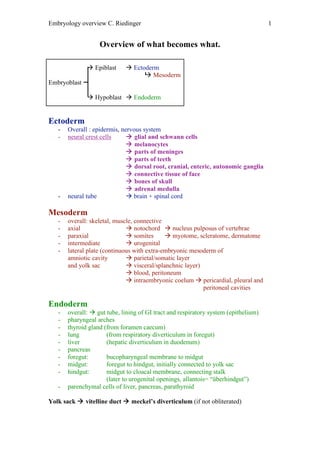
  • 1. Embryology overview C. Riedinger 1 Overview of what becomes what.  Epiblast  Ectoderm  Mesoderm Embryoblast  Hypoblast  Endoderm Ectoderm - Overall : epidermis, nervous system - neural crest cells  glial and schwann cells  melanocytes  parts of meninges  parts of teeth  dorsal root, cranial, enteric, autonomic ganglia  connective tissue of face  bones of skull  adrenal medulla - neural tube  brain + spinal cord Mesoderm - overall: skeletal, muscle, connective - axial  notochord  nucleus pulposus of vertebrae - paraxial  somites  myotome, scleratome, dermatome - intermediate  urogenital - lateral plate (continuous with extra-embryonic mesoderm of amniotic cavity  parietal/somatic layer and yolk sac  visceral/splanchnic layer)  blood, peritoneum  intraembryonic coelum  pericardial, pleural and peritoneal cavities Endoderm - overall:  gut tube, lining of GI tract and respiratory system (epithelium) - pharyngeal arches - thyroid gland (from foramen caecum) - lung (from respiratory diverticulum in foregut) - liver (hepatic diverticulum in duodenum) - pancreas - foregut: bucopharyngeal membrane to midgut - midgut: foregut to hindgut, initially connected to yolk sac - hindgut: midgut to cloacal membrane, connecting stalk (later to urogenital openings, allantois= “überhindgut”) - parenchymal cells of liver, pancreas, parathyroid Yolk sack  vitelline duct  meckel’s diverticulum (if not obliterated)
  • 2. Embryology overview C. Riedinger 2 Pharyngeal arches Pharyngeal Pharyngeal Pharyngeal Pharyngeal arches cartilages pouches (inside) clefts (outside) 1 Vc: Aliphenoid Tympanic cavity External Mastication malleolus incus Eustachian tube auditory MATT Meckel’s meatus cartilage 2 VII: Stapes Palatine tonsils facial expression styloid process POSS stylohyoid ligament Cervical sinus lesser cornu of (obliterated) hyoid 3 XI: Greater cornu of Inferior parathyroid stylopharyngeus hyoid thymus body of hyoid 4 Sup. laryngeal: Thyroid cartilage Superior cricothyroid cricoid cartilage parathyroid Middle + inferior (could also be 6) ultimo-branchial constrictor body (parafollicular calcitonin- producing cells) 6 Recurrent laryngeal: larynx Cross-talk between epithelium (endoderm) and mesenchyme (mesoderm) - mesenchyme = undifferentiated loose connective tissue derived mainly from mesoderm (some from neural crest cells though which are ectodermal) - parenchyma = functional parts of an organ in the body - stroma = structural tissue (connective, supportive) - differentiation of endodermal epithelium dictated by signals from mesoderm (mesenchyme) - stomach: gastric glands - intestines: villi - liver: hepatic cords - pancreas: - lung: branching morphogenesis branching (in bronchi) or inhibition of branching (trachea) - kidney: dichotomous branching ureteric bud induces mesoderm to become metanephric blastema /mesenchyme, which in turn induces further buds
  • 3. Embryology overview C. Riedinger 3 Endodermal derivatives Lung - lung diverticulum (from gut endoderm) grows into splanchnopleuric/visceral mesoderm - branching morphogenesis: guided by FGF10, antagonist: sonic hedgehog - stages of lung growth: embryonic, pseudoglandular, canalicular, sacuular, alveolar Stomach - thickening of foregut tube (differential growth) more on left  greater curvature less on right  lesser curvature - 90* clockwise rotation so that: left vagus  ant right  post ventral mesentery  right  lesser omentum dorsal mesentery  left  greater omentum - pylorus rises, this makes duodenum C-shaped - duodenum is half foregut half midgut Liver - diverticulum from duodenal endoderm - pushes into septum transversum  ventral mesentery - gall bladder = ventral outpouching - Pancreas - outgrowth of hepatic diverticulum - dorsal bud  accessory pancreatic duct / minor papilla - ventral bud  uncinate process, manjor papilla along with bile Small intestine - rapid enlongation of midgut causes physiological umbilical hernia - 1* rotation, then another 90*, another 180*, all anticlockwise Bladder - at cloacal membrane (no mesoderm) urogenital septum grows in to divide hindgut from allantois - urogenital septum  perineum (?) - widening of gut on allantoic side = urogenital sinus  bladder, urethra male: only prostatic and membranous urethra female: entire urethra - allantois  urachus  median umbilical ligament
  • 4. Embryology overview C. Riedinger 4 Mesodermal derivatives Development of heart ♥ from angiogenic cell clusters in extra-embryonic mesoderm ♥ Two heart tubes form single tube during folding ♥ Truncus arteriosus  spiral septum  aorta + pulmonary trunk ♥ Bulboventricular groove ♥ Bulbis cordis  conus cordis  RV / infundibulum ♥ Ventricle  LV (trabeculated part) ♥ Atrioventricular groove  atrioventricular valves  endocard. cushions sept. intermedium (septum intermedium between right and left AV canal) spiral septum eventually fuses with septum intermedium and muscular ventricular septum ♥ Atrium  auricles ♥ Sinus venosus (right sinus horn)  RA ♥ Left sinus horn  coronary sinus ♥ Septum primum has osteum primum (which closes) and then osteum secundum, septum ♥ Right/left directionality determined by nodal gene Fetal circulation: ♥ 3 shunts: o ductus venosus: closure within 5 days o foramen ovale o ductus arteriosus: closure within 10 days ♥ changes at birth: o lungs inflate, blood enters them and returns to the LA o p in LA > p in RA o foramen ovale shuts o prostaglandin levels decrease as no more flow from umbilical vein ♥ umbilical vein  ligamentum teres ♥ ductus venosus  ligamentum venosum ♥ foramen ovale  fossa ovalis ♥ ductus arteriosus  ligamentum arteriosum (left recurrent laryngeal winds around it) Blood vessels: - vasculogenesis: differentiation from within a cell mass - angiogenesis: invasion of tissue from existing blood vessels
  • 5. Embryology overview C. Riedinger 5 Septum transversum - thickened sheet of mesoderm between cardiogenic area and cranial margin of disc, later caudal and anterior to gut tube - septum transversum  central tendon of diaphragm - septum transversum also makes VENTRAL MESENTERY for caudal portion of foregut: liver, stomach, spleen - complete diaphragm develops from: o septum transversum o somatic mesoderm from body wall o mesentery of oesophagus o pleuroperitoneal membrane o myoblasts from cervical somites Kidney - from intra-embryonic intermediate mesoderm - nephric part or urogenital ridge - pronephros  regresses early, non-functional - mesonephros  functional, regresses - metanephros  definite kidney - duct from pronephros through mesonephros to urogenital sinus = mesonephric duct (Wolffian duct) - mesonephric duct outpouching/metanephric diverticulum  ureteric bud  metanephros
  • 6. Embryology overview C. Riedinger 6 Urogenital system - same origin as kidney, from from intra-embryonic intermediate mesoderm - gonadal part of urogenital ridge - migrating primordial germ cells enter and induce sex-specific differentiation = end of indifferent stage (germ cells originate from epiblast?) germ cells  spermatogonia / oocytes - SRY (XY gene product), SOX9 crucial for development of testes male: - mesonephric duct  vas deferens  epididymis  seminal vesicle - paramesonephric duct  regresses to prostratic utricle, appendix of the testes, ejaculatory duct - mesonephric mesenchyme  Leydig cells (make androgens!) - making testosterone requires 5-alpha reductase - sex cords  sertoli cells (Muellerian inhibitory substance to suppress formation of femal genitalia!) + seminiferous tubules (spermatogenesis) - gubernaculum guides descent of testes  gubernaculum  scrotal ligament - genital tubercle / urogenital folds  penis  corpora cavernosa  corpus spongiosum - labioscrotal swellings /folds  scrotum female: - mesonephric duct  regresses to Gartner’s cyst in wall of vagina - paramesonephric duct  fallopian tubes  uterus  top of vagina (inf end of vagina develops from urogenital sinus (sinovaginal bulb)) - mesonephric mesenchyme  thecal cells (make corpuls luteum to make progesterone but also androgens) - sex cords break up and condense around germ cells  primary follicles - gubernaculum  round ligament of ovary and uterus - genital tubercle  clitoris  corpus cavernosa  bulbospongiosum - urethral folds  labia minora - labioscrotal swellings  labia majora