Biologic Knee Replacement (BKR) is our approach to treating knee injuries, from trauma to arthritis, and is designed to help people delay, or even avoid, artificial knee replacement. BKR is a scientifically-proven collection of our out-patient surgical techniques and procedures and consists of any combination of meniscus transplantation, articular cartilage paste grafting, ligament replacement as explained in further detail below. Being "bone on bone" does not always mean that the joint needs to be artificially replaced, often the "bone on bone" is isolated to a portion of the knee joint and this can be repaired using Biologic Knee Replacement.
Model Call Girls In Chennai WhatsApp Booking 7427069034 call girl service 24 ...
Biologic Knee Replacement Presentation
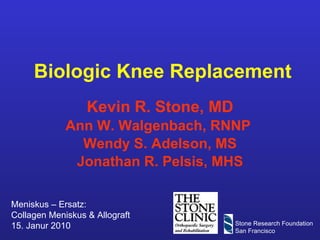
1. Biologic Knee Replacement Kevin R. Stone, MD Ann W. Walgenbach, RNNP Wendy S. Adelson, MS Jonathan R. Pelsis, MHS Meniskus – Ersatz: Collagen Meniskus & Allograft 15. Janur 2010 Stone Research Foundation San Francisco
9. Outerbridge Grading System For Cartilaginous Degeneration Outerbridge RE. The etiology of chondromalacia patellae. J Bone Joint Surg Br, 1961;43: 752-7. Grade I Soft discolored superficial fibrillation Grade II Fragmentation < 1.3 cm 2 Grade III Fragmentation > 1.3 cm 2 Grade IV Erosion to subchondral bone (eburnation)
10.
11.
12.
13.
14. The Three-Tunnel Technique Replacing the Meniscus Stone KR, Walgenbach AW. “Meniscal Allografting: the Three-Tunnel Technique.” Arthroscopy – The Journal of Arthroscopic and Related Surgery. 2003, 19(4):426-30.
102. TA: Meniscus Allograft Placement Preparation of medial meniscal allograft Placement of medial meniscal allograft Relationship of lesion to meniscus A B Movie
103.
104. TA: Revision C A Movie Revision: 8 Months Post-allograft
105. TA: Revision: Operative Images Insertion of Meniscus Allograft with Articular Cartilage Paste Grafting Joint Arthroplasty 3/2006 (38 Mo. Post Op) A B C D
Kevin R. Stone, MD Rath = severe arthritis excluded
Kevin R. Stone, MD
Kevin R. Stone, MD
Kevin R. Stone, MD
Kevin R. Stone, MD
Kevin R. Stone, MD
Kevin R. Stone, MD The procedure step by step.
Kevin R. Stone, MD
Examine the coefficients for each explanatory variable. Positive Coefficient means that the hazard is higher WORSE PROGNOSIS Negative Coefficient implies a lower hazard BETTER PROGNOSIS
Kevin R. Stone, MD Bryan Kelly
Kevin R. Stone, MD
Kevin R. Stone, MD
Kevin R. Stone, MD
Kevin R. Stone, MD
Kevin R. Stone, MD
Kevin R. Stone, MD
Kevin R. Stone, MD
Kevin R. Stone, MD
Kevin R. Stone, MD
Kevin R. Stone, MD
Kevin R. Stone, MD
Kevin R. Stone, MD A= MRI confirming articular cartilage loss of the MFC B= Long leg x-ray demonstrating varus deformity of (L-knee??? I think it should be the Right knee: see x-rays and chart notes ) of about 5-7 degrees C= PA Flexion view demonstrating medial joint space narrowing bialterally L worse than R (nearly bone on bone on the Left). 51 yo ♂ real estate broker both knees w/ problems L worse than R. He has a long hx/o degenerative changes in the medial compartment, loss of the medial meniscus and previous efforts at surgical debridement in order to relieve his medial compartment pain. Pre-operative x-rays revealed medial joint space narrowing and loss of articular cartilage. Pre-operative MRI confirmed loss of the medial meniscus and loss of the artircular cartilage of the medial compartment. He stood in varus. In view of his young age and atheletic activities he requested an effort at biological reconstruction of the medial compartment. 03/10/1999 L-med-Allo/ ArtCart-MFC & MTP/ Open high tib med wedge opening osteotomy using BionX implants and allograft bone/ chon-LFC/ debridement/ Sx: developed a “clicking soreness” on upper MFC thought to be scar tissue requested an effort at operative debridement 03/20/2002 L-knee arthros/ chon-troch/ partial (M)ectomy of Allo where at the posterior 1/3 there was a small flap tear
Kevin R. Stone, MD A= Kissing lesion, MFC, MTP w/ loss of medial meniscus B= Morcellation of the MFC & MTP lesions and loss of medial meniscus
Kevin R. Stone, MD A= Placement of medial meniscal allograft B&C= Articular cartilage paste grafting MFC.
Kevin R. Stone, MD
Kevin R. Stone, MD A= MRI (03/18/02) documenting site of medial meniscus allograft and cartilage paste graft B= Long-leg x-ray (03/14/02) demonstrating post-op alignment C= PA Flexion view (03/14/02) documenting previous osteotomy and preservation of some joint space. 03/14/02 Patient seen 3 years post-op. He noted that before surgery he was unable to do certain activities that he would like to do, and he noted that the knee just pops w/ squatting. He is otherwise quite happy. Px: He had 2 prominent bumps at the medial side of his femoral condyle that he is complaining about. He had patellofemoral crepitus. His pain level is minimal, and his activity level is high. Dx: Arthrofibrosis and bursitis of L-knee. Sx: developed a “clicking soreness” on upper MFC thought to be scar tissue requested an effort at operative debridement 03/20/2002 L-knee arthroscopy/ chond-troch/ partial (M)ectomy of Allo where at the posterior 1/3 there was a small flap tear
Kevin R. Stone, MD
Kevin R. Stone, MD A= Medial meniscus allograft 3 years S/P transplantation B= Medial meniscus allograft 3 years S/P transplantation C= Biopsy MFC 3 years S/P ArtCart
Kevin R. Stone, MD 11-06-2000 R-leg = 4 o varus L leg = 2 o varus Steve Cousins 04-23-2002 R-leg = 5 o varus L leg = 2.5 o varus 39 yo ♂ owner of a “Spicy Sports” company with a long history of injuries playing hockey and lacrosse. Symptoms since 1977 w/ knee locking on one occasion (1982) but spontaneously released without surgery. Eventually came to surgery 1999 but after skiing for 4 months pain recurred. Symptoms at time of xam: R-knee pain, swelling, instability. 11/07/2000 R-med-Allo/ ArtCart- MFC/ Mfx-MTP/ removal bucket-handle tear Developed intermittent anterior knee catching and pain for which HE requested a repeat arthroscopic evaluation and again requested that osteotomy be delayed. Physical exam: lacked final few degrees of extension – excellent flexion and stability. MRI – intact meniscus, damage on the articular cartilage surface, and anterior arthrofibrosis. X-rays- well preserved joint space. 04/02/2002 R-partial med-meniscus/ chondroplasty – trochlea/ debridement
Kevin R. Stone, MD
Kevin R. Stone, MD A= Bucket-handle tear medial meniscus, displacing into the intercondylar notch. B= Bucket-handle tear medial meniscus, displacing into the intercondylar notch.
Kevin R. Stone, MD
Kevin R. Stone, MD Placement of the medial meniscal allograft in relation to ArtCart of MFC The only other picture of this meniscus is washed out and less distinct in demonstrating the implanted meniscus.
Kevin R. Stone, MD R-lat R-med L-med L-lat 11-6-2000 8.31 mm 0.70 mm 3.89 mm 6.91 mm 04-23-2002 7.28 mm 1.83 mm 4.85 mm 6.85 mm
Kevin R. Stone, MD A= Torn posterior medial meniscus B= S/P partial medial meniscectomy Slide “C” is a movie slide – demonstrating the allograft in relation to the healed MFC ArtCart 1.5 years post-op. Developed intermittent anterior knee catching and pain for which HE requested a repeat arthroscopic evaluation and again requested that osteotomy be delayed. Px: lacked final few degrees of extension – excellent flexion and stability. MRI – intact meniscus/ damage on the articular cartilage surface, and anterior arthrofibrosis. X-rays- well preserved joint space. 04/02/2002 R-partial medial meniscectomy/ chondroplasty – trochlea/ debride
Kevin R. Stone, MD A= Torn posterior medial meniscus B= S/P partial medial meniscectomy Slide “C” is a movie slide – demonstrating the allograft in relation to the healed MFC ArtCart 1.5 years post-op. Developed intermittent anterior knee catching and pain for which HE requested a repeat arthroscopic evaluation and again requested that osteotomy be delayed. Px: lacked final few degrees of extension – excellent flexion and stability. MRI – intact meniscus/ damage on the articular cartilage surface, and anterior arthrofibrosis. X-rays- well preserved joint space. 04/02/2002 R-partial medial meniscectomy/ chondroplasty – trochlea/ debride
Kevin R. Stone, MD A= Torn posterior medial meniscus B= S/P partial medial meniscectomy Slide “C” is a movie slide – demonstrating the allograft in relation to the healed MFC ArtCart 1.5 years post-op. Developed intermittent anterior knee catching and pain for which HE requested a repeat arthroscopic evaluation and again requested that osteotomy be delayed. Px: lacked final few degrees of extension – excellent flexion and stability. MRI – intact meniscus/ damage on the articular cartilage surface, and anterior arthrofibrosis. X-rays- well preserved joint space. 04/02/2002 R-partial medial meniscectomy/ chondroplasty – trochlea/ debride
Kevin R. Stone, MD Kevin R. Stone, Biological Knee Reconstruction Annual Joint Preserving Meeting, Johns Hopkins 2004
Kevin R. Stone, MD Kevin R. Stone, Biological Knee Reconstruction Annual Joint Preserving Meeting, Johns Hopkins 2004
Kevin R. Stone, MD Kevin R. Stone, Biological Knee Reconstruction Annual Joint Preserving Meeting, Johns Hopkins 2004
Kevin R. Stone, MD Kevin R. Stone, Biological Knee Reconstruction Annual Joint Preserving Meeting, Johns Hopkins 2004
Kevin R. Stone, MD Kevin R. Stone, Biological Knee Reconstruction Annual Joint Preserving Meeting, Johns Hopkins 2004
Kevin R. Stone, MD Rhonda Topple
Kevin R. Stone, MD Kevin R. Stone, Biological Knee Reconstruction Annual Joint Preserving Meeting, Johns Hopkins 2004 RT
Kevin R. Stone, MD Kevin R. Stone, Biological Knee Reconstruction Annual Joint Preserving Meeting, Johns Hopkins 2004 RT
Kevin R. Stone, MD Kevin R. Stone, Biological Knee Reconstruction Annual Joint Preserving Meeting, Johns Hopkins 2004 RT
Kevin R. Stone, MD Kevin R. Stone, Biological Knee Reconstruction Annual Joint Preserving Meeting, Johns Hopkins 2004 RT
Kevin R. Stone, MD Kevin R. Stone, Biological Knee Reconstruction Annual Joint Preserving Meeting, Johns Hopkins 2004 RT
Kevin R. Stone, MD Kevin R. Stone, Biological Knee Reconstruction Annual Joint Preserving Meeting, Johns Hopkins 2004 RT
Kevin R. Stone, MD
Kevin R. Stone, MD
Kevin R. Stone, MD
Kevin R. Stone, MD
Kevin R. Stone, MD
Kevin R. Stone, MD
Kevin R. Stone, MD
Kevin R. Stone, MD
Kevin R. Stone, MD
Kevin R. Stone, MD
Kevin R. Stone, MD Test
Kevin R. Stone, MD TEST
Kevin R. Stone, MD Tracy Achiles A= MRI demonstrating full-thickness MFC defect B= MRI demonstrating loss of medial meniscus C= MRI demonstrating loss of articular cartilage 48 yo ♀ fitness manager and triathlon/ iron-man competitor who tripped over her dog while walking it down the driveway landing on both knees. Subsequently saw local orthop surgeon who found a R-med meniscal tear and she underwent partial (M)ectomy Apr 27, 2001. Able to return to running but Sx pain/ swelling recurred. 2 nd Surgery Jan 2002 04/02/2002 – Right knee Surg: R-med-Allo/ R-MFC-ArtCart/ R-MFC|MTP|-Mfx/ Chon – troch Two weeks post-op she swam in a pool for two hours with her legs kicking and developed immediate swelling. It was presumed that she most likely had re-torn her meniscus allograft. However, she was treated conservatively to see whether or not it would heal on its own. It failed to do so. She had recurrent swelling w/ activities and not responsive to a single effort of cortisone injection . 06/26/2002 - Right knee Surg: R- med-Allo repair/ Mfx-MFC /Chon -MTP
Kevin R. Stone, MD
Kevin R. Stone, MD A= Full thickness chondral defect MFC and loss of medial meniscus B= Full thickness chondral defect MFC and loss of medial meniscus
Kevin R. Stone, MD A= Preparation and placement of medial meniscal allograft B= Placement of medial meniscal allograft Slide “C” is a movie clip – demonstrates relationship of lesion to meniscus
Kevin R. Stone, MD A= Retained medial meniscal allograft. C= Refixation of medial meniscal allograft. Repair of the “unstable junction of meniscal capsule w/ medial meniscus allograft. Slide “B*” is a movie - demonstrates the instability of the junction of the junction of the capsule w/ the allograft
Kevin R. Stone, MD A= Retained medial meniscal allograft. C= Refixation of medial meniscal allograft. Repair of the “unstable junction of meniscal capsule w/ medial meniscus allograft. Slide “B*” is a movie - demonstrates the instability of the junction of the junction of the capsule w/ the allograft
Kevin R. Stone, MD A= Retained medial meniscal allograft. C= Refixation of medial meniscal allograft. Repair of the “unstable junction of meniscal capsule w/ medial meniscus allograft. Slide “B*” is a movie - demonstrates the instability of the junction of the junction of the capsule w/ the allograft