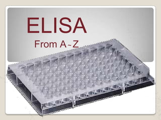
4. What is immunoassay?
The term “immunoassay” is a
combined term of “immuno”(=
immunological, practically
immunochemical antigen-antibodyreaction) and “assay” (=
determination of the purity of a
substance or the amount of any
constituent of a mixture.
5. 1. Antigen/antibody of interest is absorbed on
to plastic surface („sorbent‟).
2. Antigen is recognised by specific antibody
(„immuno‟).
3. This antibody is recognised by second
antibody („immuno‟) which has enzyme
attached („enzyme-linked‟).
4. Substrate reacts with enzyme to produce
product, usually coloured.
9. 1-Antibody (antiserum)
Antibody: proteins produced by the immune
system which help defend against antigens
SYMBOL FOR
ANTIBODY
The variable regions are though
to be the place for recognition
and binding with the antigen.
10. 2-Antigen
Any molecule that induces production of antibodies
when introduced in the body is called antigen.
OR
Any “thing”, foreign to the immune system. e.g.
bacteria, viruses, (or their parts), pollen, etc.
SYMBOL FOR ANTIGEN
11. abeling materials
In immunoassay, it is necessary to use any
marker to know the antigen-antibody
binding. For such purpose, we label either
antigen or antibody with some materials
that do not interefere with the binding.
e.g:horseradishperoxidase enzyme
substrate: trimethylbenzidine
17. Advantages of ELISA
Reagents are relatively cheap & have a long
shelf life
ELISA is highly specific and sensitive
No radiation hazards occur during labelling or
disposal of waste.
Easy to perform and quick procedures
Equipment can be inexpensive and widely
available.
ELISA can be used to a variety of infections.
18. Disadvantages of ELISA
Measurement of enzyme activity can be more complex
than measurement of activity of some type of
radioisotopes.
Enzyme activity may be affected by plasma
constituents.
Kits are commercially available, but not cheap
Very specific to a particular antigen. Won’t recognize
any other antigen
False positives/negatives possible, especially with
mutated/altered antigen
21. The direct detection method uses a labeled primary
antibody that reacts directly with the antigen. Direct
detection can be performed with antigen that is directly
immobilized on the assay plate . Direct detection is not
widely used in ELISA but is quite common for
immunohistochemical staining of tissues and cells.
22. The indirect ELISA utilizes an unlabeled
primary antibody in conjunction with a
labeled secondary antibody.The
secondary antibody has specificity for
the primary antibody
23.
24. The sandwich measures the amount of antigen between two
layers of antibodies.
Sandwich are especially useful if the concentration of
antigens is low or they are contained in a mix of high
concentrations of contaminating protein
To utilize this assay, one antibody (capture) is bound to a
microtiter plate well. Antigen is then added and bound to the
antibody. Unbound products are then removed, and 2ry
antibody is added (detection), then add the 3rd labeled
antibody to complete the sandwich
Major advantages of this technique are that the antigen does
not need to be purified prior to use, due to its high specificity.
25.
26. In this Unlabeled antibody is incubated in the
presence of its antigen. These bound
antibody/antigen complexes are then added to
an antigen coated well. The plate is washed
unbound antibody is removed. The secondary
antibody, specific to the primary antibody is
added. This second antibody is coupled to the
enzyme. A substrate is added, and remaining
enzymes elicit a chromogenic or fluorescent
signal. For competitive ELISA, the higher the
original antigen concentration, the weaker the
eventual signal.
27.
28. A newer technique uses an solid phase made up of an
immuno-sorbent polystyrene rod with 8-12 protruding
ogives.
The entire device is immersed in a test tube containing
the collected sample and the following steps (washing,
incubation in conjugate and incubation in chromogenous
) are carried out by dipping the ogives in microwells of
standard microplates pre-filled with reagents
31. APPLICATIONS
:
1-HIV-1 and HIV-2 (presence of anti-HIV antibodies).
hepatitis C (presence of antibodies).
2-hepatitis B (testing for both antibodies and a viral
antigen) .
3-Measuring hormone levels HCG (as a test for
pregnancy).
4-LH (determining the time of ovulation). TSH, T3
and T4 (for thyroid function).
32.
33.
34. 1. Coating of Wells with Antibody
100 μL of antibody diluted in buffer is added to each well.
Cover the plate and incubate at 4 °C overnight.
2. Washing
wash manually 3 times as follows:Empty the plate by inversion over a
sink. Tap the inverted plate against some layers of soft paper tissue to
remove residual liquid. Wash the plate by filling the wells by
immersion in buffer B. Leave on the table for 3 minutes. Empty the
plate as described above and repeat washing two more times.
35. 2-A concentrated solution of non-interacting protein, such as bovine
serum albumin (BSA) or casein, is added to all plate wells.This step
is known as blocking,because the serum proteins block nonspecific
adsorption of other proteins to the plate.
36. 3. Incubation with Test Samples.
100 μL of test sample or standard diluted in buffer is added
per well.
Cover the plate and incubate at room temperature for 2
hours.
4. Wash as described in step 2.
5. Incubation with enzyme- Conjugated Antibody.
100 μL of enzyme-conjugated antibody diluted in buffer is
added to each well.
Cover the plate and incubate at room temperature for 1 hour.
The enzyme-conjugated antibody should be directed against
the antigen to be determined.
37. 6. Wash as described in step 2.
7. Colour Development
100 μL of chromogenic substrate is added to each well.
Cover the plate and incubate for 15 minutes, or until a suitable
colour has developed. The plate should preferably be
protected against light during this incubation.
8. Stopping the Colour Development
Stop the reaction by adding 100 μL 0.5 M H2SO4 to each well.
9. Reading of Results
Read results directly through the bottom of the microwell
plate using an automated or semiautomated
photometer (ELISA-reader). The subtraction of the
absorbance at a reference wavelength (between 620 and
650 nm) is recommended.
45. 7-HOW TO TREAT WITH THE REAGENTS?
Use reservoir for each
reagent
Label the reservoi
46. Don‟t use the same
reservoir for multiple
regents
Don‟t return the
reagents to the stock
47. 8. Shaking of the well-plate
for mixing
Place the plate on the flat and smooth surface
of a laboratory table, hold the plate and move
the plate roundly to draw circles rapidly for
approx. 10 seconds while lightly pressing the
plate on the surface. Repeat 3 times.
48. Important points in
performing ELISA and
improvement of assay
performance
1-Sample treatment.
3-Stability of assay samples.
2-Infleunce of humidity and air stream.
49. Important points in performing ELISA and
improvement of assay performance
1. Sampling and treatments of samples
Serum or plasma
50. sampling
In general, we recommend using
When getting
heparin is most often used as an anticoagulant
Use of fluoride must be avoided because fluoride
ion is a potent inhibitor of peroxidase.
51. An important phenomenon with frozen plasma is
that an insoluble substance (fibrin) will be formed
when thawed. In this case, the sample must be
mixed and centrifuged, then the insoluble cluster
flowing in the plasma should be taken out by a thin
wire needle sharply bent at an end. If such fibrin
remains in the sample, it may clog the tip of a
pipette and influences assay variability
53. pH Of the sample
Serum or plasma, when fresh, shows
pH near neutral, however, it very
quickly goes to alkaline more than pH 8
by losing CO2.
In alkaline pH, the antigenantibody reaction is
interfered. resulting in
cancellation of the assay or
giving inaccurate assay values.
54. Storage temperature and freezingthawing.
Sample storage temperature is better to be lower
than -35 C. Ultra-low temperature such as -80 C is
recommended for a long-term storage.
Repeated freezing and thawing is also harmful to
the protein, and may cause inactivation.
55. When samples
are taken out
from the freezer
and thawed,
never forget to
mix these
samples because
the solution after
thawing is not
homogeneous,
and the bottom
area contains
more solute
56. 2-Stability of assay samples.
In assay, the problem of sample stability, i.e. how long the
substance to be measured can keep its immunoreactivity,
in serum or plasma, is very important. Blood samples also
contain enzymes to destroy peptides or proteins, and
stability against those enzymes differs from substance to
substance.
Freezer of –20C is not trustable for the constancy of
temperature but use of a freezer of –35 C or lower
temperature is recommended.
58. 3-infeluence of humidity and air stream
During all the incubation process, the well-plate
should be covered using the attached plate cover.
Plate cover is effective only under the most suitable
condition, i.e. room temperature, humidity more than
50%, and air stream of less than 0.2m/sec.
N.B
It is recommend to get a small
semi-transparent plastic box, and put moistened
paper towel on the bottom .
60. 1-Poor or no coloration after the last step
1) The standard or samples might not be added.
2) Reagents necessary for coloration might not be added.
3) Wrong reagents related to coloration might have been
added.
4) Influence of the temperature under which the kits
had been stored.
5) Excessive hard washing of the well plate.
61. 2)The standard curve obtained was not smooth.
There might be some mistake in the serial dilution of
the original standard solution.
3)Flat standard curve.
Standard solutions are not added.
62. 4-Big variation between two wells in duplicated assay was
observed.
1) Scratching the bottom of the well by aspirator tip
during aspiration of washing buffer.
2) Scratching the bottom of the well by pipette tip
during addition of standards, samples, or reagents.
3)Assay might be started while the well-plate was still
cooler than room temperature.
63. 4) Air stream, warmer or cooler than room temperature
5) Air stream from air conditioner or other instruments
might dry wells.
6) Insufficient removal of washing buffer from the wells
might dilute reagent solution added in the following step
of the procedure.
7)Big variation would be obtained if the sample is not
homogeneous.
64. Shapes of standard curves depending on scales
in X-and Y-axes.
Standard curve of ELISA prepared by plotting
standard concentration on X-axis and
absorbance on Y-axis, both in normal scale,
looks like a linear line except for lower
concentration area.