1. So What’s in a Hand? : A Multimodality
Pictorial Review
of Congenital Hand Anomalies
Crapp SJ, Kan JH, Martus JE
Educational Goals/Teaching Points
•Discuss imaging approach to a variety of congenital hand anomalies
Abstract •Review the imaging findings of congenital hand anomalies and associated
syndromes when relevant
When children with idiopathic or congenital anomalies of the hand are
referred for imaging, it is important for the radiologist to succinctly and
•Provide relevant orthopaedic surgical perspective
accurately describe these findings, as well as have a basic understanding
of the clinical or surgical significance of these findings. Congenital
anomalies of the hand have been historically classified by systems
adopted by the International Federation of Societies for Surgery of the
Hand (IFSSH) based on Swanson’s original classification system which
was first proposed in 1964. As the molecular pathogenesis as a basis for
development of these anomalies has become more clear in recent
years, newer classification systems based on the dysmorphology of each
entity have been proposed. (1) Although many types of congenital hand B
anomalies occur in isolation, up to one fifth of encountered anomalies B
have an association with an identifiable syndrome. (2) This pictorial A
review will illustrate various common and rare anomalies of the hand
C D
including but not limited to brachydactyly, longitudinal epiphyseal
bracket
deformity, symphalangism, syndactyly, polydactyly, syndactyly, and
clubhand. Orthopaedic surgical perspective of these findings will also be A C
provided when relevant.
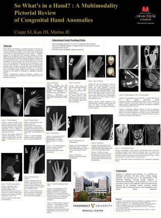
Figure 7. Apert’s Syndrome.
Figure 5. Phocomelia. Figure 6. Phocomelia
Findings: PA hand (A) Syndactyly of right 2nd
Findings: Absence of the
ulna, anterior displaced radius and Findings: Absent radius and through 4th rays. Aplasia/hypoplasia of 2nd-5th
single rudimentary digit. Most ulna with single digit emanating middle and distal phalanges. Post-axial
commonly associated with from the right upper polysyndactyly with duplication beginning at the
thalidomide embryopathy but can be extremity, with dysplasia of the level of the middle and distal phalanx. Delta
sporadic or associated w/ phalanx of the proximal phalanx of the first digit.
TAR, Roberts and Grebe syndromes distal humerus.
Surgical: Prosthetic fitting may 3D CT skull (B&C) Bony synostosis of the
Surgical: Observation is coronal sutures bilaterally with a brachycephalic Figure 12. Bracket epiphysis of the 1st proximal phalanx.
recommended as this child will adapt be pursued however many
effectively to the differences of this children will prefer use of their skull. Consistent with clinical features of Apert’s
limb. own sensate limb. This child is (not genetically proven). Findings: PA radiograph(A) and Coronal GRE, T1 and PD MRI (B,C &
able to manipulate objects with D). Most common associations include
the remaining digit. Surgical: Priorities include correction of the syndactyly, polydactyly, symphalangism, clubfoot, Apert's syndrome, &
thumb deformity, creation of a functional first web Poland's syndrome. Curved physis (green arrows) of the 1st proximal
space, and release of finger syndactylies / phalanx makes a C-shaped band extending and surrounding the metaphyses
synostosis. Ablation of the postaxial polydactyly with resultant deformity of the bone.
may be considered.
Surgical: Longitudinal epiphyseal bracket can lead to progressive
deformity via asymmetric growth. Resection of the abnormal central
physis with fat graft interposition can allow for more normal growth.
Transverse corrective osteotomy may be combined with bracket excision to
provide acute correction of the deformity.
Figure 1. Thumb hypoplasia. Figure 2. Radial deficiency.
Findings: Complete absence of the Findings: Complete absence of the
thumb in a patient with Type V thumb radius in patient with
hypoplasia and genetically confirmed thrombocytopenia absent radius
Holt-Oram Syndrome. Note the mild (TAR) syndrome. Note the radial
clinodactyly of the 2nd through 5th angulation of the hand at the level of
digits and radial deviation of the hand the wrist and near normal appearance
at the level of the wrist. of the thumb, a unique finding seen in
TAR. Figure 8. Brachydactyly.
Surgical: Index finger pollicization
would be recommended. This is a Surgical: A carpal centralization Findings: Foreshortening of the 4th
complex procedure where the index procedure would be considered to digit and deformed 4th and 5th
finger is reconstructed to act as a improve function and appearance. middle phalanges. The middle
thumb. phalanges of the 2nd -5th rays are Figure 9. Brachydactyly and
short. symphalangism.
Figure 10. Polydactyly.
Findings: Fusion of the capitate and Findings: Preaxial polydactyly with A B C
Surgical: 4th /5th
middle phalangeal
osteotomy could be considered. hamate bones. Short 1st MC bone. hypoplastic radial digit which has two
Absent middle phalanges from the 2nd ossification centers and articulates with Figure 13. Lunotriquetral coalition.
Otherwise, if the hand is
functional, observation would be -5th rays & clinodactyly of the 5th a bifid 1st MC head (yellow arrow). Findings: PA radiograph (A) and coronal T1 & PD FS MRI (B & C) of the hand.
recommended. finger. Surgical: This Wassel 4 thumb Incomplete separation of the lunate and triquetral bones in a patient with
duplication would require ablation of lunotriquetral coalition. This is the most common carpal coalition. Common
Surgical: If the hand is the radial digit, reconstruction of the associated syndromes include Ellis-van Crevald, Holt-Oram and Turner. Occurs
functional, observation would be lateral collateral ligament of the MCP more commonly in females and those of African descent.
recommended. joint, and repair of the insertion of the
Surgical: Observation would be recommended for this asymptomatic radiographic
thumb intrinsics.
finding.
Conclusion
Imaging of congenital hand anomalies is a challenge for
radiologists unfamiliar with these entities. Various imaging
Figure 4. Constriction band
syndrome. modalities and techniques aid in the determination of the
underlying pathology allowing accurate diagnosis. A
Findings: Amputation of the 2nd
through 5th rays at the level of the
fundamental understanding of these anomalies as well as their
Figure 3. Amniotic band syndrome. proximal middle phalanges with Figure 11. Isolated Triphalangeal thumb associated syndromes when applicable, allows early and accurate
Findings: Amputation of the 1st
multiple soft tissue constrictions (blue (TPT). detection by the radiologist, thereby providing valuable
arrows). Syndactyly of the 3rd and 4th diagnostic data to referring physicians in the management of this
through 4th digits at the level
rays. The thumb is intact. Findings: Extra middle phalanx of the unique patient population.
proximal phalanges with
Surgical: Constriction ring release thumb. The middle phalanx may be
characteristic soft tissue constriction
may be considered if of functional triangular, trapezoidal, or rectangular.
(red arrows) related to amniotic band
and cosmetic benefit. Syndactyly Isolated TPT occurs in opposable & non-
syndrome. Note soft tissue syndactyly
opposable forms. TPT has associations with
between the 3rd and 4th rays. release would be recommended.
a number of syndromes including many
Surgical: Syndactyly release would with hand & foot anomalies and AD
References:
be recommended. inheritance.
1. Oberg et al, “Developmental Biology and Classification of Congenital Anomalies of
Surgical: Priorities include 1) adequate 1st the Hand and Upper Extremity”, J Hand Surgery (2010) 35A:2066–2076.
2. Watts, A.C., Hooper, G., “(iii) Congenital hand anomalies” from Mini-Symposium:
web space → web deepening 2) lack of
Children’s Orthopedic Surgery in Current Orthopaedics (2006) 20:266–273
opposition → tendon transfer to restore 3. Linder et al, “Congenital Anomalies of the Hand: An Overview”, J Craniofacial
opposition 3) angular deformity → Surgery (2009) 20:999-1004
osteotomy and / or fusion of the abnormal 4. Chavan et al, “Twenty classic hand radiographs that lead to diagnosis.” Pediatric
middle phalanx to the proximal or distal Radiology (2010) 40:747–761
phalanx.