Compact, High-Frequency Tablet Ultrasound for Small Animal Imaging
•Als PPTX, PDF herunterladen•
0 gefällt mir•143 views

Hands-on demonstration of the Prospect T1, high-frequency ultrasound system During this live demonstration we demonstrated the animal preparation steps and positioning for both mice and rats on the Prospect T1. We also showed the following cardiac views on both species: Long Axis, in B-Mode Short Axis, in B-Mode and M-Mode Pulmonary Artery, in B-Mode, Color Doppler, Pulsed Wave Doppler Apical 4-Chamber to Measure Mitral Valve Flow, in B-Mode, Color Doppler, Pulsed Wave Doppler, and Tissue Doppler We also examined the aortic arch, carotid arteries, abdominal aorta, and other abdominal organs throughout the demonstration.

Empfohlen
Empfohlen
Weitere ähnliche Inhalte
Ähnlich wie Compact, High-Frequency Tablet Ultrasound for Small Animal Imaging
Ähnlich wie Compact, High-Frequency Tablet Ultrasound for Small Animal Imaging (20)
Mehr von Scintica Instrumentation
Mehr von Scintica Instrumentation (20)
Kürzlich hochgeladen
Kürzlich hochgeladen (20)
Compact, High-Frequency Tablet Ultrasound for Small Animal Imaging
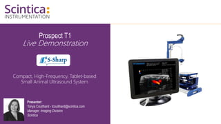
- 1. Prospect T1 Live Demonstration Compact, High-Frequency, Tablet-based Small Animal Ultrasound System Presenter: Tonya Coulthard - tcoulthard@scintica.com Manager, Imaging Division Scintica
- 2. WWW.SCINTICA.COM Topics of Discussion • Prospect T1 System Overview • Pre-Recorded System Demonstration • Example Images and Sample Measurements 2
- 3. WWW.SCINTICA.COM Prospect T1 System Overview • System components and standard configuration • Add-on hardware and software components 3
- 4. WWW.SCINTICA.COM Prospect T1 System Components • The Prospect T1 is the first tablet based high-frequency ultrasound system specifically designed for pre-clinical imaging of small animals • System components: • Tablet • Probe • Scanning Platform 4
- 5. WWW.SCINTICA.COM Prospect T1 System Components: Tablet • The powerful tablet reduces the footprint of the system, taking up less lab space, making it easy to move when necessary • The intuitive workflow and touch screen allow researchers to start acquire images and generating data quickly • Multiple data formats exist for either still, cine loop, or RAW data storage • Offline software analysis is possible to preserve time on the system for imaging 5
- 6. WWW.SCINTICA.COM Prospect T1 System Components: Probes • Three single element probes are available: • 20 MHz (user selectable between 15-30 MHz) • Primarily used for rat imaging, as well as harmonic contrast imaging • 40 MHz (user selectable between 30-50 MHz) • Primarily used for mouse imaging, and superficial anatomical targets in larger species like rats • 50 MHz (user selectable between 30-50 MHz) • Primarily used for superficial anatomical targets in both mice and rats 6
- 7. WWW.SCINTICA.COM Prospect T1 System Components: Scanning Platform • The platform is compact in design, again to limit the footprint of the system • The scanning platform has been designed for ergonomical positioning of the probe • Animal beds have integrated heating, and ECG and respiratory monitoring • Interchangeable beds are available for mice or rats • Animal beds can be precisely adjusted in the X, Y, and Z axis 7
- 8. WWW.SCINTICA.COM Standard System Configuration • Standard system configuration for mouse • B-Mode • M-Mode • Pulsed Wave / Color / Power / Tissue Doppler Mode • Contrast Mode • Comprehensive Measurement and Analysis Tools • Scanning Platform – with mouse bed • 40 MHz probe • Standard system configuration for rat • B-Mode • M-Mode • Pulsed Wave / Color / Power / Tissue Doppler Mode • Contrast Mode • Comprehensive Measurement and Analysis Tools • Scanning Platform – with rat bed • 20 MHz probe 8
- 9. WWW.SCINTICA.COM Add-On Hardware: 3D Motor • The 3D motor expands the capabilities of the Prospect T1 to acquire 3D B- mode images • Add-on includes the software analysis package to view the 3D images and perform volume calculations 9
- 10. WWW.SCINTICA.COM Add-On Hardware: Image Guided Needle Injection • The image guided needle injection mount integrates with probe • Injections may be performed with a regular syringe and steel needle, or pulled glass capillary needle • Injections may be made into developing embryos, adult myocardium, or abdominal/muscle targets 10 E15.5 mouse embryo Adult mouse myocardium
- 11. WWW.SCINTICA.COM Add-On Hardware: Shear Wave Elastography • Shear wave elastography is used to quantify mechanical and elastic properties of tissues • The acoustic radiation force is generated by a push probe mounted on the side of the imaging probe • The software analysis generates a colored elastogram which is overlaid on a B-mode image 11
- 12. WWW.SCINTICA.COM Add-On Hardware: Integrated Sonoporation • Sonoporation is the controlled cavitation or bursting of microbubbles with the intention of increasing the permeability of the cell membrane or to open to blood brain barrier • Sonoporation is performed by a secondary, non-imaging, probe directed at the anatomical target • Software integration and control of the sonoporation probe is included with this add-on 12
- 13. WWW.SCINTICA.COM Key Research Applications • Cardiovascular Research • Cancer Biology • Abdominal & Anatomical Imaging • Developmental Biology • Ophthalmology • Other Animal Models – Zebrafish, Chick Embryos 13
- 14. 14 • Cardiovascular Research • Systolic function • Diastolic function • Left Ventricle, Right Ventricle • Aorta, Pulmonary Artery • Mitral Valve, Tricuspid Valve • Peripheral Vessels • Image Guided Needle Injections
- 15. WWW.SCINTICA.COM Cardiovascular Research: Mouse Systolic Function B-Mode 15 Long Axis View IVS LV LVPW AO LA PM Mitral valve Short Axis View IVS LV LVPW LVAW PM LV : left ventricle LVAW: left ventricular anterior wall LVPW : left ventricular posterior wall PM: papillary muscle VS : interventricular septum AO : aortic orifice LA : left atrium
- 16. WWW.SCINTICA.COM Cardiovascular Research: Mouse Systolic Function ECG Gated Kilohertz Visualization (EKV) Mode 16 B-Mode Frame Rate = 30fps ECG Gated Kilohertz Visualization = 30fps
- 17. WWW.SCINTICA.COM Cardiovascular Research: Mouse Systolic Function B-Mode; Area Length Measurement • End diastolic; End systolic volume • Stroke volume • Ejection fraction • Fractional area change (from short axis) • Fractional shortening • Left ventricular mass • Left ventricular mass index 17
- 18. WWW.SCINTICA.COM Cardiovascular Research: Mouse Systolic Function B-Mode; LV Volume Integral Technique • End diastolic; End systolic volume • Stroke volume • Ejection fraction 18
- 19. WWW.SCINTICA.COM Cardiovascular Research: Mouse Systolic Function B-Mode; Teichholz Formula • Can be done on either the long or short axis B-mode image • LV mass • LV mass index • Fractional shortening • End diastolic volume; end systolic volume • Stroke volume • Ejection fraction 19
- 20. WWW.SCINTICA.COM Cardiovascular Research: Mouse Systolic Function B-Mode; Modified Simpson’s Rule • Fractional shortening • End diastolic volume; end systolic volume • Stroke volume • Ejection fraction • Fractional area change 20
- 21. WWW.SCINTICA.COM Cardiovascular Research: Mouse Systolic Function M-Mode 21
- 22. WWW.SCINTICA.COM Cardiovascular Research: Mouse Systolic Function M-Mode • Can be done on either the long or short axis M-mode image • LV mass • LV mass index • Fractional shortening • End diastolic volume; end systolic volume • Stroke volume • Ejection fraction • Cardiac output 22
- 23. WWW.SCINTICA.COM Cardiovascular Research: Mouse Systolic Function B-Mode and PW Doppler – Ascending Aorta • Stroke Volume is calculated as a function of the Velocity Time Interval (VTI) and vessel diameter • Cardiac Output is simply stroke volume x heart rate • VTI can be manually or automatically traced on the PW Doppler: • Peak velocity • Peak pressure gradient • Mean velocity • Mean pressure gradient • Acceleration & Deceleration 23
- 24. WWW.SCINTICA.COM Cardiovascular Research: Mouse Systolic Function B-Mode and PW Doppler – Pulmonary Artery • Stroke Volume is calculated as a function of the Velocity Time Interval (VTI) and vessel diameter • Cardiac Output is simply stroke volume x heart rate • VTI can be manually or automatically traced on the PW Doppler: • Peak velocity • Peak pressure gradient • Mean velocity • Mean pressure gradient • Acceleration & Deceleration 24
- 25. WWW.SCINTICA.COM Cardiovascular Research: Mouse Diastolic Function Color and PW Doppler 25 Mitral Valve Tricuspid Valve LV RV LA RA MV TV LV : left ventricle RV: right ventricle LA: left atrium RA: right atrium MV: mitral valve TV: tricuspid valve
- 26. WWW.SCINTICA.COM Cardiovascular Research: Mouse Diastolic Function Color and PW Doppler • Acceleration rate of E wave • Peak velocity of E & A waves • Deceleration time of E wave • E:A ratio • Isovolumic relaxation/contraction time (IVRT & IVCT) • Ejection time • Myocardial performance index (Tei index) 26
- 27. WWW.SCINTICA.COM Cardiovascular Research: Mouse Diastolic Function Tissue Doppler • Peak velocity of E & A waves • Isovolumic relaxation/contraction time (IVRT & IVCT) • Ejection time • Filling time 27
- 28. WWW.SCINTICA.COM Cardiovascular Research: Mouse Aortic Arch 28 RPA IA LCCA LSCA AAr AAr AAo IA LCCA LSCA AAr : Aortic Arch RPA : Right Pulmonary Artery IA: Innominate Artery LCCA : Left Common Carotid Artery LSCA : Left Subclavian Artery
- 29. WWW.SCINTICA.COM Cardiovascular Research: Mouse Carotid Arteries B-Mode 29
- 30. WWW.SCINTICA.COM Cardiovascular Research: Mouse Carotid Arteries M-Mode, Color and PW Doppler 30 Vessel diameter through systole and diastole may be measured using M-mode. Color Doppler is used to visualize direction of flow within vessels. Aliasing shows highest velocity flow at bifurcation of carotid artery. Resistive and pulsatility indices may be measured on peripheral vessels using PW Doppler.
- 31. WWW.SCINTICA.COM Cardiovascular Research: Rat Systolic Function B-Mode 31 380g rat 600g rat
- 32. WWW.SCINTICA.COM Cardiovascular Research: Rat Carotid Arteries B-Mode 32
- 33. WWW.SCINTICA.COM Cardiovascular Research: Image Guided Needle Injection – Adult Mouse Myocardium • Image guided injection may be done into the myocardium or other anatomical target • Stem cells or other therapy may be injected into the myocardium to study the effect on myocardial infarction lesion size, for example 33
- 34. 34 • Cancer Biology • Tumor detection • 3D volume measurements • Surrounding tissue investigation – i.e. lymph nodes • Blood flow monitoring • Power Doppler • Linear contrast agent imaging • Non-linear contrast agent imaging
- 35. WWW.SCINTICA.COM Cancer Research – Preclinical Solid Tumor Models • A number of different types of solid tumor models are used commonly in preclinical research: • Cell line-derived models • Patient Derived Xenograft (PDX) models • Environmentally induced models • Genetically Engineered Mouse (GEM) models 35 Figure from Gengenbacher et al. Nature Reviews Cancer (2017) 17:751-765.
- 36. WWW.SCINTICA.COM Cancer Research – Tumor Detection and Monitoring • Tumors of all types are visible, whether subcutaneous or orthotopic • Standard 2D B-mode imaging is used to provide a greyscale image, tumors often show up as a different echogenicity than the surrounding tissue • 2D measurements can be done to measure linear or area measurements of tumor size 36 Subcutaneous tumor model Transgenic liver tumor model
- 37. WWW.SCINTICA.COM Cancer Research – Tumor Detection and Monitoring • Complex tumor models can also be investigated using ultrasound • Normal tissues must be identified, followed by the identification of abnormal tissues • Changes in nearby tissues may also be investigated 37 IP injection of ovarian tumor cells (SKOV-3) Tumour Stomach Kidney Splenic Vein Tumour Intestine
- 38. WWW.SCINTICA.COM Cancer Research – 3D Tumor Volume Measurements • 3D volume measurements can be made on any visualized tumor using the 3D motor add-on • Volume measurements may be used to follow the same tumor over a longitudinal study to monitor tumor progression or therapeutic response 38 Orthotopic Mammary Fat Pad Tumor (MDA-MB-231) Volume = 263mm3
- 39. WWW.SCINTICA.COM Cancer Research – 3D Tumor Volume Measurements • Complex tumor structures can be visualized in 3D • Longitudinal imaging to monitor tumor progression or therapeutic response 39 IP injection of ovarian tumor cells (SKOV-3) Tumor
- 40. WWW.SCINTICA.COM Cancer Research – 3D Tumor Volume Measurements • Due to the complex nature of this tumor model the images were analyzed in VivoQuant • Red Tumor = 4.7mm3 • Green Tumor = 16.0mm3 40 IP injection of ovarian tumor cells (SKOV-3)
- 41. WWW.SCINTICA.COM Cancer Research – Surrounding Tissue Investigation • Surrounding tissues may be investigated, as can other tissues and organs which may be affected • Lymph nodes may show signs of involvement, and may indicated spread of the disease • The spleen may be involved and show an altered appearance or larger size 41 Lymph Node Skeletal Muscle Spleen
- 42. WWW.SCINTICA.COM Cancer Research – Blood Flow Monitoring Power Doppler • Surrounding tissues may be investigated, as can other tissues and organs which may be affected • Lymph nodes may show signs of involvement, and may indicated spread of the disease • The spleen may be involved and show an altered appearance or larger size 42
- 43. WWW.SCINTICA.COM Cancer Research – Blood Flow Monitoring Linear Contrast Agent Imaging • Microbubble contrast agents are injected i.v. to study microvascular perfusion • Microbubbles are typically 2-3µm in diameter, and mimic red blood cells when they are non-targeted; may also be targeted to bind to biomarkers • Linear contrast agent imaging uses reference subtraction to create a green overlay, and is available on all probes 43
- 44. WWW.SCINTICA.COM Cancer Research – Blood Flow Monitoring Non-Linear (Harmonic) Contrast Agent Imaging • Harmonic imaging is used to acquire a more specific signal coming from the microbubbles • Using only the 20MHz probe, the system will transmit at 20MHz, but listen for the 1st harmonic – 40MHz. In doing this, tissue signal is removed, and only microbubbles are visualized • Time vs. Intensity curves can be created for numerous ROIs in both modes 44
- 45. 45 • Abdominal & Anatomical Imaging • Organ visualization • 3D volume measurements • Surrounding tissue investigation, including visualization of oedema • Blood flow monitoring • PW Doppler • Color & Power Doppler • Linear contrast agent imaging • Non-linear contrast agent imaging
- 46. WWW.SCINTICA.COM Abdominal Imaging – Liver and Gallbladder (Mouse) 46 Liver Liver vessels Gallbladder
- 47. WWW.SCINTICA.COM Abdominal Imaging – Spleen and Pancreas (Mouse) 47 Spleen Splenic vein Pancreas Spleen Kidney
- 48. WWW.SCINTICA.COM Abdominal Imaging – Kidney (Mouse) 48 Kidney
- 49. WWW.SCINTICA.COM Abdominal Imaging – Uterine Horn and Testis (Mouse) 49 Bladder Right Horn Left Horn Testis Caput Epididymis
- 50. 50 • Developmental Biology • Confirmation of pregnancy • Embryo counting • Monitoring embryonic developmental stages • Monitoring cardiac function and flow within the embryos • Placental and umbilical cord flow measurements • Image guided needles injections into externalized embryos
- 51. WWW.SCINTICA.COM Developmental Biology – Development (Mouse) 51 Embryos_E7.5 Embryonic brain_E12.5 Embryonic spinal cord_E12.5 Embryonic head and forelimb_E14.5 Embryonic heart and neural tube _E9.5
- 52. WWW.SCINTICA.COM Developmental Biology – Cardiac Function in Pups (Mouse) 52 Mitral Valve Umbilical cord Dorsal Aorta
- 53. WWW.SCINTICA.COM Developmental Biology – Image Guided Injection (Mouse) 53 • Image guided injection may be done into a variety of anatomical targets within the embryo • The uterine horn is exposed from the dame and injections done into the exposed embryos
- 54. 54 • Other Animal Models • Same types of analysis can be completed with all of the standard modes, as long as the ultrasound signal can penetrate the imaging subject • Examples of animal models • Zebrafish • Chick embryo • Other small animals • Ultrasound gel or water may be used to allow for imaging – i.e. zebrafish in their tank water with an anesthetic
- 55. WWW.SCINTICA.COM Other Animal Models - Zebrafish 55 Gills Fin Eye Spinal Cord Ventricular Inflow E A
- 56. WWW.SCINTICA.COM Other Animal Models – Chick Embyro 56 5 Day 7 Day 7 Day
- 57. Globally linking scientists with precision tools for research through expertise in science, engineering and support
- 58. Please contact info@scintica.com for additional information