SlideShare ist ein Scribd-Unternehmen logo
1 von 48
Tonya Coulthard, MSc.
Manager, Imaging Division
Scintica Instrumentation
Phone: +1 (519) 914 5495
tcoulthard@scintica.com
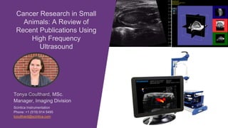
Cancer Research in Small
Animals: A Review of
Recent Publications Using
High Frequency
Ultrasound
• Preclinical Cancer Research
• Imaging in Preclinical Cancer Research
• Recent Publications
Topics of Discussion
Preclinical Cancer Research
• Preclinical Solid Tumor Model Overview
• Cell Line Derived / Transplantable Tumor Models
• Genetically Engineered Mouse Tumor Models
• Patient Derived Xenograft Tumor Models
• Preclinical Metastatic Tumor Models
• Preclinical cancer research focuses on the following:
• identify biological pathways and mechanisms of
tumorgenesis, growth and metastasis
• screen potential therapeutic compounds or
approaches for their efficacy
• The focus is on the translational aspects of bringing
novel therapeutic approaches to the clinic
Begley et al. Nature (2012) 483(7391): 531-533.
Preclinical Cancer Research
• A number of different types of solid tumor models are used
commonly in preclinical research:
• Cell line-derived models
• Patient Derived Xenograft (PDX) models
• Environmentally induced models
• Genetically Engineered Mouse (GEM) models
Gengenbacher et al. Nature Reviews Cancer (2017) 17:751-765.
Preclinical Solid Tumor Models
• A review of publications from
2016 found that most work was
being done using a single type
of model, on a single tumor
type
• Breast, lung, colorectal,
melanoma, pancreas, brain and
liver were the top solid tumor
types being studied
• There is a clear preference for
cell line derived models, with
varying levels of PDX, GEM, and
environmental models depending
on the tumor type
Gengenbacher et al. Nature Reviews Cancer (2017) 17:751-765.
Preclinical Solid Tumor Models
• Findings may not correlate to clinical outcomes – cell lines represent only a single population of cancer patients
Dranoff, Glenn Nature Reviews Immunology (2012) 12:61-66.
Cell Line Derived / Transplantable Tumor Models
• Work horse in cancer research – propagated
easily
• Subcutaneous injection
• Orthotopic injection – relevant
microenvironment may be present
• IV injection for metastasis
• May be genetically engineered to contain
luciferase or other compounds to allow them to be
imaged – i.e. bioluminescence
Dranoff, Glenn Nature Reviews Immunology (2012) 12:61-66.
Genetically Engineered Mouse Tumor Models
• Mice with genetically engineered/humanized immune
systems
• used to study specific parts of the immune system
• may cause enhanced tumor susceptibility
• Tissue-specific and/or temporally controlled
expression of oncogenes or loss of tumor
suppressor genes
• spontaneous tumor development
• more closely mimic the human disease
• Tumor formation is variable and takes longer to
develop
• Model the multiple stages in the development of cancer, and importance of the interaction between neoplastic cells
and the tissue microenvironment
Lai et al. Journal of Hematology & Oncology (2017) 10:106-.
Patient Derived Xenograft Tumor Models
• Resected tumor tissue is implanted into
immunodeficient mice; may be placed
subcutaneously or orthotopically
• Can be expanded in vivo through limited
passages in mice
• Only model system that allows for the vast
inter-patient and intra-tumor
heterogeneity that is inherent to human
cancer
• PDX models can be used to identify and test efficacy of therapeutic compounds in vivo and in vitro
• The actual patient may enroll in a specific trial, simultaneously avatar mice can be used in parallel to explore
multiple therapeutic options
• When used, primary tumor may be engrafted
ectopically or orthotopically, and in some studies
both methods were employed
• Metastatic studies make up only about 25% of the
cancer research being performed
• Metastasis can be induced in two ways
• IV injection of tumor cells either in the tail
vein, intracardiac injection, splenic vein
injection or intraperitoneal injection
• By spontaneous metastasis from a
primary tumor – at times the primary tumor
is resected to ensure survival of the animal
Gengenbacher et al. Nature Reviews Cancer (2017) 17:751-765.
Preclinical Metastatic Tumor Models
Audience Poll #1
Imaging in Preclinical Cancer Research
• Imaging Modality Review
• High Frequency Ultrasound – Prospect T1 Imaging Capabilities
• Tumor detection/monitoring and surrounding tissue imaging
• Injections for orthotopic placement or metastatic tumor models
• Tumor vasculature and biomarker detection
• Advanced techniques for drug delivery and early lesion detection
Li et al. Cancers (2019) 11:1800.
Imaging in Cancer Research
• Each modality has specific strengths/weaknesses as they
relate to cancer research
• CT provides good resolution of bones and lung, while
exposing the imaging subject to radiation
• PET requires the use of radioactive tracers to visualize
the tumors, and should be coregistered with another
imaging modality to provide anatomical context, but is
highly sensitive to metastasis and early pathological
changes
• Bioluminescence requires the tumor that express
luciferase; tumor volume measurements are dependent
on expression of the luciferase enzyme and delivery of
the luciferin substrate to the tumor cells, which may
change throughout disease progression or regression
Li et al. Cancers (2019) 11:1800.
Imaging in Cancer Research
• MRI provides excellent soft tissue resolution to
visualize normal and abnormal tissue structures, including
tumors; acquisition times vary from 2-5 minutes, but
provides good resolution (150-300µm) images of the
whole body. Contrast agents can be used to study
perfusion and specific biomarkers.
• Ultrasound (high frequency) provides high-resolution
real-time images with a strong soft tissue contrast to
visualize the tumor; contrast agents may be used to
study tumor perfusion or molecular expression of specific
targets
• Ultrasound is a non-invasive imaging technique which does not required the use of ionizing radiation,
instead it uses sound waves
• The transducer both sends and receives the ultrasound waves and the computer interprets the returned signal
into an image
Note – these images were not taken
with the Prospect T1 system
Ultrasound Imaging
• High-frequency ultrasound waves are necessary to resolve the small anatomical targets in preclinical
research
• Compromise is shorter penetration depth
Ultrasound Imaging
Prospect T1
• High-frequency ultrasound system specifically small animal imaging
• Up to 30µm resolution - small tumors, early pathological changes
• B-mode imaging to identify tumors and investigate surrounding
tissue
• 3D imaging to allow for tumor volume quantification
• Image guided needle injections for orthotopic tumor models
• Power Doppler imaging to study tumor vasculature
• Contrast agent imaging for tumor perfusion and biomarker detection
• Integrated sonoporation for drug delivery and cell membrane
permeability applications
• Shear wave elastography for early lesion detection
Tumor Detection and Monitoring
• Tumors of all types are visible, whether
subcutaneous or orthotopic
• Standard 2D B-mode imaging is used to
provide a greyscale image, tumors often
show up as a different echogenicity than
the surrounding tissue
• 2D measurements can be done to
measure linear or area measurements of
tumor size
Surrounding Tissue Imaging
• Normal structures have a characteristic look
for that specific tissue
• Abnormal changes can be identified – for
example cysts located in the ovaries
• It may be important to look at tissues such as
the lymph node and spleen to identify
systemic changes
Lymph Node
Skeletal Muscle Liver
Spleen
Cyst
Ovary
3D B-Mode Imaging
• The 3D motor expands the capabilities of the Prospect T1
to acquire 3D B-mode images
• Add-on includes the software analysis package to view
the 3D images and perform volume calculations
3D B-Mode Imaging
• Multiple slices are acquired
to cover the entire tumor
volume
• Two areas are drawn
perpendicular to one
another
• Software performs edge
detection and creates a
volume
• 39.0mm3
Image Guided Needle Injection
• The image guided needle injection mount integrates with probe
• Injections may be performed with a regular syringe and steel
needle, or pulled glass capillary needle
• Injections may be made into developing embryos, adult
myocardium, or any number of abdominal organ/muscle targets
Adult mouse
myocardium
Power Doppler – Tumor Vasculature
• Power Doppler is used to visualize the tumor
vasculature
• Larger vasculature is detected and displayed as a
color overlay on the B-mode image
Contrast Imaging – Tumor Microvasculature
• Contrast agents used to detect the
microvasculature – microbubbles are 2-3µm
• Imaging can be done using reference
subtraction or subharmonic imaging
techniques
• Reference subtraction imaging provides a
color overlay to highlight the inflowing
microbubbles
• Multiple regions of interest can be drawn to
Contrast Imaging – Tumor Microvasculature
• Subharmonic imaging
isolates the signal coming only
from the microbubbles –
listening only for the 1st
harmonic signal, tissue signal
is removed
• Multiple regions of interest
can be drawn to create time
vs. intensity curves
Integrated Sonoporation
• Sonoporation is the controlled cavitation or
bursting of microbubbles with the intention of
increasing the permeability of the cell
membrane, to open to blood brain barrier, or to
facilitate drug delivery
• Sonoporation is performed by a secondary, non-
imaging, probe directed at the anatomical target
• Software integration and control of the
sonoporation probe is included with this add-on
Shear Wave Elastography
• Shear wave elastography is used to quantify mechanical/elastic properties of
tissues
• The acoustic radiation force is generated by a push probe mounted on the side of
the imaging probe
• The software analysis generates a colored elastogram which is overlaid on a B-
mode image
Audience Poll #2
Recent Publications
• Targeted tumor theranostic agent was created with selective
tumor accumulation with diagnostic capabilities, as well as
increased retention, and drug release to effect tumor growth
• Trimodal imaging capabilities were included in the design,
with folate on the surface for binding to tumor cells, along
with 5-fluorouracil included as the therapeutic agent which
blocks DNA synthesis
• Fluorescence
• IR-780 in the
nanoparticle shell
• MRI
• Gd conjugated to
the 5-FU in shell
• Ultrasound
• gas filled core
Li et al. NPG Asia Materials (2018) 10: 1046-1060
• In vitro – nanobubbles were clearly visible in a gel mold
• In vivo – clear accumulation of the targeted nanobubbles was observed using
all three imaging modalities
• Ultrasound - red arrows below show accumulation within the tumor at the
same location over time
Li et al. NPG Asia Materials (2018) 10: 1046-1060
• 5-FU was released from the nanobubble
surface in an acidic pH environment and with
laser irradiation
• Various combined therapies were applied to the
mice; only the nanobubbles with laser resulted
in a decrease in tumor volumes. Other
therapies slowed the growth, but tumors remained
Li et al. NPG Asia Materials (2018) 10: 1046-1060
• Hepatocellular carcinoma (HCC) is one of the most
common malignant tumors, causing significant mortality
• Synergistic effect of chemotherapy and gene therapy
• Oxaliplatin – mechanism is not fully understood, but
believed to act on DNA synthesis pathways
• Recombinant human adenovirus Aspp2 (apoptosis
stimulating protein of p53-2) – Aspp2 binds to p53 and
inhibits cell growth and induces apoptosis
• in vivo and in vitro studies on HCC cell lines and xenograft
models in nude mice
Liu et al. International Journal of Oncology (2017) 51:1291-1299.
• Relative tumor volume decreased with all treatments,
however, was only significant when both chemotherapy and
gene therapy were combined
• This group was also able to use Power Doppler to assess
the small vessels within the tumors – they noted that vessel
quantity decreased in all three treatment groups (data not
shown)
Liu et al. International Journal of Oncology (2017) 51:1291-1299.
Want to see the system in action? Join us for a virtual demo
• Images were acquired by implanting a small portion of a blackberry into clear gelatin. The inner structure of the flesh of
the berry as well as the seeds are clearly visible through the clear gelatin mold
Audience Poll #3
• Targeted ultrasound contrast agents combine the advantages of
ultrasound imaging with molecular imaging to visualize molecular
biomarkers with high sensitivity and specificity in vivo
• Gas filled nanobubbles may be functionalized with specific cell
targets
• VEGFR2 (vascular endothelial grown factor receptor 2) –
expressed by endothelial cells of newly formed blood vessels
• HER2 (human epidermal growth factor 2) – has been identified as
a key target to improve detection and diagnosis of breast cancer
• Traditional microbubbles are typically blood pool agents as they are
too large to fit through the leaky tumor vasculature limiting their targets
to endothelial cell surface markers (VEGFR2); nanobubbles however
are able to extravasate from the vasculature and bind to tumor cell
surface markers (HER2)
Du et al. Scientific Reports (2018) 8:3887.
• Both in vitro and in vivo studies showed enhanced binding of dual-targeted (VEGFR2 & HER2) nanobubbles compared
to single targeted (VEGFR2 or HER2) nanobubbles, or untargeted (no antibody) nanobubbles alone
• Sustained increased greyscale intensity was seen as far out as 6-minute post injection, showing retention of the
nanobubbles within the tumor
• Fluorescent in vivo imaging and other histological techniques were used to confirm these results
Du et al. Scientific Reports (2018) 8:3887.
• Theranostic agents combine imaging capabilities with therapeutic
function into a single agent
• Multiwalled carbon nanotubes targeted to prostate stem cell
antigen (PSCA) are used to both detect tumors but also to deliver a
therapeutic compound – doxorubicin
• PSCA is only expressed at very low levels in normal prostate tissue,
but is highly expressed in all forms of prostate cancer, and even
more so in all metastatic prostate tumors
• Subcutaneous prostate tumors were used to study the
characteristics of this contrast agent
Wu et al. Biomaterials (2014) 35:5369-5380.
• Targeted carbon nanotubes were found to
accumulate in the tumors over time; non-
targeted and antibody blocked nanotubes
did not show the same level of binding
within the tumor
• Further studies showed the therapeutic
efficacy of the drug delivery component of
the nanotubes, as well as the reduced
overall toxicity of doxorubicin when
delivered in a targeted manner
Wu et al. Biomaterials (2014) 35:5369-5380.
• Preclinical Cancer Research
• Imaging in Preclinical Cancer Research
• Recent Publications
Topics of Discussion
Tonya Coulthard, MSc.
Manager, Imaging Division
Scintica Instrumentation
Phone: +1 (519) 914 5495
tcoulthard@scintica.com
Q&A
SESSION:
To ask a question, click the Q&A Button,
type your question and click send. Any
questions that are not addressed during
the live webinar will be answered
following the event.
Thank you for participating!
• Molecular imaging, using targeted ultrasound contrast agents, allows
for subtle changes to be detected non-invasively, prior to any
phenotypical changes which may be observed without a contrast agent
• Ultrasound imaging is advantageous for frequent monitoring as there is
no ionizing radiation
• Dual targeted gold nanoshelled poly(lactic-co-glycolic acid) [PLGA]
nanocapsules were created targeting both VEGFR2 (vascular
endothelial growth factor receptor 2) and and p53
Xu et al. International Journal of Nanomedicine (2018): 13:1791-1807.
• Orthotopic breast tumors
were used to study
diagnostic capabilities of
this contrast agent
• Ultrasound results showed that dual-targeted nanocapsules
showed higher signal enhancement then either of the single
targeted agents (VEGFR2 or p53), and that non-targeted and
dual-targeted nanocapsules that were pre-treated with
antibodies before injection showed the lowest signal intensity,
similar to that seen in the hind limb adductor muscle
Xu et al. International Journal of Nanomedicine (2018): 13:1791-1807.
• Hepatocellular carcinoma is characterized by chronic inflammation and
an immune-suppressive tumor microenvironment
• Immuno-oncology is a new area of drug development focused on
enhancing the host’s effective anti-cancer immune response
• Orthotopic liver tumors expressing programmed death ligand 1 (PD-L1)
to explore the growth characteristics, regulation of the immune
microenvironment, and potential association with anti-PD1 therapy
• Tumor perfusion was assessed using an intravenous microbubble
injection, measured using time-intensity curves as well as vascular
index curves
Ou et al. Liver Cancer (2019) 8:155-171.
• Expression of PD-L1 in the tumor cells did not effect
• Survival
• Tumor growth
• Tumor perfusion
• Anti-PD1 antibodies, as immunotherapy re-sensitized the PD-L1 expressing
tumors to sorafenib (conventional chemotherapy)
• Anti-CD8+ antibodies were used to deplete the CD8+ T cells from the host
 neither anti-PD1 nor sorafenib were effective treatments when these T
cells were removed from hosts immune system response
Ou et al. Liver Cancer (2019) 8:155-171.
• Exosomes are created by a number of different cell types; natural
killer (NK) cells play a central role in the immune response against
cancer
• Exosomes from NK cells could be used to exploit the antitumor
properties of NK cells
• Melanoma is an aggressive type of skin cancer with a poor prognosis,
and is often resistant to multimodal treatment approaches (resection,
chemo, radiation)
• Ultrasound and bioluminescence was used to assess tumor volumes
in this study
Zhu et al. Theranostics (2017) 7(10):2732-45.
• Bioluminescence and ultrasound both showed the effectiveness of the
NK derived exosomes and controlling tumor growth; these results
correlated well with the final tumor weights taken at the end of the
study
Zhu et al. Theranostics (2017) 7(10):2732-45.

Weitere ähnliche Inhalte

Was ist angesagt?

Bilateral basal ganglia abnormalities - MRI
Bilateral basal ganglia abnormalities - MRIBilateral basal ganglia abnormalities - MRI
Bilateral basal ganglia abnormalities - MRIRoshan Valentine
 
Sebastian Lattuga M.D. - Lumbar Spinal Stenosis
Sebastian Lattuga M.D. - Lumbar Spinal StenosisSebastian Lattuga M.D. - Lumbar Spinal Stenosis
Sebastian Lattuga M.D. - Lumbar Spinal StenosisSebastian Lattuga
 
Conus medullaris and cauda equina syndromes
Conus medullaris and cauda equina syndromesConus medullaris and cauda equina syndromes
Conus medullaris and cauda equina syndromesshaishav_bhagat
 
Cranio vertebral junction (CV Junction) - Dr Sameep Koshti (Consultant Neuros...
Cranio vertebral junction (CV Junction) - Dr Sameep Koshti (Consultant Neuros...Cranio vertebral junction (CV Junction) - Dr Sameep Koshti (Consultant Neuros...
Cranio vertebral junction (CV Junction) - Dr Sameep Koshti (Consultant Neuros...Sameep Koshti
 
Imaging of infection of brain and its linings
Imaging of infection of brain and its liningsImaging of infection of brain and its linings
Imaging of infection of brain and its liningscharusmita chaudhary
 
Role of head and neck imaging in trigeminal neuralgia
Role of head and neck imaging in trigeminal neuralgiaRole of head and neck imaging in trigeminal neuralgia
Role of head and neck imaging in trigeminal neuralgiaIndian dental academy
 
Image interpretation (for radiology residents)
Image interpretation (for radiology residents)Image interpretation (for radiology residents)
Image interpretation (for radiology residents)Riham Dessouky
 
Presentation1, artifact and pitfalls of the knee, hip and ankle joints.
Presentation1, artifact and pitfalls of the knee, hip and ankle joints.Presentation1, artifact and pitfalls of the knee, hip and ankle joints.
Presentation1, artifact and pitfalls of the knee, hip and ankle joints.Abdellah Nazeer
 
Imaging in degenerative diseases of spine
Imaging in degenerative diseases of spineImaging in degenerative diseases of spine
Imaging in degenerative diseases of spineGirendra Shankar
 
Presentation1, radiological film reading of the sholder joint.
Presentation1,  radiological film reading of the sholder joint.Presentation1,  radiological film reading of the sholder joint.
Presentation1, radiological film reading of the sholder joint.Abdellah Nazeer
 
Coma, vegetative and locked in syndrome
Coma, vegetative and locked in syndromeComa, vegetative and locked in syndrome
Coma, vegetative and locked in syndromeSandesh Dahal
 
Basics of Neuroradiology
Basics of NeuroradiologyBasics of Neuroradiology
Basics of NeuroradiologyAmr Hassan
 
Fungal infections of CNS
Fungal infections of CNS Fungal infections of CNS
Fungal infections of CNS NeurologyKota
 

Was ist angesagt? (20)

Brain anatomy on ct & amp; mri
Brain anatomy on ct & amp; mriBrain anatomy on ct & amp; mri
Brain anatomy on ct & amp; mri
 
Spinal tuberculosis
Spinal tuberculosisSpinal tuberculosis
Spinal tuberculosis
 
Bilateral basal ganglia abnormalities - MRI
Bilateral basal ganglia abnormalities - MRIBilateral basal ganglia abnormalities - MRI
Bilateral basal ganglia abnormalities - MRI
 
Moyamoya disease
Moyamoya diseaseMoyamoya disease
Moyamoya disease
 
Hypothalamic Gliomas
Hypothalamic GliomasHypothalamic Gliomas
Hypothalamic Gliomas
 
Sebastian Lattuga M.D. - Lumbar Spinal Stenosis
Sebastian Lattuga M.D. - Lumbar Spinal StenosisSebastian Lattuga M.D. - Lumbar Spinal Stenosis
Sebastian Lattuga M.D. - Lumbar Spinal Stenosis
 
Conus medullaris and cauda equina syndromes
Conus medullaris and cauda equina syndromesConus medullaris and cauda equina syndromes
Conus medullaris and cauda equina syndromes
 
Cranio vertebral junction (CV Junction) - Dr Sameep Koshti (Consultant Neuros...
Cranio vertebral junction (CV Junction) - Dr Sameep Koshti (Consultant Neuros...Cranio vertebral junction (CV Junction) - Dr Sameep Koshti (Consultant Neuros...
Cranio vertebral junction (CV Junction) - Dr Sameep Koshti (Consultant Neuros...
 
Imaging of infection of brain and its linings
Imaging of infection of brain and its liningsImaging of infection of brain and its linings
Imaging of infection of brain and its linings
 
Role of head and neck imaging in trigeminal neuralgia
Role of head and neck imaging in trigeminal neuralgiaRole of head and neck imaging in trigeminal neuralgia
Role of head and neck imaging in trigeminal neuralgia
 
Image interpretation (for radiology residents)
Image interpretation (for radiology residents)Image interpretation (for radiology residents)
Image interpretation (for radiology residents)
 
MRI of the shoulder
MRI of the shoulderMRI of the shoulder
MRI of the shoulder
 
Presentation1, artifact and pitfalls of the knee, hip and ankle joints.
Presentation1, artifact and pitfalls of the knee, hip and ankle joints.Presentation1, artifact and pitfalls of the knee, hip and ankle joints.
Presentation1, artifact and pitfalls of the knee, hip and ankle joints.
 
Imaging in degenerative diseases of spine
Imaging in degenerative diseases of spineImaging in degenerative diseases of spine
Imaging in degenerative diseases of spine
 
Presentation1, radiological film reading of the sholder joint.
Presentation1,  radiological film reading of the sholder joint.Presentation1,  radiological film reading of the sholder joint.
Presentation1, radiological film reading of the sholder joint.
 
Coma, vegetative and locked in syndrome
Coma, vegetative and locked in syndromeComa, vegetative and locked in syndrome
Coma, vegetative and locked in syndrome
 
Basics of Neuroradiology
Basics of NeuroradiologyBasics of Neuroradiology
Basics of Neuroradiology
 
Fungal infections of CNS
Fungal infections of CNS Fungal infections of CNS
Fungal infections of CNS
 
Degenerative diseases of spine : Imaging
Degenerative diseases of spine : ImagingDegenerative diseases of spine : Imaging
Degenerative diseases of spine : Imaging
 
CNS infections
CNS infectionsCNS infections
CNS infections
 

Ähnlich wie Cancer Research in Small Animals: A Review of Recent Publications Using High Frequency Ultrasound

seminar on new technologies of cell and molecular biology
seminar on new technologies of cell and molecular biologyseminar on new technologies of cell and molecular biology
seminar on new technologies of cell and molecular biologyBiswajit Deka
 
Cervical Cancer [ Incidence, Prevalence, Research Study, Introduction About R...
Cervical Cancer [ Incidence, Prevalence, Research Study, Introduction About R...Cervical Cancer [ Incidence, Prevalence, Research Study, Introduction About R...
Cervical Cancer [ Incidence, Prevalence, Research Study, Introduction About R...DR .PALLAVI PATHANIA
 
14. Brain Tumour.pptx
14. Brain  Tumour.pptx14. Brain  Tumour.pptx
14. Brain Tumour.pptxMs.Elizabeth
 
Metastatic bone disease
Metastatic bone disease Metastatic bone disease
Metastatic bone disease marcell wijaya
 
Cancer diagnosis and treatment-1.pptx
Cancer diagnosis and treatment-1.pptxCancer diagnosis and treatment-1.pptx
Cancer diagnosis and treatment-1.pptxRohanRo11
 
Localization of non palpable breast tumor for surgery
Localization of non palpable breast tumor for surgeryLocalization of non palpable breast tumor for surgery
Localization of non palpable breast tumor for surgeryGowtham Krishnan
 
(March 13, 2024) Overview of Preclinical Small Animal and Multimodal Imaging
(March 13, 2024) Overview of Preclinical Small Animal and Multimodal Imaging(March 13, 2024) Overview of Preclinical Small Animal and Multimodal Imaging
(March 13, 2024) Overview of Preclinical Small Animal and Multimodal ImagingScintica Instrumentation
 
The innovation of breast scanning
The innovation of breast scanningThe innovation of breast scanning
The innovation of breast scanningKtn Aye
 
diagnosis of cancer
diagnosis of cancer diagnosis of cancer
diagnosis of cancer milan mistry
 
RADIOLOGY and US Imaging for Protozoal Diseases.pptx
RADIOLOGY and US Imaging for Protozoal Diseases.pptxRADIOLOGY and US Imaging for Protozoal Diseases.pptx
RADIOLOGY and US Imaging for Protozoal Diseases.pptxIbrahimAboAlasaad
 
Diagnosis of cancer
Diagnosis of cancerDiagnosis of cancer
Diagnosis of cancerSmart Karthi
 
Extreme Computing, Clinical Medicine and GPUs or Can GPUs Cure Cancer
Extreme Computing, Clinical Medicine and GPUs or Can GPUs Cure CancerExtreme Computing, Clinical Medicine and GPUs or Can GPUs Cure Cancer
Extreme Computing, Clinical Medicine and GPUs or Can GPUs Cure CancerJoel Saltz
 
Imaging RADIOKLOGY and US for Protozoal Diseases.pptx
Imaging RADIOKLOGY and US for Protozoal Diseases.pptxImaging RADIOKLOGY and US for Protozoal Diseases.pptx
Imaging RADIOKLOGY and US for Protozoal Diseases.pptxIbrahimAboAlasaad
 

Ähnlich wie Cancer Research in Small Animals: A Review of Recent Publications Using High Frequency Ultrasound (20)

Fusion prostatic biopsy
Fusion prostatic biopsyFusion prostatic biopsy
Fusion prostatic biopsy
 
seminar on new technologies of cell and molecular biology
seminar on new technologies of cell and molecular biologyseminar on new technologies of cell and molecular biology
seminar on new technologies of cell and molecular biology
 
Cervical Cancer [ Incidence, Prevalence, Research Study, Introduction About R...
Cervical Cancer [ Incidence, Prevalence, Research Study, Introduction About R...Cervical Cancer [ Incidence, Prevalence, Research Study, Introduction About R...
Cervical Cancer [ Incidence, Prevalence, Research Study, Introduction About R...
 
Tumor markers
Tumor markersTumor markers
Tumor markers
 
14. Brain Tumour.pptx
14. Brain  Tumour.pptx14. Brain  Tumour.pptx
14. Brain Tumour.pptx
 
Metastatic bone disease
Metastatic bone disease Metastatic bone disease
Metastatic bone disease
 
Cancer diagnosis and treatment-1.pptx
Cancer diagnosis and treatment-1.pptxCancer diagnosis and treatment-1.pptx
Cancer diagnosis and treatment-1.pptx
 
MALIGNANT TUMOR.pptx
MALIGNANT TUMOR.pptxMALIGNANT TUMOR.pptx
MALIGNANT TUMOR.pptx
 
Localization of non palpable breast tumor for surgery
Localization of non palpable breast tumor for surgeryLocalization of non palpable breast tumor for surgery
Localization of non palpable breast tumor for surgery
 
(March 13, 2024) Overview of Preclinical Small Animal and Multimodal Imaging
(March 13, 2024) Overview of Preclinical Small Animal and Multimodal Imaging(March 13, 2024) Overview of Preclinical Small Animal and Multimodal Imaging
(March 13, 2024) Overview of Preclinical Small Animal and Multimodal Imaging
 
Diagnosis of cancer
Diagnosis of cancerDiagnosis of cancer
Diagnosis of cancer
 
The innovation of breast scanning
The innovation of breast scanningThe innovation of breast scanning
The innovation of breast scanning
 
Diagnosis of cancer
Diagnosis of cancerDiagnosis of cancer
Diagnosis of cancer
 
diagnosis of cancer
diagnosis of cancer diagnosis of cancer
diagnosis of cancer
 
RADIOLOGY and US Imaging for Protozoal Diseases.pptx
RADIOLOGY and US Imaging for Protozoal Diseases.pptxRADIOLOGY and US Imaging for Protozoal Diseases.pptx
RADIOLOGY and US Imaging for Protozoal Diseases.pptx
 
Asnr
AsnrAsnr
Asnr
 
Principle of oncology
Principle of oncologyPrinciple of oncology
Principle of oncology
 
Diagnosis of cancer
Diagnosis of cancerDiagnosis of cancer
Diagnosis of cancer
 
Extreme Computing, Clinical Medicine and GPUs or Can GPUs Cure Cancer
Extreme Computing, Clinical Medicine and GPUs or Can GPUs Cure CancerExtreme Computing, Clinical Medicine and GPUs or Can GPUs Cure Cancer
Extreme Computing, Clinical Medicine and GPUs or Can GPUs Cure Cancer
 
Imaging RADIOKLOGY and US for Protozoal Diseases.pptx
Imaging RADIOKLOGY and US for Protozoal Diseases.pptxImaging RADIOKLOGY and US for Protozoal Diseases.pptx
Imaging RADIOKLOGY and US for Protozoal Diseases.pptx
 

Mehr von Scintica Instrumentation

(May 9, 2024) Enhanced Ultrafast Vector Flow Imaging (VFI) Using Multi-Angle ...
(May 9, 2024) Enhanced Ultrafast Vector Flow Imaging (VFI) Using Multi-Angle ...(May 9, 2024) Enhanced Ultrafast Vector Flow Imaging (VFI) Using Multi-Angle ...
(May 9, 2024) Enhanced Ultrafast Vector Flow Imaging (VFI) Using Multi-Angle ...Scintica Instrumentation
 
Accelerating the Delivery of New Treatments for Children with Neuroblastoma 2...
Accelerating the Delivery of New Treatments for Children with Neuroblastoma 2...Accelerating the Delivery of New Treatments for Children with Neuroblastoma 2...
Accelerating the Delivery of New Treatments for Children with Neuroblastoma 2...Scintica Instrumentation
 
(March 14, 2024) Webinar: Validation of DEXA for Longitudinal Quantification ...
(March 14, 2024) Webinar: Validation of DEXA for Longitudinal Quantification ...(March 14, 2024) Webinar: Validation of DEXA for Longitudinal Quantification ...
(March 14, 2024) Webinar: Validation of DEXA for Longitudinal Quantification ...Scintica Instrumentation
 
(September 20, 2023) Webinar: An Introduction to Photoacoustic Imaging
(September 20, 2023) Webinar: An Introduction to Photoacoustic Imaging(September 20, 2023) Webinar: An Introduction to Photoacoustic Imaging
(September 20, 2023) Webinar: An Introduction to Photoacoustic ImagingScintica Instrumentation
 
(September 13, 2023) Webinar: Seeing Double: Preclinical Multiplexed PET for...
 (September 13, 2023) Webinar: Seeing Double: Preclinical Multiplexed PET for... (September 13, 2023) Webinar: Seeing Double: Preclinical Multiplexed PET for...
(September 13, 2023) Webinar: Seeing Double: Preclinical Multiplexed PET for...Scintica Instrumentation
 
(June 29, 2023) Webinar: Designer and Targeted Contrast Agent for Photoacoust...
(June 29, 2023) Webinar: Designer and Targeted Contrast Agent for Photoacoust...(June 29, 2023) Webinar: Designer and Targeted Contrast Agent for Photoacoust...
(June 29, 2023) Webinar: Designer and Targeted Contrast Agent for Photoacoust...Scintica Instrumentation
 
(June 22, 2023) Webinar: iNSiGHT & PIXImus: A Tale of Two Systems​
(June 22, 2023) Webinar: iNSiGHT & PIXImus: A Tale of Two Systems​(June 22, 2023) Webinar: iNSiGHT & PIXImus: A Tale of Two Systems​
(June 22, 2023) Webinar: iNSiGHT & PIXImus: A Tale of Two Systems​Scintica Instrumentation
 
(May 3, 2023) Webinar: Exploring a Novel NIR-2 Photoacoustic Agent to Improve...
(May 3, 2023) Webinar: Exploring a Novel NIR-2 Photoacoustic Agent to Improve...(May 3, 2023) Webinar: Exploring a Novel NIR-2 Photoacoustic Agent to Improve...
(May 3, 2023) Webinar: Exploring a Novel NIR-2 Photoacoustic Agent to Improve...Scintica Instrumentation
 
(April 5, 2023) Webinar: Prodigy Open-Platform Research Ultrasound System Ov...
 (April 5, 2023) Webinar: Prodigy Open-Platform Research Ultrasound System Ov... (April 5, 2023) Webinar: Prodigy Open-Platform Research Ultrasound System Ov...
(April 5, 2023) Webinar: Prodigy Open-Platform Research Ultrasound System Ov...Scintica Instrumentation
 
(April 4, 2023) Overview of Preclinical Small Animal Imaging Modalities & Mul...
(April 4, 2023) Overview of Preclinical Small Animal Imaging Modalities & Mul...(April 4, 2023) Overview of Preclinical Small Animal Imaging Modalities & Mul...
(April 4, 2023) Overview of Preclinical Small Animal Imaging Modalities & Mul...Scintica Instrumentation
 
(March 29, 2023) Webinar: Evaluating Intracerebral Injections of Radiation Na...
(March 29, 2023) Webinar: Evaluating Intracerebral Injections of Radiation Na...(March 29, 2023) Webinar: Evaluating Intracerebral Injections of Radiation Na...
(March 29, 2023) Webinar: Evaluating Intracerebral Injections of Radiation Na...Scintica Instrumentation
 
(February 16, 2023) Webinar: Intracerebral Transplantation of Autologous Bone...
(February 16, 2023) Webinar: Intracerebral Transplantation of Autologous Bone...(February 16, 2023) Webinar: Intracerebral Transplantation of Autologous Bone...
(February 16, 2023) Webinar: Intracerebral Transplantation of Autologous Bone...Scintica Instrumentation
 
(December 7, 2022) Webinar: Low-Dose Lithium Supplementation Promotes Adipose...
(December 7, 2022) Webinar: Low-Dose Lithium Supplementation Promotes Adipose...(December 7, 2022) Webinar: Low-Dose Lithium Supplementation Promotes Adipose...
(December 7, 2022) Webinar: Low-Dose Lithium Supplementation Promotes Adipose...Scintica Instrumentation
 
(November 30, 2022) Webinar: Molecular Mechanisms Behind Lameness in Meat Chi...
(November 30, 2022) Webinar: Molecular Mechanisms Behind Lameness in Meat Chi...(November 30, 2022) Webinar: Molecular Mechanisms Behind Lameness in Meat Chi...
(November 30, 2022) Webinar: Molecular Mechanisms Behind Lameness in Meat Chi...Scintica Instrumentation
 
(December 2, 2021) The Bench to Bedside Series: Preclinical Cancer Research w...
(December 2, 2021) The Bench to Bedside Series: Preclinical Cancer Research w...(December 2, 2021) The Bench to Bedside Series: Preclinical Cancer Research w...
(December 2, 2021) The Bench to Bedside Series: Preclinical Cancer Research w...Scintica Instrumentation
 
(October 27, 2021) Webinar: GSK3 Inhibition and Muscle Wasting Disorders: Mus...
(October 27, 2021) Webinar: GSK3 Inhibition and Muscle Wasting Disorders: Mus...(October 27, 2021) Webinar: GSK3 Inhibition and Muscle Wasting Disorders: Mus...
(October 27, 2021) Webinar: GSK3 Inhibition and Muscle Wasting Disorders: Mus...Scintica Instrumentation
 
(October 12, 2021) Webinar: Clinical Field MRI As A Measurement Instrument fo...
(October 12, 2021) Webinar: Clinical Field MRI As A Measurement Instrument fo...(October 12, 2021) Webinar: Clinical Field MRI As A Measurement Instrument fo...
(October 12, 2021) Webinar: Clinical Field MRI As A Measurement Instrument fo...Scintica Instrumentation
 
Webinar #4 Live Virtual Demonstration of the IVM System
Webinar #4 Live Virtual Demonstration of the IVM SystemWebinar #4 Live Virtual Demonstration of the IVM System
Webinar #4 Live Virtual Demonstration of the IVM SystemScintica Instrumentation
 
(June 22nd, 2021) Introducing Vilber’s Product Line of Gel Imaging Systems
(June 22nd, 2021) Introducing Vilber’s Product Line of Gel Imaging Systems(June 22nd, 2021) Introducing Vilber’s Product Line of Gel Imaging Systems
(June 22nd, 2021) Introducing Vilber’s Product Line of Gel Imaging SystemsScintica Instrumentation
 

Mehr von Scintica Instrumentation (20)

(May 9, 2024) Enhanced Ultrafast Vector Flow Imaging (VFI) Using Multi-Angle ...
(May 9, 2024) Enhanced Ultrafast Vector Flow Imaging (VFI) Using Multi-Angle ...(May 9, 2024) Enhanced Ultrafast Vector Flow Imaging (VFI) Using Multi-Angle ...
(May 9, 2024) Enhanced Ultrafast Vector Flow Imaging (VFI) Using Multi-Angle ...
 
Accelerating the Delivery of New Treatments for Children with Neuroblastoma 2...
Accelerating the Delivery of New Treatments for Children with Neuroblastoma 2...Accelerating the Delivery of New Treatments for Children with Neuroblastoma 2...
Accelerating the Delivery of New Treatments for Children with Neuroblastoma 2...
 
(March 14, 2024) Webinar: Validation of DEXA for Longitudinal Quantification ...
(March 14, 2024) Webinar: Validation of DEXA for Longitudinal Quantification ...(March 14, 2024) Webinar: Validation of DEXA for Longitudinal Quantification ...
(March 14, 2024) Webinar: Validation of DEXA for Longitudinal Quantification ...
 
(September 20, 2023) Webinar: An Introduction to Photoacoustic Imaging
(September 20, 2023) Webinar: An Introduction to Photoacoustic Imaging(September 20, 2023) Webinar: An Introduction to Photoacoustic Imaging
(September 20, 2023) Webinar: An Introduction to Photoacoustic Imaging
 
(September 13, 2023) Webinar: Seeing Double: Preclinical Multiplexed PET for...
 (September 13, 2023) Webinar: Seeing Double: Preclinical Multiplexed PET for... (September 13, 2023) Webinar: Seeing Double: Preclinical Multiplexed PET for...
(September 13, 2023) Webinar: Seeing Double: Preclinical Multiplexed PET for...
 
(June 29, 2023) Webinar: Designer and Targeted Contrast Agent for Photoacoust...
(June 29, 2023) Webinar: Designer and Targeted Contrast Agent for Photoacoust...(June 29, 2023) Webinar: Designer and Targeted Contrast Agent for Photoacoust...
(June 29, 2023) Webinar: Designer and Targeted Contrast Agent for Photoacoust...
 
(June 22, 2023) Webinar: iNSiGHT & PIXImus: A Tale of Two Systems​
(June 22, 2023) Webinar: iNSiGHT & PIXImus: A Tale of Two Systems​(June 22, 2023) Webinar: iNSiGHT & PIXImus: A Tale of Two Systems​
(June 22, 2023) Webinar: iNSiGHT & PIXImus: A Tale of Two Systems​
 
(May 3, 2023) Webinar: Exploring a Novel NIR-2 Photoacoustic Agent to Improve...
(May 3, 2023) Webinar: Exploring a Novel NIR-2 Photoacoustic Agent to Improve...(May 3, 2023) Webinar: Exploring a Novel NIR-2 Photoacoustic Agent to Improve...
(May 3, 2023) Webinar: Exploring a Novel NIR-2 Photoacoustic Agent to Improve...
 
(April 5, 2023) Webinar: Prodigy Open-Platform Research Ultrasound System Ov...
 (April 5, 2023) Webinar: Prodigy Open-Platform Research Ultrasound System Ov... (April 5, 2023) Webinar: Prodigy Open-Platform Research Ultrasound System Ov...
(April 5, 2023) Webinar: Prodigy Open-Platform Research Ultrasound System Ov...
 
(April 4, 2023) Overview of Preclinical Small Animal Imaging Modalities & Mul...
(April 4, 2023) Overview of Preclinical Small Animal Imaging Modalities & Mul...(April 4, 2023) Overview of Preclinical Small Animal Imaging Modalities & Mul...
(April 4, 2023) Overview of Preclinical Small Animal Imaging Modalities & Mul...
 
(March 29, 2023) Webinar: Evaluating Intracerebral Injections of Radiation Na...
(March 29, 2023) Webinar: Evaluating Intracerebral Injections of Radiation Na...(March 29, 2023) Webinar: Evaluating Intracerebral Injections of Radiation Na...
(March 29, 2023) Webinar: Evaluating Intracerebral Injections of Radiation Na...
 
(February 16, 2023) Webinar: Intracerebral Transplantation of Autologous Bone...
(February 16, 2023) Webinar: Intracerebral Transplantation of Autologous Bone...(February 16, 2023) Webinar: Intracerebral Transplantation of Autologous Bone...
(February 16, 2023) Webinar: Intracerebral Transplantation of Autologous Bone...
 
(December 7, 2022) Webinar: Low-Dose Lithium Supplementation Promotes Adipose...
(December 7, 2022) Webinar: Low-Dose Lithium Supplementation Promotes Adipose...(December 7, 2022) Webinar: Low-Dose Lithium Supplementation Promotes Adipose...
(December 7, 2022) Webinar: Low-Dose Lithium Supplementation Promotes Adipose...
 
(November 30, 2022) Webinar: Molecular Mechanisms Behind Lameness in Meat Chi...
(November 30, 2022) Webinar: Molecular Mechanisms Behind Lameness in Meat Chi...(November 30, 2022) Webinar: Molecular Mechanisms Behind Lameness in Meat Chi...
(November 30, 2022) Webinar: Molecular Mechanisms Behind Lameness in Meat Chi...
 
Imaging Hypoxia Webinar
Imaging Hypoxia WebinarImaging Hypoxia Webinar
Imaging Hypoxia Webinar
 
(December 2, 2021) The Bench to Bedside Series: Preclinical Cancer Research w...
(December 2, 2021) The Bench to Bedside Series: Preclinical Cancer Research w...(December 2, 2021) The Bench to Bedside Series: Preclinical Cancer Research w...
(December 2, 2021) The Bench to Bedside Series: Preclinical Cancer Research w...
 
(October 27, 2021) Webinar: GSK3 Inhibition and Muscle Wasting Disorders: Mus...
(October 27, 2021) Webinar: GSK3 Inhibition and Muscle Wasting Disorders: Mus...(October 27, 2021) Webinar: GSK3 Inhibition and Muscle Wasting Disorders: Mus...
(October 27, 2021) Webinar: GSK3 Inhibition and Muscle Wasting Disorders: Mus...
 
(October 12, 2021) Webinar: Clinical Field MRI As A Measurement Instrument fo...
(October 12, 2021) Webinar: Clinical Field MRI As A Measurement Instrument fo...(October 12, 2021) Webinar: Clinical Field MRI As A Measurement Instrument fo...
(October 12, 2021) Webinar: Clinical Field MRI As A Measurement Instrument fo...
 
Webinar #4 Live Virtual Demonstration of the IVM System
Webinar #4 Live Virtual Demonstration of the IVM SystemWebinar #4 Live Virtual Demonstration of the IVM System
Webinar #4 Live Virtual Demonstration of the IVM System
 
(June 22nd, 2021) Introducing Vilber’s Product Line of Gel Imaging Systems
(June 22nd, 2021) Introducing Vilber’s Product Line of Gel Imaging Systems(June 22nd, 2021) Introducing Vilber’s Product Line of Gel Imaging Systems
(June 22nd, 2021) Introducing Vilber’s Product Line of Gel Imaging Systems
 

Kürzlich hochgeladen

Measures of Central Tendency: Mean, Median and Mode
Measures of Central Tendency: Mean, Median and ModeMeasures of Central Tendency: Mean, Median and Mode
Measures of Central Tendency: Mean, Median and ModeThiyagu K
 
The basics of sentences session 3pptx.pptx
The basics of sentences session 3pptx.pptxThe basics of sentences session 3pptx.pptx
The basics of sentences session 3pptx.pptxheathfieldcps1
 
How to Give a Domain for a Field in Odoo 17
How to Give a Domain for a Field in Odoo 17How to Give a Domain for a Field in Odoo 17
How to Give a Domain for a Field in Odoo 17Celine George
 
microwave assisted reaction. General introduction
microwave assisted reaction. General introductionmicrowave assisted reaction. General introduction
microwave assisted reaction. General introductionMaksud Ahmed
 
Holdier Curriculum Vitae (April 2024).pdf
Holdier Curriculum Vitae (April 2024).pdfHoldier Curriculum Vitae (April 2024).pdf
Holdier Curriculum Vitae (April 2024).pdfagholdier
 
Class 11th Physics NEET formula sheet pdf
Class 11th Physics NEET formula sheet pdfClass 11th Physics NEET formula sheet pdf
Class 11th Physics NEET formula sheet pdfAyushMahapatra5
 
2024-NATIONAL-LEARNING-CAMP-AND-OTHER.pptx
2024-NATIONAL-LEARNING-CAMP-AND-OTHER.pptx2024-NATIONAL-LEARNING-CAMP-AND-OTHER.pptx
2024-NATIONAL-LEARNING-CAMP-AND-OTHER.pptxMaritesTamaniVerdade
 
Explore beautiful and ugly buildings. Mathematics helps us create beautiful d...
Explore beautiful and ugly buildings. Mathematics helps us create beautiful d...Explore beautiful and ugly buildings. Mathematics helps us create beautiful d...
Explore beautiful and ugly buildings. Mathematics helps us create beautiful d...christianmathematics
 
Sociology 101 Demonstration of Learning Exhibit
Sociology 101 Demonstration of Learning ExhibitSociology 101 Demonstration of Learning Exhibit
Sociology 101 Demonstration of Learning Exhibitjbellavia9
 
Advanced Views - Calendar View in Odoo 17
Advanced Views - Calendar View in Odoo 17Advanced Views - Calendar View in Odoo 17
Advanced Views - Calendar View in Odoo 17Celine George
 
This PowerPoint helps students to consider the concept of infinity.
This PowerPoint helps students to consider the concept of infinity.This PowerPoint helps students to consider the concept of infinity.
This PowerPoint helps students to consider the concept of infinity.christianmathematics
 
Unit-V; Pricing (Pharma Marketing Management).pptx
Unit-V; Pricing (Pharma Marketing Management).pptxUnit-V; Pricing (Pharma Marketing Management).pptx
Unit-V; Pricing (Pharma Marketing Management).pptxVishalSingh1417
 
Unit-IV- Pharma. Marketing Channels.pptx
Unit-IV- Pharma. Marketing Channels.pptxUnit-IV- Pharma. Marketing Channels.pptx
Unit-IV- Pharma. Marketing Channels.pptxVishalSingh1417
 
1029-Danh muc Sach Giao Khoa khoi 6.pdf
1029-Danh muc Sach Giao Khoa khoi  6.pdf1029-Danh muc Sach Giao Khoa khoi  6.pdf
1029-Danh muc Sach Giao Khoa khoi 6.pdfQucHHunhnh
 
Seal of Good Local Governance (SGLG) 2024Final.pptx
Seal of Good Local Governance (SGLG) 2024Final.pptxSeal of Good Local Governance (SGLG) 2024Final.pptx
Seal of Good Local Governance (SGLG) 2024Final.pptxnegromaestrong
 
1029 - Danh muc Sach Giao Khoa 10 . pdf
1029 -  Danh muc Sach Giao Khoa 10 . pdf1029 -  Danh muc Sach Giao Khoa 10 . pdf
1029 - Danh muc Sach Giao Khoa 10 . pdfQucHHunhnh
 
Making and Justifying Mathematical Decisions.pdf
Making and Justifying Mathematical Decisions.pdfMaking and Justifying Mathematical Decisions.pdf
Making and Justifying Mathematical Decisions.pdfChris Hunter
 
ICT Role in 21st Century Education & its Challenges.pptx
ICT Role in 21st Century Education & its Challenges.pptxICT Role in 21st Century Education & its Challenges.pptx
ICT Role in 21st Century Education & its Challenges.pptxAreebaZafar22
 
PROCESS RECORDING FORMAT.docx
PROCESS      RECORDING        FORMAT.docxPROCESS      RECORDING        FORMAT.docx
PROCESS RECORDING FORMAT.docxPoojaSen20
 

Kürzlich hochgeladen (20)

Measures of Central Tendency: Mean, Median and Mode
Measures of Central Tendency: Mean, Median and ModeMeasures of Central Tendency: Mean, Median and Mode
Measures of Central Tendency: Mean, Median and Mode
 
The basics of sentences session 3pptx.pptx
The basics of sentences session 3pptx.pptxThe basics of sentences session 3pptx.pptx
The basics of sentences session 3pptx.pptx
 
How to Give a Domain for a Field in Odoo 17
How to Give a Domain for a Field in Odoo 17How to Give a Domain for a Field in Odoo 17
How to Give a Domain for a Field in Odoo 17
 
INDIA QUIZ 2024 RLAC DELHI UNIVERSITY.pptx
INDIA QUIZ 2024 RLAC DELHI UNIVERSITY.pptxINDIA QUIZ 2024 RLAC DELHI UNIVERSITY.pptx
INDIA QUIZ 2024 RLAC DELHI UNIVERSITY.pptx
 
microwave assisted reaction. General introduction
microwave assisted reaction. General introductionmicrowave assisted reaction. General introduction
microwave assisted reaction. General introduction
 
Holdier Curriculum Vitae (April 2024).pdf
Holdier Curriculum Vitae (April 2024).pdfHoldier Curriculum Vitae (April 2024).pdf
Holdier Curriculum Vitae (April 2024).pdf
 
Class 11th Physics NEET formula sheet pdf
Class 11th Physics NEET formula sheet pdfClass 11th Physics NEET formula sheet pdf
Class 11th Physics NEET formula sheet pdf
 
2024-NATIONAL-LEARNING-CAMP-AND-OTHER.pptx
2024-NATIONAL-LEARNING-CAMP-AND-OTHER.pptx2024-NATIONAL-LEARNING-CAMP-AND-OTHER.pptx
2024-NATIONAL-LEARNING-CAMP-AND-OTHER.pptx
 
Explore beautiful and ugly buildings. Mathematics helps us create beautiful d...
Explore beautiful and ugly buildings. Mathematics helps us create beautiful d...Explore beautiful and ugly buildings. Mathematics helps us create beautiful d...
Explore beautiful and ugly buildings. Mathematics helps us create beautiful d...
 
Sociology 101 Demonstration of Learning Exhibit
Sociology 101 Demonstration of Learning ExhibitSociology 101 Demonstration of Learning Exhibit
Sociology 101 Demonstration of Learning Exhibit
 
Advanced Views - Calendar View in Odoo 17
Advanced Views - Calendar View in Odoo 17Advanced Views - Calendar View in Odoo 17
Advanced Views - Calendar View in Odoo 17
 
This PowerPoint helps students to consider the concept of infinity.
This PowerPoint helps students to consider the concept of infinity.This PowerPoint helps students to consider the concept of infinity.
This PowerPoint helps students to consider the concept of infinity.
 
Unit-V; Pricing (Pharma Marketing Management).pptx
Unit-V; Pricing (Pharma Marketing Management).pptxUnit-V; Pricing (Pharma Marketing Management).pptx
Unit-V; Pricing (Pharma Marketing Management).pptx
 
Unit-IV- Pharma. Marketing Channels.pptx
Unit-IV- Pharma. Marketing Channels.pptxUnit-IV- Pharma. Marketing Channels.pptx
Unit-IV- Pharma. Marketing Channels.pptx
 
1029-Danh muc Sach Giao Khoa khoi 6.pdf
1029-Danh muc Sach Giao Khoa khoi  6.pdf1029-Danh muc Sach Giao Khoa khoi  6.pdf
1029-Danh muc Sach Giao Khoa khoi 6.pdf
 
Seal of Good Local Governance (SGLG) 2024Final.pptx
Seal of Good Local Governance (SGLG) 2024Final.pptxSeal of Good Local Governance (SGLG) 2024Final.pptx
Seal of Good Local Governance (SGLG) 2024Final.pptx
 
1029 - Danh muc Sach Giao Khoa 10 . pdf
1029 -  Danh muc Sach Giao Khoa 10 . pdf1029 -  Danh muc Sach Giao Khoa 10 . pdf
1029 - Danh muc Sach Giao Khoa 10 . pdf
 
Making and Justifying Mathematical Decisions.pdf
Making and Justifying Mathematical Decisions.pdfMaking and Justifying Mathematical Decisions.pdf
Making and Justifying Mathematical Decisions.pdf
 
ICT Role in 21st Century Education & its Challenges.pptx
ICT Role in 21st Century Education & its Challenges.pptxICT Role in 21st Century Education & its Challenges.pptx
ICT Role in 21st Century Education & its Challenges.pptx
 
PROCESS RECORDING FORMAT.docx
PROCESS      RECORDING        FORMAT.docxPROCESS      RECORDING        FORMAT.docx
PROCESS RECORDING FORMAT.docx
 

Cancer Research in Small Animals: A Review of Recent Publications Using High Frequency Ultrasound

  • 1. Tonya Coulthard, MSc. Manager, Imaging Division Scintica Instrumentation Phone: +1 (519) 914 5495 tcoulthard@scintica.com Cancer Research in Small Animals: A Review of Recent Publications Using High Frequency Ultrasound
  • 2. • Preclinical Cancer Research • Imaging in Preclinical Cancer Research • Recent Publications Topics of Discussion
  • 3. Preclinical Cancer Research • Preclinical Solid Tumor Model Overview • Cell Line Derived / Transplantable Tumor Models • Genetically Engineered Mouse Tumor Models • Patient Derived Xenograft Tumor Models • Preclinical Metastatic Tumor Models
  • 4. • Preclinical cancer research focuses on the following: • identify biological pathways and mechanisms of tumorgenesis, growth and metastasis • screen potential therapeutic compounds or approaches for their efficacy • The focus is on the translational aspects of bringing novel therapeutic approaches to the clinic Begley et al. Nature (2012) 483(7391): 531-533. Preclinical Cancer Research
  • 5. • A number of different types of solid tumor models are used commonly in preclinical research: • Cell line-derived models • Patient Derived Xenograft (PDX) models • Environmentally induced models • Genetically Engineered Mouse (GEM) models Gengenbacher et al. Nature Reviews Cancer (2017) 17:751-765. Preclinical Solid Tumor Models
  • 6. • A review of publications from 2016 found that most work was being done using a single type of model, on a single tumor type • Breast, lung, colorectal, melanoma, pancreas, brain and liver were the top solid tumor types being studied • There is a clear preference for cell line derived models, with varying levels of PDX, GEM, and environmental models depending on the tumor type Gengenbacher et al. Nature Reviews Cancer (2017) 17:751-765. Preclinical Solid Tumor Models
  • 7. • Findings may not correlate to clinical outcomes – cell lines represent only a single population of cancer patients Dranoff, Glenn Nature Reviews Immunology (2012) 12:61-66. Cell Line Derived / Transplantable Tumor Models • Work horse in cancer research – propagated easily • Subcutaneous injection • Orthotopic injection – relevant microenvironment may be present • IV injection for metastasis • May be genetically engineered to contain luciferase or other compounds to allow them to be imaged – i.e. bioluminescence
  • 8. Dranoff, Glenn Nature Reviews Immunology (2012) 12:61-66. Genetically Engineered Mouse Tumor Models • Mice with genetically engineered/humanized immune systems • used to study specific parts of the immune system • may cause enhanced tumor susceptibility • Tissue-specific and/or temporally controlled expression of oncogenes or loss of tumor suppressor genes • spontaneous tumor development • more closely mimic the human disease • Tumor formation is variable and takes longer to develop • Model the multiple stages in the development of cancer, and importance of the interaction between neoplastic cells and the tissue microenvironment
  • 9. Lai et al. Journal of Hematology & Oncology (2017) 10:106-. Patient Derived Xenograft Tumor Models • Resected tumor tissue is implanted into immunodeficient mice; may be placed subcutaneously or orthotopically • Can be expanded in vivo through limited passages in mice • Only model system that allows for the vast inter-patient and intra-tumor heterogeneity that is inherent to human cancer • PDX models can be used to identify and test efficacy of therapeutic compounds in vivo and in vitro • The actual patient may enroll in a specific trial, simultaneously avatar mice can be used in parallel to explore multiple therapeutic options
  • 10. • When used, primary tumor may be engrafted ectopically or orthotopically, and in some studies both methods were employed • Metastatic studies make up only about 25% of the cancer research being performed • Metastasis can be induced in two ways • IV injection of tumor cells either in the tail vein, intracardiac injection, splenic vein injection or intraperitoneal injection • By spontaneous metastasis from a primary tumor – at times the primary tumor is resected to ensure survival of the animal Gengenbacher et al. Nature Reviews Cancer (2017) 17:751-765. Preclinical Metastatic Tumor Models
  • 12. Imaging in Preclinical Cancer Research • Imaging Modality Review • High Frequency Ultrasound – Prospect T1 Imaging Capabilities • Tumor detection/monitoring and surrounding tissue imaging • Injections for orthotopic placement or metastatic tumor models • Tumor vasculature and biomarker detection • Advanced techniques for drug delivery and early lesion detection
  • 13. Li et al. Cancers (2019) 11:1800. Imaging in Cancer Research • Each modality has specific strengths/weaknesses as they relate to cancer research • CT provides good resolution of bones and lung, while exposing the imaging subject to radiation • PET requires the use of radioactive tracers to visualize the tumors, and should be coregistered with another imaging modality to provide anatomical context, but is highly sensitive to metastasis and early pathological changes • Bioluminescence requires the tumor that express luciferase; tumor volume measurements are dependent on expression of the luciferase enzyme and delivery of the luciferin substrate to the tumor cells, which may change throughout disease progression or regression
  • 14. Li et al. Cancers (2019) 11:1800. Imaging in Cancer Research • MRI provides excellent soft tissue resolution to visualize normal and abnormal tissue structures, including tumors; acquisition times vary from 2-5 minutes, but provides good resolution (150-300µm) images of the whole body. Contrast agents can be used to study perfusion and specific biomarkers. • Ultrasound (high frequency) provides high-resolution real-time images with a strong soft tissue contrast to visualize the tumor; contrast agents may be used to study tumor perfusion or molecular expression of specific targets
  • 15. • Ultrasound is a non-invasive imaging technique which does not required the use of ionizing radiation, instead it uses sound waves • The transducer both sends and receives the ultrasound waves and the computer interprets the returned signal into an image Note – these images were not taken with the Prospect T1 system Ultrasound Imaging
  • 16. • High-frequency ultrasound waves are necessary to resolve the small anatomical targets in preclinical research • Compromise is shorter penetration depth Ultrasound Imaging
  • 17. Prospect T1 • High-frequency ultrasound system specifically small animal imaging • Up to 30µm resolution - small tumors, early pathological changes • B-mode imaging to identify tumors and investigate surrounding tissue • 3D imaging to allow for tumor volume quantification • Image guided needle injections for orthotopic tumor models • Power Doppler imaging to study tumor vasculature • Contrast agent imaging for tumor perfusion and biomarker detection • Integrated sonoporation for drug delivery and cell membrane permeability applications • Shear wave elastography for early lesion detection
  • 18. Tumor Detection and Monitoring • Tumors of all types are visible, whether subcutaneous or orthotopic • Standard 2D B-mode imaging is used to provide a greyscale image, tumors often show up as a different echogenicity than the surrounding tissue • 2D measurements can be done to measure linear or area measurements of tumor size
  • 19. Surrounding Tissue Imaging • Normal structures have a characteristic look for that specific tissue • Abnormal changes can be identified – for example cysts located in the ovaries • It may be important to look at tissues such as the lymph node and spleen to identify systemic changes Lymph Node Skeletal Muscle Liver Spleen Cyst Ovary
  • 20. 3D B-Mode Imaging • The 3D motor expands the capabilities of the Prospect T1 to acquire 3D B-mode images • Add-on includes the software analysis package to view the 3D images and perform volume calculations
  • 21. 3D B-Mode Imaging • Multiple slices are acquired to cover the entire tumor volume • Two areas are drawn perpendicular to one another • Software performs edge detection and creates a volume • 39.0mm3
  • 22. Image Guided Needle Injection • The image guided needle injection mount integrates with probe • Injections may be performed with a regular syringe and steel needle, or pulled glass capillary needle • Injections may be made into developing embryos, adult myocardium, or any number of abdominal organ/muscle targets Adult mouse myocardium
  • 23. Power Doppler – Tumor Vasculature • Power Doppler is used to visualize the tumor vasculature • Larger vasculature is detected and displayed as a color overlay on the B-mode image
  • 24. Contrast Imaging – Tumor Microvasculature • Contrast agents used to detect the microvasculature – microbubbles are 2-3µm • Imaging can be done using reference subtraction or subharmonic imaging techniques • Reference subtraction imaging provides a color overlay to highlight the inflowing microbubbles • Multiple regions of interest can be drawn to
  • 25. Contrast Imaging – Tumor Microvasculature • Subharmonic imaging isolates the signal coming only from the microbubbles – listening only for the 1st harmonic signal, tissue signal is removed • Multiple regions of interest can be drawn to create time vs. intensity curves
  • 26. Integrated Sonoporation • Sonoporation is the controlled cavitation or bursting of microbubbles with the intention of increasing the permeability of the cell membrane, to open to blood brain barrier, or to facilitate drug delivery • Sonoporation is performed by a secondary, non- imaging, probe directed at the anatomical target • Software integration and control of the sonoporation probe is included with this add-on
  • 27. Shear Wave Elastography • Shear wave elastography is used to quantify mechanical/elastic properties of tissues • The acoustic radiation force is generated by a push probe mounted on the side of the imaging probe • The software analysis generates a colored elastogram which is overlaid on a B- mode image
  • 30. • Targeted tumor theranostic agent was created with selective tumor accumulation with diagnostic capabilities, as well as increased retention, and drug release to effect tumor growth • Trimodal imaging capabilities were included in the design, with folate on the surface for binding to tumor cells, along with 5-fluorouracil included as the therapeutic agent which blocks DNA synthesis • Fluorescence • IR-780 in the nanoparticle shell • MRI • Gd conjugated to the 5-FU in shell • Ultrasound • gas filled core Li et al. NPG Asia Materials (2018) 10: 1046-1060
  • 31. • In vitro – nanobubbles were clearly visible in a gel mold • In vivo – clear accumulation of the targeted nanobubbles was observed using all three imaging modalities • Ultrasound - red arrows below show accumulation within the tumor at the same location over time Li et al. NPG Asia Materials (2018) 10: 1046-1060
  • 32. • 5-FU was released from the nanobubble surface in an acidic pH environment and with laser irradiation • Various combined therapies were applied to the mice; only the nanobubbles with laser resulted in a decrease in tumor volumes. Other therapies slowed the growth, but tumors remained Li et al. NPG Asia Materials (2018) 10: 1046-1060
  • 33. • Hepatocellular carcinoma (HCC) is one of the most common malignant tumors, causing significant mortality • Synergistic effect of chemotherapy and gene therapy • Oxaliplatin – mechanism is not fully understood, but believed to act on DNA synthesis pathways • Recombinant human adenovirus Aspp2 (apoptosis stimulating protein of p53-2) – Aspp2 binds to p53 and inhibits cell growth and induces apoptosis • in vivo and in vitro studies on HCC cell lines and xenograft models in nude mice Liu et al. International Journal of Oncology (2017) 51:1291-1299.
  • 34. • Relative tumor volume decreased with all treatments, however, was only significant when both chemotherapy and gene therapy were combined • This group was also able to use Power Doppler to assess the small vessels within the tumors – they noted that vessel quantity decreased in all three treatment groups (data not shown) Liu et al. International Journal of Oncology (2017) 51:1291-1299.
  • 35. Want to see the system in action? Join us for a virtual demo • Images were acquired by implanting a small portion of a blackberry into clear gelatin. The inner structure of the flesh of the berry as well as the seeds are clearly visible through the clear gelatin mold
  • 37. • Targeted ultrasound contrast agents combine the advantages of ultrasound imaging with molecular imaging to visualize molecular biomarkers with high sensitivity and specificity in vivo • Gas filled nanobubbles may be functionalized with specific cell targets • VEGFR2 (vascular endothelial grown factor receptor 2) – expressed by endothelial cells of newly formed blood vessels • HER2 (human epidermal growth factor 2) – has been identified as a key target to improve detection and diagnosis of breast cancer • Traditional microbubbles are typically blood pool agents as they are too large to fit through the leaky tumor vasculature limiting their targets to endothelial cell surface markers (VEGFR2); nanobubbles however are able to extravasate from the vasculature and bind to tumor cell surface markers (HER2) Du et al. Scientific Reports (2018) 8:3887.
  • 38. • Both in vitro and in vivo studies showed enhanced binding of dual-targeted (VEGFR2 & HER2) nanobubbles compared to single targeted (VEGFR2 or HER2) nanobubbles, or untargeted (no antibody) nanobubbles alone • Sustained increased greyscale intensity was seen as far out as 6-minute post injection, showing retention of the nanobubbles within the tumor • Fluorescent in vivo imaging and other histological techniques were used to confirm these results Du et al. Scientific Reports (2018) 8:3887.
  • 39. • Theranostic agents combine imaging capabilities with therapeutic function into a single agent • Multiwalled carbon nanotubes targeted to prostate stem cell antigen (PSCA) are used to both detect tumors but also to deliver a therapeutic compound – doxorubicin • PSCA is only expressed at very low levels in normal prostate tissue, but is highly expressed in all forms of prostate cancer, and even more so in all metastatic prostate tumors • Subcutaneous prostate tumors were used to study the characteristics of this contrast agent Wu et al. Biomaterials (2014) 35:5369-5380.
  • 40. • Targeted carbon nanotubes were found to accumulate in the tumors over time; non- targeted and antibody blocked nanotubes did not show the same level of binding within the tumor • Further studies showed the therapeutic efficacy of the drug delivery component of the nanotubes, as well as the reduced overall toxicity of doxorubicin when delivered in a targeted manner Wu et al. Biomaterials (2014) 35:5369-5380.
  • 41. • Preclinical Cancer Research • Imaging in Preclinical Cancer Research • Recent Publications Topics of Discussion
  • 42. Tonya Coulthard, MSc. Manager, Imaging Division Scintica Instrumentation Phone: +1 (519) 914 5495 tcoulthard@scintica.com Q&A SESSION: To ask a question, click the Q&A Button, type your question and click send. Any questions that are not addressed during the live webinar will be answered following the event. Thank you for participating!
  • 43. • Molecular imaging, using targeted ultrasound contrast agents, allows for subtle changes to be detected non-invasively, prior to any phenotypical changes which may be observed without a contrast agent • Ultrasound imaging is advantageous for frequent monitoring as there is no ionizing radiation • Dual targeted gold nanoshelled poly(lactic-co-glycolic acid) [PLGA] nanocapsules were created targeting both VEGFR2 (vascular endothelial growth factor receptor 2) and and p53 Xu et al. International Journal of Nanomedicine (2018): 13:1791-1807. • Orthotopic breast tumors were used to study diagnostic capabilities of this contrast agent
  • 44. • Ultrasound results showed that dual-targeted nanocapsules showed higher signal enhancement then either of the single targeted agents (VEGFR2 or p53), and that non-targeted and dual-targeted nanocapsules that were pre-treated with antibodies before injection showed the lowest signal intensity, similar to that seen in the hind limb adductor muscle Xu et al. International Journal of Nanomedicine (2018): 13:1791-1807.
  • 45. • Hepatocellular carcinoma is characterized by chronic inflammation and an immune-suppressive tumor microenvironment • Immuno-oncology is a new area of drug development focused on enhancing the host’s effective anti-cancer immune response • Orthotopic liver tumors expressing programmed death ligand 1 (PD-L1) to explore the growth characteristics, regulation of the immune microenvironment, and potential association with anti-PD1 therapy • Tumor perfusion was assessed using an intravenous microbubble injection, measured using time-intensity curves as well as vascular index curves Ou et al. Liver Cancer (2019) 8:155-171.
  • 46. • Expression of PD-L1 in the tumor cells did not effect • Survival • Tumor growth • Tumor perfusion • Anti-PD1 antibodies, as immunotherapy re-sensitized the PD-L1 expressing tumors to sorafenib (conventional chemotherapy) • Anti-CD8+ antibodies were used to deplete the CD8+ T cells from the host  neither anti-PD1 nor sorafenib were effective treatments when these T cells were removed from hosts immune system response Ou et al. Liver Cancer (2019) 8:155-171.
  • 47. • Exosomes are created by a number of different cell types; natural killer (NK) cells play a central role in the immune response against cancer • Exosomes from NK cells could be used to exploit the antitumor properties of NK cells • Melanoma is an aggressive type of skin cancer with a poor prognosis, and is often resistant to multimodal treatment approaches (resection, chemo, radiation) • Ultrasound and bioluminescence was used to assess tumor volumes in this study Zhu et al. Theranostics (2017) 7(10):2732-45.
  • 48. • Bioluminescence and ultrasound both showed the effectiveness of the NK derived exosomes and controlling tumor growth; these results correlated well with the final tumor weights taken at the end of the study Zhu et al. Theranostics (2017) 7(10):2732-45.